Human Anatomy Part 1 - K. A. Dyubenko, A. K. Kolomiytsev, Y. B. Chaykovsky 2002
Special Part
Reproductive Systems, systemata genitalia — Female Reproductive System, systema genitale femininum
External Female Genital Organs, organa genitalia feminina externa — Development of the Reproductive Systems
Gonadal development. The Gonads (sex glands) develop alongside the excretory Organs, specifically the primitive Kidneys (mesonephros). On their medioventral surface, mesenchymal elevations (genital or gonadal ridges) appear, covered by mesothelium. The latter is a simple squamous epithelium that develops from the ventral mesoderm and contributes to the serous membranes.
The mesothelium penetrates the mesenchyme of the gonadal ridges and forms cords of Cells—the sex cords. At this stage of development, The Structure of the gonadal primordia is identical regardless of the embryo's sex (the indifferent stage of gonad development). The transformation of the gonads into Testes or Ovaries begins after the invasion of primordial Germ Cells—gonocytes. According to modern views, the latter originate from the hypoblast material, subsequently appear within the primitive streak, and from there migrate into the endoderm of the yolk sac. From this site, via Blood Vessels (or through amoeboid movement), they migrate through the dorsal mesentery into the gonadal primordia. Once the gonocytes invade the gonads, they begin to proliferate actively. In male development, they form spermatogonia, whereas in female development, they form oogonia. The transformation of the gonadal primordium into testes depends on the Y chromosome. During female development, the gonads transform into ovaries.
By the end of the 2nd month, during The Development of the testes, the sex cords form the convoluted seminiferous tubules, tubuli semeniferi convoluti, and the rete Testis. In The process of ovarian development, the sex cords are separated by the mesenchyme into isolated islands—the egg-bearing layers.
Thus, the epithelium of the convoluted seminiferous tubules and ovarian follicles is formed by the cellular elements of the sex cords, i.e., from the coelomic epithelium. Spermatogonia and oogonia, which give rise to spermatozoa or oocytes, are derivatives of gonocytes.
The interstitial Cells of the testes and ovaries, as well as the Connective Tissue forming the stroma of these organs, are mesenchymal derivatives. As noted, the epithelium externally covering the capsule (tunica albuginea) of the testes and ovaries is derived from the mesothelium covering the gonadal ridges.
Müllerian duct. During the sixth week of Embryogenesis, paired Müllerian ducts arise, formed from the material of the mesonephric duct. Initially, it is located lateral to the Wolffian duct, and subsequently assumes a medial position relative to it. In the cranial Regions of the Müllerian duct, funnel-shaped expansions open into the coelom, while their ventral ends fuse with each other to form a single canal that opens into the cloaca.
Indifferent stage of Urogenital System development. At approximately the seventh week of embryogenesis, the STRUCTURE OF THE internal genital organs is identical regardless of the embryo's sex (Fig. 337).
The definitive kidneys (metanephroi) are located most cranially. Ureters, which formed from a diverticulum (outpouching) of the mesonephric duct, extend from them. The ureteral orifices open onto the posterior wall of the Urinary Bladder. Due to the uneven growth of the bladder, they are shifted (cranially) away from the opening of the mesonephric and Müllerian ducts. Somewhat caudal to the metanephroi lie the primary kidneys, on the Medial surface of which the gonadal primordia (testes or ovaries, depending on the embryo's sex) are identified. The mesonephric and Müllerian ducts converge toward the urogenital sinus and form the common Müllerian duct. Having entered the lesser pelvis, all four ducts unite into a single bundle, forming the so-called genital cord surrounded by connective tissue. On a transverse section, it is visible that the mesonephric ducts occupy the anterolateral position within the genital cord, whereas the Müllerian ducts occupy the posteromedial position (see Fig. 337).
Near the anterior margin of the cloaca, which is closed by the cloacal membrane, the primordia of the external genitalia develop. During male development in the region of the tunica albuginea of the Mediastinum, the tubules of the rete testis connect with the efferent ductules, which originate from the cranial segments of the mesonephric Kidney. Through these, the seminiferous tubules communicate with the Wolffian duct. The latter gives rise to the duct of the Epididymis, the Ductus deferens, and the ejaculatory duct. The caudal segments of the mesonephroi undergo regression, with the exception of a small portion.
Class="center">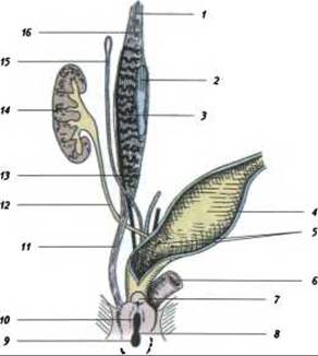
Fig. 337. Urogenital system at an early stage, prior to sex differentiation (after Hertwig):
1 - diaphragmatic ligament of the mesonephros; 2 - gonad; 3 - mesonephric tubule; 4 - urinary bladder; 5 - ureteral orifices; 6 - rectum; 7 - genital tubercle; 8 - urorectal fold; 9 - anal part of the cloacal opening; 10 - urogenital part of the cloacal opening;
11 - inguinal ligament of the mesonephros; 12 - metanephric duct; 13 - mesonephric duct; 14 - kidney; 15 - Müllerian duct; 16 - degenerating mesonephric tubules
The Müllerian ducts regress almost entirely. However, in adults, certain vestiges and the prostatic utricle persist as their remnants; the latter arises from the fused caudal sections of the ducts and appears as a small tube located near the Prostate Gland between the openings of the two ejaculatory ducts, as well as the extremity of the Müllerian duct (the so-called Appendix testis), which forms from the cranial sections of the ducts and is a vesicular structure located on the epididymis.
The epithelial component of the seminal passages is formed from the material of the Wolffian ducts. The connective and Smooth Muscle Tissues of these organs develop from the mesenchyme.
As embryonic development progresses, the testis and its associated remnants migrate caudally from the Abdominal cavity (Fig. 338). Initially, they are located at the level of the lumbar spine, at the third month—in the greater pelvis, and at the fifth and sixth months they lie against the inner aspect of the anterior abdominal wall near the inguinal ring. The migration of the testes is associated with The formation of the inguinal ligament, gubernaculum. It appears As a result of the enlarging primary kidney (mesonephros) bulging into the coelom and displacing the Peritoneum covering the organ. The peritoneum cranial and caudal to the mesonephros gathers into folds. The upper diaphragmatic fold extends upward toward the Diaphragm, whereas the lower fold, acquiring a fibrous structure, extends toward the lower part of the coelom and penetrates the Skin of the inguinal region.

Fig. 338. Scheme of male urogenital system development (after Hertwig and B. Patten):
1 - diaphragmatic ligament; 2 - epididymis; 3 - testis (before descent); 4 - urinary bladder; 5 - ureteral orifice; 6 - prostatic sinus; 7 - prostate; 8 - Bulbourethral gland; 9 - Urethra; 10 - Scrotum; 11 - testis after descent; 12 - opening of the ejaculatory duct; 13 - inguinal ligament; 14 - mesonephric duct; 15 - Müllerian duct; 16 - kidney
During the involution of the mesonephros and the growth of the testes, the peritoneum covers the latter, and the inguinal ligament becomes the gubernaculum testis. In the inguinal region on each side of the body, at the site of attachment of the caudal ends of the gubernaculum testis, coelomic wall outpouchings (vaginal processes) are formed, which serve as the primordia of the scrotum. As a result of the contraction of the gubernaculum and the rapid growth of the pelvic and inguinal regions of the embryo, the length of the inguinal fold remains insignificant. The testes shift caudally and descend into the scrotum. In the process, the testes, covered by peritoneum, enter the scrotum. This layer of peritoneum is visceral, whereas the parietal layer lies anterior to it. The canal formed as a result of the Displacement of the serous membrane into the scrotum becomes obliterated through the fusion of its margins, leaving the testes enclosed within a sac formed by the portion of the serous membrane that separates from the peritoneum. Typically, by the eighth month of development, the testes are located in the scrotum.
The aforementioned process of testicular descent into the scrotum explains the structure of its tunics, which originate from the abdominal wall. The inner tunic of the scrotum, corresponding to the peritoneum, is designated as the tunica vaginalis propria. It comprises both the visceral layer (covering the testis) and the parietal layer. External to the tunica vaginalis propria lies the tunica vaginalis communis, which represents a thin layer of Muscles AND FASCIAE of the abdominal wall. The outermost layer of the scrotum, the scrotum itself, is represented by skin.
The prostate gland [glandula prostatica] develops from the urethral epithelium, from which cords grow into the surrounding connective tissue. These cords form groups of glands corresponding to the lobes of the gland. During the last three months of embryonic development, smooth muscle-containing septa form within the gland.
The Bulbourethral Glands [glandulae bulbo-urethrales] arise from epithelial outgrowths of the proximal segment of the cavernous urethra. During the Formation of the corpus cavernosum urethrae, the ducts of these glands become incorporated into its bulbous part.
During the Development of the Male Reproductive System, the endocrine function of the testes is initiated earlier than the generative function (production of germ cells). The male sex hormone (testosterone) begins to be produced at 8–10 weeks of embryogenesis. However, even before testosterone production commences, Peptide Hormones are produced in the embryonic testes.
First, already at the indifferent stage of development, a hormone is secreted that causes the regression of the paramesonephric (Müllerian) duct and initiates the development of the reproductive system primordium along the male pathway. Under METABOLISM/18.html">The Influence of testosterone, the mesonephric ducts transform, as noted, into the deferent ducts, and the prostate gland and Seminal Vesicles are formed.
From approximately the middle of Pregnancy, the rete testis epithelial cells begin to produce a second hormone, inhibin, which suppresses the proliferation of gonocytes within the seminiferous tubules and inhibits the secretion of follicle-stimulating hormone (FSH) by the anterior Pituitary Gland.
Transformation under female sex development
Under female sex development, the mesonephric (Wolffian) ducts undergo regression. The gonad primordia differentiate into ovaries (Fig. 339). From the anterior margin of the mesonephros—which regresses and gives rise to the epididymis in male development—the epoophoron (paraovarium) develops in female embryos. It is located within the broad ligament of the Uterus between the Ovary and the paramesonephric (Mullerian) duct. The posterior segment of the mesonephros, which forms the paradidymis and ductuli aberrantes in males, also regresses in females, leaving behind the paraoophoron situated medially to the epoophoron.
Similar to the testes, the ovaries descend from the abdominal cavity into the pelvis. In newborns, they lie near the pelvic inlet. The descent of the testes is presumably facilitated by the aforementioned inguinal ligament of the mesonephros. Its lower segment forms the round ligament of the uterus, lig. teres uteri. It extends to the inguinal region and, passing through the abdominal wall, terminates in the skin of the Labia Majora. The subsequent migration of the ovaries into the lesser pelvis is presumably associated with their attachment to the uterus via the ovarian ligament, lig. ovarii.
The fallopian tubes with their infundibula develop from the portions of the paramesonephric (Mullerian) ducts located along the margin of the broad ligament of the uterus (Fig. 339). Part of the upper segment of the Mullerian duct regresses, while its remaining part transforms into the vesicular appendix (hydatid of Morgagni). Furthermore, the uterus and Vagina are formed by the fusion of the portions of the Mullerian duct incorporated into the genital cord, which occurs as early as the second month of embryonic development.
Last update: 08/08/2026
Editorial and Educational Adaptation: This material has been compiled based on the primary/original source text. The project team performed an editorial review, corrected technical inaccuracies, structured sections, and adapted the content for an educational format.
What was processed:
- elimination of formatting defects (OCR errors, structural breaks, corrupted characters);
- editorial organization of content;
- standardization of terminology in accordance with academic sources;
- verification of factual statements against the original source text.
All mentions of the author, publication year, and origin of the primary text have been preserved in accordance with the source.