Human Anatomy Part 1 - K. A. Dyubenko, A. K. Kolomiytsev, Yu. B. Chaykovsky 2002
Special Part
Reproductive Systems, systemata genitalia — Female Reproductive System, systema genitale femininum
External Female Genital Organs, organa genitalia feminina externa — Development of the Urinary and Reproductive Systems
The primary source for The Development of the URINARY AND REPRODUCTIVE systems is the intermediate mesoderm. The only exceptions are certain sections of the Urinary Tract, which originate from the caudal part of the hindgut (cloaca). The fact that A number of Organs belonging to both the excretory and reproductive systems develop from common primordia is a key factor in grouping them into a single unified system.
During the Prenatal period of ontogeny in mammals and humans, three successive generations of Kidney systems develop (pronephros, mesonephros, and metanephros), recapitulating the phylogenetic evolution of these organs in vertebrates. The pronephros (HEAD kidney) represents the most primitive type of excretory organ. It Functions in certain lower fish, as well as in the larvae of fish and amphibians. In higher fish and amphibians, the pronephros degenerates, and excretory function is taken over by the mesonephros (middle kidney), located more caudally. In birds and mammals, the mesonephros is subsequently replaced by the metanephros, or definitive kidney. All of these kidney types are paired organs situated in a retroperitoneal position near the dorsolateral body wall.
The pronephros originates from the cranially positioned Regions of the nephrotome—the intermediate mesoderm—which forms tubule-like structures. Its ventral opening, which communicates with the coelomic cavity, is termed the nephrostome. It is surrounded by a ciliated funnel-shaped expansion. The dorsal ends of several tubules fuse to form the pronephric duct, which grows caudally beneath the ectoderm and eventually opens into the cloaca. Initially, this duct appears as a solid cord of Cells that subsequently canalizes. As the pronephric duct elongates, additional tubules, also derived from the intermediate mesoderm, empty into it. In humans, the pronephros is a rudimentary organ with a transient existence. Pronephric tubules appear at the end of the third week of development and begin to degenerate by the end of the fourth week. Thus, The formation of the pronephros serves as an instance of recapitulation, reflecting a specific stage in the phylogeny of excretory organs.
Consequently, to fully understand the morphofunctional Organization OF THE pronephros, it is helpful to examine its Structure in animals where it functions permanently or temporarily as an excretory organ. For instance, in amphibian larvae, a ciliated funnel develops around the opening (nephrostome) located at the ventral ends of the tubules that open into the coelom. As noted above, the dorsal end of the tubule connects with the pronephric duct. The tubule bends in an S-shape, with its convex side directed toward the dorsal mesoderm.
Vascular branches extend from the aorta toward the medial wall of the coelom, forming glomeruli that invaginate the coelomic wall near the nephrostomes. Thus, during pronephric development, the vascular glomerulus forms within the wall of the secondary body cavity rather than within the wall of the urinary tubule. As a result, Urine Formation depends directly on the coelom. Filtration occurs into the coelomic cavity, from which fluid enters the tubules and subsequently the pronephric duct through the action of the ciliated epithelium lining the nephrostomal funnels. Therefore, the pronephros lacks a direct connection between the tubules and the vascular glomeruli. A diagram illustrating the MICROSTRUCTURE OF THE pronephros in cross and longitudinal section is shown in Fig. 336.
The primary developmental features that determine the Structure and function of the pronephros are:
- development derived from the intermediate mesoderm of the head segments;
- clearly defined metameric features;
- the absence of a direct vascular connection between the tubules and Blood Vessels.
Naturally, due to the absence of true nephrons in the pronephros, its concentrating ability is negligible. For animals that live permanently in Water, this is less critical than for animals that evolved to transition from water to land, since aquatic organisms rely heavily on the Skin, gills, and mucous membranes—and, in marine fish, other specialized organs—for excretion. In most fish and amphibians, the mesonephros becomes the primary excretory organ. In the embryos of higher vertebrates, the pronephros functions during early Embryogenesis before undergoing regression, with the exception of certain ducts that are incorporated into the Male Reproductive System.
The mesonephric tubules develop from regions of the intermediate mesoderm located caudal to the pronephros. At the stage of tubule formation, the mesoderm is unsegmented and appears as a continuous cord connecting the dorsal mesoderm (somites) to the ventral mesoderm. Consequently, this unsegmented intermediate mesoderm is referred to as the nephrogenic cord. As with the pronephros, these tubules lose their connection with the dorsal mesoderm and instead open into the pronephric duct, which thus becomes the duct of the primitive kidney (the mesonephric or Wolffian duct). In avian embryos, some of the anterior mesonephric tubules open into the coelom, forming rudimentary nephrostomes. At the same time, new structural features emerge in the primitive kidney, driven by the establishment of a direct connection between the tubules and The Vascular System. Outgrowths extend from the lateral walls of the tubules toward the aorta, terminating in expanded double-walled cup structures known as Bowman's capsules (or Malpighian capsules). A tuft of capillaries—the glomerulus—invaginates into the capsule, together forming the renal corpuscle. In higher vertebrates, the tubules of the greater part of the mesonephros lack nephrostomes. The presence of coelom-connected tubules within the mesonephros illustrates the gradual evolutionary transition from a simpler excretory organ (the pronephros) to a more complex one (the mesonephros). It is likely that this evolutionarily older mode of excreting fluid via nephrostomes persists until true renal tubules and nephrons are fully formed.
Class="center">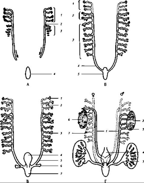
Fig. 336. Schematic diagram showing the relationship between the pronephros, mesonephros, and metanephros, and the chronological sequence of their formation during embryonic development (after B. Patten): A: 1 - pronephric tubules; 2 - pronephric duct; 3 - developing tubules; 4 - cloaca; B: 1 - pronephric tubules (degenerating); 2 - mesonephric tubules with openings; 3 - tubules without openings; 4 - mesonephric duct; 5 - cloaca; C: 1 - pronephric tubules (degenerating); 2 - mesonephric tubules with openings; 3 - mesonephric tubules without openings; 4 - allantois; 5 - mesonephric duct; 6 - ureteric bud (metanephric duct); D: 1 - mesonephric ducts; 2 — Testes; 3 - mesonephric tubules; 4 - allantois; 5 - Ureter (metanephric duct); 6 - Ovary; 7 - degenerating mesonephric tubules and duct
Nephrostomes do not form in the mesonephric tubules of mammalian embryos, and urine production relies entirely on Glomerular Filtration. The mesonephric tubules elongate and take on an S-shape. Outgrowths from these tubules form new tubular segments complete with renal corpuscles, causing the overall volume of the primitive kidney to increase substantially. The considerable length of the mesonephric tubules and their connection with blood capillaries enhance the concentrating capacity of the primitive kidney compared to the pronephros, as ions and various other substances are reabsorbed from the blood filtrate produced in the renal corpuscles as it passes through the tubules. Nevertheless, the primitive kidney lacks or has a poorly developed medulla, resulting in a lower capacity to concentrate urine compared to the metanephros. This limitation is related to the lesser Development of the countercurrent multiplier system, which enables water conservation within the body. Such a mechanism is not critical for an embryo developing in an aquatic environment or for permanently aquatic animals such as fish and amphibians.
The mesonephros reaches its maximum development by the end of the second month of embryonic life. The developmental features that determine its Structure and Functional capabilities are:
- the participation of A large number of tubules in kidney formation, which originate from the intermediate mesoderm spanning from the 14th to the 26th somites (second sacral segment);
- the establishment of a direct vascular connection between the tubules and blood vessels, leading to the formation of renal corpuscles and nephrons;
- the retention of metameric characteristics during early developmental stages, reflected not only in the segmental arrangement of the tubules but also in the blood supply, as the mesonephros receives numerous arterial branches from the aorta;
- the formation of a poorly developed medulla, which, as noted, accounts for the lower concentrating ability of the mesonephros relative to the metanephros.
Development of the metanephros (definitive kidney). The Emergence of the definitive kidney begins at the end of the fourth week of embryogenesis with the formation of an outgrowth from the primitive kidney duct (the Wolffian duct)—known as the metanephric diverticulum (ureteric bud). This diverticulum originates just cranial to the opening of the mesonephric duct into the cloaca and extends its expanded distal end into the intermediate mesoderm in the region between the 20th and 28th somites. This unsegmented region of the intermediate mesoderm is called the metanephric blastema. The parenchyma of the definitive kidney derives from two distinct sources. The renal tubules and Bowman's capsules, which form part of the renal cortex, originate from the metanephric blastema, whereas the medulla—consisting primarily of collecting ducts and papillary tubules—develops from the metanephric diverticulum. The epithelium of the minor calyces, renal pelvis, and Ureters also develops from the latter. The metanephric diverticulum grows dorsally and then cranially, and its blind end expands (see Fig. 336 C, D); this expansion gives rise to the epithelium of the renal pelvis, calyces, papillary ducts, and collecting tubules. The narrower, proximal stalk of the diverticulum, connecting to the cloaca, forms the epithelium of the ureter.
The epithelial component of the nephrons is derived from the metanephric blastema, while the Connective Tissue of the Kidneys and the urinary ducts originates from mesenchyme.
A distinctive feature of metanephric diverticulum development is its repeated dichotomous branching (up to 14–15 times) after invading the metanephric blastema. This extensive branching results in the formation of a well-developed renal medulla. Each medullary pyramid corresponds to a renal lobe associated with a minor calyx.
The terminal ends of the branching ducts stimulate the formation of nephron tubules and Bowman's capsules within the blastema. The capsules form when a vascular glomerulus pushes into the blind-ended terminal portion of a tubule derived from the metanephric blastema, invaginating its wall to create the capsule and renal corpuscle. Driven by intensive growth, the nephron tubules become convoluted and form loops of Henle that penetrate the peripheral regions of the medulla.
As nephron formation continues, the volume of the renal cortex increases. However, by the time of birth, undifferentiated areas of the metanephric blastema still persist within the cortex, serving as the source for new generations of nephrons that form postnatally.
As noted, the metanephros forms opposite the 26th to 28th somites (fourth to fifth lumbar segments). By the time of birth, the kidney shifts cranially to the level of the first lumbar or twelfth thoracic vertebra, occupying a retroperitoneal position dorsal to the mesonephros. This upward displacement is primarily driven by the accelerated growth of the embryonic body wall cranial to the kidneys' initial Location. Concurrently, the kidneys undergo a 90° rotation, shifting their originally dorsal convex borders into a lateral orientation.
Initially, the metanephros consists of separate lobes separated by sulci. In humans, this lobulation eventually disappears.
Thus, the key developmental Features of the metanephros that determine the Main principles of its morphofunctional organization include:
- the absence of metamerism signs (the definitive kidney typically receives a single artery);
- the Formation of the organ parenchyma from two sources: the cortex and medullary rays originating from the metanephrogenic blastema, and the medulla originating from the metanephric diverticulum. The presence of a well-developed medulla in the metanephros, which facilitates the formation of long loops of Henle, endows it (as observed in birds and mammals) with a very high concentration capacity.
Formation of the Urinary Bladder. As noted in the section «EMBRYOGENESIS OF THE Face, Digestive, and Respiratory Systems», an outpocketing called the allantois extends from the anterior wall of the hindgut (cloaca). Mesenchyme located in the space between the allantois and the cloaca forms a fold known as the urorectal septum. Expanding in size and reaching the cloacal membrane, the septum divides the cloaca into a posterior part, the rectum, and an anterior part, the urogenital sinus. Following this division, due to the thickening of the urogenital sinus, the allantois becomes its direct continuation. The proximal part of the allantois expands to form the urinary bladder. Initially, the orifices of the metanephric and mesonephric ducts open into the urogenital sinus close to each other. Later, presumably due to the cranial migration of the kidney, the openings of the metanephric ducts shift laterally and cranially relative to the mesonephric duct orifices, entering the region of the urogenital sinus that will subsequently contribute to the urinary bladder. Meanwhile, the mesonephric ducts open into the narrowed portion of the urogenital sinus, which gives rise to the Urethra.
The segment of the allantois extending from the urinary bladder to the umbilicus forms the urachus, which normally obliterates by the end of prenatal development. After birth, it transforms into the median umbilical ligament (while the medial umbilical ligaments are formed from the obliterated portions of the umbilical Arteries).
The site of contact between the urorectal septum and the cloacal membrane serves as the primordium of the Perineum. Concurrently with the division of the cloaca into the rectum and the urogenital sinus, the cloacal membrane ruptures, opening both PARTS OF THE cloaca to the exterior via the anus and the urogenital aperture, respectively.
Last update: 08/08/2026
Editorial and Educational Adaptation: This material has been compiled based on the primary/original source text. The project team performed an editorial review, corrected technical inaccuracies, structured sections, and adapted the content for an educational format.
What was processed:
- elimination of formatting defects (OCR errors, structural breaks, corrupted characters);
- editorial organization of content;
- standardization of terminology in accordance with academic sources;
- verification of factual statements against the original source text.
All mentions of the author, publication year, and origin of the primary text have been preserved in accordance with the source.