PLANT BIOPHYSICS - Y. I. Posudin - 2004
II. TRANSPORT PROCESSES IN THE SOIL-PLANT-ATMOSPHERE SYSTEM
12. EFFECT OF ENVIRONMENTAL FACTORS ON PLANTS
12.2. PHOTOBIOLOGICAL REACTIONS OF PLANTS
Class="center">12.2.1. Classification of plant photobiological reactions
For proper development, plants require sunlight, carbon dioxide, Water, and nutrients. These drive plant development—defined as the qualitative Changes in the Structure and Functional activity of the plant and its parts (Cells, Tissues, and Organs) throughout its life cycle—as well as plant growth, which is the irreversible increase in the size of the plant or its organs resulting from The formation of new cells, organs, or their individual elements. Light is the most critical environmental factor for numerous Plant Growth and developmental processes. Sunlight exposure triggers photobiological reactions, which are physical or chemical alterations within plant systems. All photobiological reactions can be divided into four main groups [Konev and Volotovsky, 1979]:
1. Energetic reactions: Processes in which light energy is transformed into chemical energy through the synthesis of new organic molecules. Photosynthesis is a prime example of such reactions.
2. Informational reactions: Processes in which light acts as a regulatory signal that triggers the formation of photoproducts via specialized mechanisms and provides environmental information. Photomovement, photomorphogenesis, phototropism, and Photoperiodism fall into this category.
3. Biosynthetic reactions: Processes in which a complex chain of sequential steps in the synthesis of organic molecules incorporates specific photochemical stages that occur exclusively under METABOLISM/18.html">The Influence of light. These include chlorophyll Cell/9.html">Biosynthesis and the induced synthesis of pigments and Vitamins.
4. Destructive-modifying reactions: Processes associated with light-induced damage to biological substrate molecules, leading to lethal or mutational consequences. This type of reaction includes photosensitivity, photoreactivation, and the response of biological systems to ultraviolet radiation.
Let us examine several photobiological reactions of major importance to higher and lower plants.
12.2.2. Photosynthesis
The primary functional units driving photosynthetic activity in plants and Algae are Photosystems PSI and PSII, which contain approximately 250–400 pigment molecules. While all of these pigments can absorb light quanta, only a single chlorophyll molecule within a given photosystem is capable of converting the absorbed energy into photochemical reactions. This particular chlorophyll molecule is designated as the reaction center of the photosystem, whereas the others function as antenna pigments. Following the absorption of a light quantum by the antenna pigments, the captured Energy is transferred to the photosystem reaction centers. This process excites the chlorophyll molecule P and elevates its electrons to a higher energy level. In its excited state, the P*680 molecule transfers electrons to an acceptor—pheophytin (Ph)—followed by the primary quinone acceptor QA and the secondary quinone acceptor QB. Subsequently, the electrons are sequentially passed to a pool of plastoquinone (PQ) molecules—which shuttle electrons and protons across the lipid phase of the membrane—an iron-sulfur protein FeSR, cytochrome $f$, and plastocyanin (PC). The electrons then travel along the Electron Transport Chain to PSI, where light energy transfers them from the P700 chlorophyll molecule to the P430 electron acceptor, after which they undergo further transfer via ferredoxin (Fd) to the coenzyme NADP (Fig. 12.5).
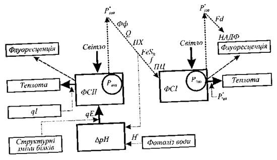
Fig. 12.5. Diagram illustrating energy conversion processes into fluorescence and heat during non-Cyclic electron transport.
Alternatively, the absorbed energy may be released as heat or dissipated through a radiative process such as fluorescence. At normal temperatures, chlorophyll fluorescence originates from the PSII antenna pigments; THE CONTRIBUTION OF PSI to chlorophyll fluorescence occurs exclusively in the far-red region of the spectrum (740 nm). Variable fluorescence primarily stems from PSII, and excitation transfer to PSI can be interpreted as an additional competing pathway of PSII de-excitation. During the operation of the photosynthetic apparatus, chlorophyll a fluorescence emission accounts for 2–5% of the absorbed energy and varies depending on chloroplast type, plant age, physiological state, stress conditions, and the intensity and wavelength of the exciting radiation.
Electron flow along The electron transport chain is accompanied by a decrease (quenching) in chlorophyll fluorescence. This reduction occurs due to The oxidation of the acceptor, which consists of a complex of pheophytin and Quinones. When the acceptor is oxidized via electron transfer to NADP and ultimately to CO2, fluorescence diminishes. This phenomenon is termed "photochemical quenching" and is characterized by the photochemical quenching coefficient qP. Concurrently, other quenching mechanisms of a non-chemical nature—known as "non-photochemical quenching" and characterized by the non-photochemical quenching coefficient qN—are also at play. The primary quenching processes include energy-dependent quenching (associated with the proton gradient induced across the thylakoid membrane) and photoinhibition-induced quenching caused by excessive irradiation. Consequently, fluorescence is a complementary process to photochemical and thermal pathways: fluorescence yield increases as energy losses to photochemical reactions or heat decrease.
12.2.3. Photomovement in algae
The movement or change in movement of organisms induced by light is termed photomovement. Photomovement is the outcome of movement photoregulation—a set of elementary processes triggered by a light stimulus, namely: photoreception, sensory Transduction of the light stimulus into a physiological signal that directs the motor apparatus, and the execution of the Organism's photoorientation. Light is a vital environmental factor utilized by motile organisms (primarily algae) via photomovement to locate optimal living conditions.
Photoreception. Let us examine the principal mechanisms of algal photoreception [Posudin, 1989; Masyuk and Posudin, 1991].
Periodic illumination and shading of the photoreceptor by the stigma. As The Cell rotates around its longitudinal axis, amplitude modulation of the light striking the photoreceptor occurs. The magnitude of this modulation depends on the direction of cell movement relative to the propagation vector of the stimulating light. A specialized organelle, the stigma (eyespot), acts as the modulator. This modulation mechanism is characteristic of representatives of Euglenophyceae.
Dichroic mechanism. This mechanism is based on the anisotropy of Light absorption by dipolar photoreceptor molecules oriented in a specific manner relative to the cell's longitudinal axis. Maximum light absorption takes place when the cell moves parallel to the direction of light propagation. This mechanism has been identified in the euglenoid alga Euglena gracilis, where a combination of modulation and dichroic mechanisms cannot be ruled out.
Waveguide mechanism. This involves the propagation of side light—perpendicular to the cell's longitudinal axis—through paired thylakoids oriented perpendicular to the plane of the pigmented stigma layer and the cell's longitudinal axis. These thylakoids, possessing a high refractive index relative to the intervening spaces, act as a specialized light guide for the propagating light. Such a mechanism is found in cryptophyte algae such as Chroomonas hansenii and Cryptomonas species.
Ocelloid mechanism. Photoreception occurs via a specialized organelle known as an ocelloid, which Functions as a distinct focusing system. Light striking the ocelloid is focused by a cuticular lens onto the laminar layer of a retinoid body. A similar mechanism is characteristic of dinoflagellates belonging to the family Warnowiaceae.
Interference mechanism. The stigma, composed of multiple pigmented and unpigmented layers and potentially located beneath the photoreceptor, acts as a multilayer quarter-wave plate. Due to light reflection from each layer, optical interference occurs; if the thickness of the layers and the intervals between them equal one-quarter of the light wavelength, the light reaching the photoreceptor is amplified. This mechanism is observed in representatives of Chlorophyceae, Prasinophyceae, and certain Dinophyceae species.
Diffraction mechanism. In many green algae, the stigma contains only a single layer of pigmented globules. It is suggested that the pigmented globules of the stigma form a periodic structure that functions as a diffraction grating. When light interacts with such a structure, light diffraction occurs, accompanied by the formation of diffraction maxima. The intensity and spatial position of these maxima depend on the geometry of the periodic structure, the angle of incidence of the light, and the wavelength. Depending on whether a diffraction magnitude coincides with the Location OF THE receptor, the magnitude of the light signal hitting the photoreceptor changes.
The structure of some algal photoreceptor systems is illustrated in Fig. 12.6.
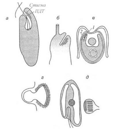
Fig. 12.6. Structure of various algal photoreceptor systems: a - stigma and paraxial body of the euglenophyte Euglena gracilis; b - paraflagellar Swelling in yellow-green algae; c - stigma (1) and putative receptor (2) of green algae; d - cryptophyte receptor; e - ocelloid (family Warnowiaceae).
Sensory transduction. Following the absorption of a light quantum by the photoreceptor, a sequence of events takes place that leads to The conversion of the light stimulus into a physiological signal controlling the algal motor apparatus—a process known as sensory transduction. There are numerous methodological and experimental approaches aimed at elucidating the key stages of sensory transduction, such as the application of specific metabolic agents (chemical element ions, calcium channel blockers, ionophores, ionotropic drugs, etc.). Unfortunately, to this day, The pathway of the light signal from the photoreceptor to the motor apparatus remains a sort of "black box," as many aspects of this problem are yet to be resolved.
12.2.4. Chloroplast Photomovement
Light-induced movement of Chloroplasts within a cell occurs via two mechanisms. First, chloroplasts can participate in general cytoplasmic streaming (translational, rotational, or circulatory); here, light affecting a portion of the Cytoplasm alters its streaming velocity, while the spatial arrangement of the chloroplasts relative to one another remains unchanged. This light-dependent chloroplast response is called photokinesis. Second, chloroplasts are capable of reorienting themselves within the cell; their resulting distribution depends on the pattern of light absorption by the cell, which is determined by the direction of light propagation. This reaction is known as photorotaxis (or chloroplast orientation). While the stimulus in the first case is light intensity, In the second case it is the direction of light propagation.
Photokinesis is exhibited by the chloroplasts of Elodea and Vallisneria. Changes in the cytoplasmic streaming velocity in epidermal cells of Elodea and in mesophyll and epidermal cells of Vallisneria under the influence of light induce chloroplast displacement. According to one hypothesis, light controls the activation of Actin-like microfilaments and changes in the ATP level. Blue light-induced increases in Oxidative Phosphorylation mediated by flavins are considered responsible for the altered passive mobility of the cytoplasm at low streaming speeds. At medium speeds, competition from photosynthesis begins; at high speeds, when photosynthesis reaches saturation, there is an excess of ATP due to cyclic Photophosphorylation, which, together with light-induced ATP oxidation, leads to Actomyosin activation. This hypothesis is supported by experiments using uncouplers or inhibitors of photosynthetic electron transport.
Regarding chloroplast orientation, several patterns can be distinguished: under lateral low-intensity illumination, chloroplasts move to the cell walls and position themselves perpendicular to the direction of light propagation (epistrophe); with increasing light intensity, chloroplasts orient themselves along the lateral cell walls parallel to the direction of light propagation (parastrophe). In the dark, chloroplasts are distributed randomly (apostrophe). These types of chloroplast orientation are schematically shown in Fig. 12.7. The orientation pattern is influenced by light scattering and absorption within the cell, which generate light gradients, as well as "lens" effects that lead to light focusing. One of the main (and far from simple) aspects of chloroplast photomovement is determining the structure, pigment composition, and localization of the photoreceptor. Some organisms (Lemna, Vallisneria, Vaucheria, Mesotaenium, Selaginella, Mougeotia, Hormidium, Spirodela) exhibit a chloroplast response to blue light, for which Flavoproteins may serve as receptors. Chloroplast movement upon irradiation with intense red light can be observed in Selaginella and Vallisneria, and under low-intensity red light in Hormidium. Red light reception is carried out by Photosynthetic Pigments, as evidenced by experiments with compounds affecting photosynthesis. A red-light response is demonstrated by the chloroplasts of Mesotaenium and Mougeotia; in this case, the photoreceptor pigment is Phytochrome. It should also be noted that photoreceptors exhibit diverse locations—they can reside not only in chloroplasts but also in the cytoplasm or The Plasma Membrane.
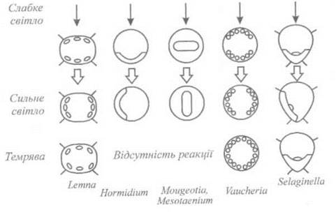
Fig. 12.7. Types of chloroplast photoorientation.
12.2.5. Photomorphogenesis
This category of photobiological reactions encompasses all light-controlled processes associated with Plant GROWTH AND DEVELOPMENT, where the plant's response is not directed relative to the light source, and the light stimulus lacks spatial directionality and periodicity.
Photomorphogenic reactions include seed germination, stem elongation, leaf formation, chloroplast development, and others.
Most plant photomorphogenic reactions proceed through the action of Three types of photoreceptors: phytochrome, and cryptochrome—the receptor for blue and ultraviolet UV-A (320-400 nm) radiation. Phytochrome consists of a protein moiety and a light-absorbing chromophore, a linear tetrapyrrole (phytochromobilin). Phytochrome can exist in two interconvertible forms: Pr (the physiologically inactive red-absorbing form) and Pfr (the physiologically active far-red-absorbing form). When a Pr molecule absorbs a red photon with a wavelength of 660 nm, it converts into Pfr; conversely, when a Pfr molecule absorbs a far-red photon at 730 nm, it converts back into Pr. Such light-driven transitions are called photoconversions. The involvement of phytochrome in morphogenetic processes is confirmed by Experiments on the reversible stimulation and inhibition of morphogenesis by red (660 nm) and far-red (730 nm) light. Phytochrome is localized in plant membranes; its primary MECHANISM OF ACTION is considered to be The regulation of Membrane Functions through conformational changes that affect membrane permeability, active Ion transport across the membrane, and The activity of membrane-bound phytohormones and Proteins. Cryptochrome is responsible for Light absorption in the blue (400-500 nm) and ultraviolet UV-A Regions of the spectrum; its functions can be fulfilled by flavins or carotenoids. Cryptochrome consists of a protein bound to two chromophores. Its action spectrum spans the 390-480 nm region with a maximum at 450 nm. Cryptochrome is responsible for the inhibition of stem elongation, leaf development, photoperiodic flowering, etc.
12.2.6. Phototropism
The light-induced movement of a part of a fixed plant organism is called phototropism. This photobiological response represents a plant's reaction to a light gradient, which can be formed due to the absorption or scattering of light passing through the plant organ. Positive phototropism (movement toward the light source) and negative phototropism (movement away from it) are distinguished. SHOOT tips typically exhibit positive phototropism, whereas roots show negative phototropism. If, for instance, an oat coleoptile is illuminated with low-intensity light from one side, or with varying intensities from different sides, the organ bends toward the brighter light. The typical action spectrum of phototropism (Fig. 12.8) features maxima in the visible region at 420 nm, 450 nm, and 475 nm, along with a maximum at 370 nm in the ultraviolet region. Thus, the action spectrum indicates that only blue light and a portion of ultraviolet radiation induce bending.
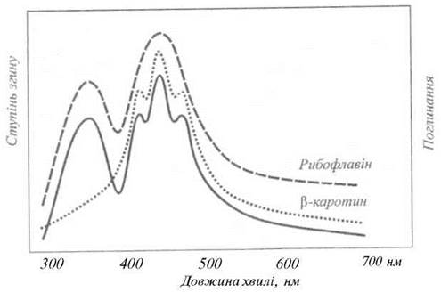
Fig. 12.8. Typical action spectrum of phototropism.
The probable chromophores responsible for phototropic responses are carotenoids and flavins (or flavoproteins), as evidenced by the close match between the action spectrum of phototropism and the absorption spectra of these pigments.
It should be noted that the exact nature of these chromophores is still far from being definitively resolved. Phototropic bending of a plant organ is associated with the asymmetric distribution within that organ of auxin—a physiologically active substance produced in plant cells that regulates growth processes. Light induces organ bending by affecting the lateral distribution of auxin. Differences in auxin concentration, in turn, cause unequal growth rates on the Two Sides of the organ, resulting in curvature.
The Mechanism of auxin action is related to two factors [Hapston et al., 1983]: the extensibility of cell walls and the turgor pressure of the cell sap acting upon the wall. Auxin increases longitudinal cell expansion, which is facilitated by the helical arrangement of Cellulose microfibrils in The Cell wall and the ability of auxin to enhance wall extensibility. This auxin-induced increase in plastic extensibility occurs only in living cells. According to one hypothesis, to allow the relative sliding of cellulose chains, cross-links between cellulose molecules must be severed; these functions are performed by a yet unidentified enzyme whose activity depends on the intracellular pH. When hydrogen ions are pumped into the cell wall under the influence of auxin present in the cell, the enzyme is activated and breaks the cross-links, freeing the cellulose chains to slide past one another. Such sliding is driven by the turgor pressure of the cell sap, leading to wall stretching and cell enlargement.
12.2.7. Photoperiodism
The rhythmic variations in various morphological, biochemical, and physiological Properties and functions of organisms in response to the alternation and duration of light and dark intervals are called photoperiodism.
Based on their photoperiodic responses, plants can be divided into three groups: short-day plants (flowering and fruiting occur when the day length is shortened to 8-12 hours); long-day plants (the same effect is achieved when the day length is extended to 16-20 hours); and day-neutral plants (in which changes in day length cause no noticeable alterations throughout their life cycle). Short-day plants include cotton, coffee, strawberry, millet, rice, soybean, tobacco, and sugarcane; long-day plants include rye, flax, lupine, oats, wheat, radish, sugar beet, spinach, and barley; Examples of day-neutral plants are pea, buckwheat, and sunflower.
Processes such as flowering, bud dormancy, leaf shedding, seed germination, bulb and tuber formation, and others depend on the photoperiod. The Nature of the photoperiodic response is related to the geographical Origin of the plant and has evolved over time. The lack of precise data on photoperiodic action spectra prevents any definitive Conclusions regarding the Nature of the photoreceptor responsible for the photoperiodic response. Among the existing hypotheses [Hrodzynskyi, 1972; Konev and Volotovsky, 1979], notable ones include the phytochrome hypothesis (which links the photobiological response to the phototransformations of phytochrome), the hormonal hypothesis (where the photobiological effect is caused by The production of a specific hormone, florigen), the endogenous rhythm hypothesis (based on the interaction of biological and astronomical clocks), and the inhibitor hypothesis (which suggests the formation of specific flowering inhibitors in the plant under unfavorable photoperiodic conditions). None of these hypotheses are yet capable of explaining the full complexity and diversity of plant photoperiodic responses.
12.2.8. Photosensitization
The phenomenon of photosensitization. In 1900, Oscar Raab studied the effects of light and acridine Dyes on the vital activity of Protozoa (Paramecium). He observed that neither light alone nor the dye had any effect on the infusoria, whereas the combined action of these two factors led to their death. The phenomenon of photosensitization refers to processes in which light energy absorbed by molecules containing chromophores (sensitizers) is transferred to other molecules that are incapable of absorbing light on their own.
This phenomenon is widespread in nature—it is known that certain wild plants contain potent chemical compounds; animals that graze on these plants become sensitive to light in unpigmented areas of the Skin, especially around the eyes, Mouth, ears, and hooves where the Hair coat is sparse. The main symptoms include itching, the formation of pea-sized blisters (which subsequently rupture), Conjunctivitis, stomatitis, fever, inflammation (even in the Brain), and agitation or depression. The animal begins to rub itself, introduces infection, and may die within 8–10 hours.
Basic mechanisms of photosensitization. We can distinguish the processes that characterize the interaction between light, the sensitizer, and the biological substrate, namely: light absorption by the sensitizer molecule and its transition to an excited state; intersystem crossing to the triplet state; energy transfer from the triplet state of the sensitizer to the triplet state of oxygen; and energy transfer from the triplet state of the sensitizer to a substrate molecule resulting in the formation of radicals. The reactive properties of singlet oxygen and chemical radicals cause specific alterations in the substrate molecules.
MAIN TYPES OF photosensitization. All diseases caused by photosensitization can be divided into three classes.
Primary photosensitivity. Chemical compounds that increase an animal's sensitivity to light (photosensitizers) include hypericin and fagopyrin. Hypericin is a red pigment found in the leaves, stems, and flowers of members of the family Guttiferae, specifically: Hypericum perforatum (St. John's wort), H. crispum, H. pulchrum, H. leucoptycodes, and H. maculatum. The resulting condition is collectively known as hypericism. Animals that consume plants containing hypericin become sensitive to light. Typically, sheep, horses, and cattle are affected by hypericin, although goats and pigs can also fall victim to its photosensitizing effects. Sheep that graze on Hypericum perforatum show no symptoms while kept in a dark enclosure. However, when exposed to light, the animals exhibit photosensitivity that persists for several weeks. Interestingly, many insects avoid plants containing hypericins. An exception is the beetle Chrysolina gemellata, which feeds on the nectar of Hypericum perforatum. However, it avoids other plants, thereby regulating hypericin production in certain areas.
Fagopyrism is caused by fagopyrin, a plant pigment related to hypericin that is present in members of the genus Fagopyrum (F. esculentum or Polygonum fagopyrum), commonly known as common buckwheat. Fagopyrin also exerts a photosensitizing effect on pigs, cattle, and chickens. Sheep, pigs, and horses that consume buckwheat, millet, or alsike clover in the sunshine may develop cutaneous exanthema, which is absent on pigmented areas of the body; sheep, horses, and cattle with white markings develop dermatitis, stomatitis, and Burns on unpigmented areas.
Photosensitivity associated with impaired pigment synthesis. Among diseases of this type, congenital porphyria stands out, caused by endogenous Porphyrins—uroporphyrin and coproporphyrin. The former is excreted in the urine, while the latter is deposited in the bones and Teeth (hence the alternative name of the disease, pink teeth). There is a hypothesis suggesting that these porphyrins arise from a disruption in the synthesis of protoporphyrin, which is a component of the Hemoglobin molecule. Overall, however, the exact origin of porphyria remains unknown.
Hepatogenic photosensitivity. Attention should be drawn to the photosensitizing properties of phylloerythrin, a porphyrin derived from chlorophyll through the action of Bacteria present in the intestines of various animals, by the removal of the phytyl group that otherwise prevents chlorophyll from crossing cell membranes. Under normal conditions, the herbivorous organism rids itself of phylloerythrin via Bile; however, when the animal consumes certain grasses that block the bile duct, phylloerythrin enters the Blood and skin. In this case, it begins to act as a sensitizer upon exposure of the animal to visible or ultraviolet light. The skin turns yellow and swells. Phylloerythrin can be isolated from the blood of sick animals and crystallized. When injected into animals that have been feeding on green forage, phylloerythrin induces symptoms of jaundice. The ingestion of certain green feeds thus renders the animal sensitive to light.
Among the plants that cause animal diseases are members of the genus Tribulus (hence the name of the disease, tribolism), specifically T. terrestris and T. dubius (family Zygophyllaceae); certain species of Lippia: L. rhennana, L. pretoriensis (family Verbenaceae); and grasses of the genus Panicum: P. laevifolium and P. coloratum (family Poaceae). In our latitudes, photosensitization is caused by clover (Trifolium L.) and tall hedge mustard (Sisymbrium altissimum L.).
Disease Prevention involves changing pastures and keeping livestock in shaded areas.
Last update: 07/08/2026
Editorial and Educational Adaptation: This material has been compiled based on the primary/original source text. The project team performed an editorial review, corrected technical inaccuracies, structured sections, and adapted the content for an educational format.
What was processed:
- elimination of formatting defects (OCR errors, structural breaks, corrupted characters);
- editorial organization of content;
- standardization of terminology in accordance with academic sources;
- verification of factual statements against the original source text.
All mentions of the author, publication year, and origin of the primary text have been preserved in accordance with the source.