Algology - Kostikov I.Yu. - 2009-2013
Chapter 24. Green Algae – Chlorophyta
24.2. The Chlorophytic Lineage
The chlorophytic Lineage comprises green algal classes whose monadoid stages are characterized by a cruciate Cytoskeleton and closed or semi-open mitosis. Cytokinesis occurs via a phycoplast or with the participation of a Cleavage furrow (with the exception of the order Trentepohliales). This lineage is represented by four classes: Chlorophyceae, Trebouxiophyceae, Ulvophyceae, and Siphonophyceae.
24.2.1. Class Chlorophyceae
This Class includes about 4,000 species of Algae exhibiting unicellular, multicellular, or coenocytic Organization. Chlorophyceans inhabit primarily fresh continental waters, and are less commonly found in terrestrial biotopes. Representatives of the class are diverse in their flagellar apparatus Structure, mitosis, Morphology, reproduction, and life cycles. Chlorophyceae represents a line of green algae in which cytokinesis involves a phycoplast, and the flagellar ROOT system is cruciate with basal bodies positioned opposite one another or shifted clockwise.
Features and CHARACTERISTICS OF THE Class
Biochemical Features
The composition of pigments and assimilation products is typical of green algae in general (chlorophylls a and b, all types of carotenes, xanthophylls of the lutein series, and starch as the primary assimilation product).
Cytological Features
Cell Coverings. The Cells of certain unicellular chlorophycean algae (e.g., Dunaliella) are bounded solely by a Plasmalemma. However, the vast majority of species possess true cell walls with a characteristic trilaminar structure. In monadoid forms, these walls are generally glycoproteinaceous, whereas in coccoid, multicellular, and coenocytic forms, they are cellulosic-pectic. Occasionally, an additional sporopollenin layer is located between the middle and inner wall layers, imparting mechanical strength and chemical resistance to The Cell coverings.
Submicroscopic scales are absent from both the cell surface and the flagella in chlorophycean algae.
Nuclear Apparatus and Features of Mitosis and Cytokinesis. Monadoid forms lack centrioles, their function being performed by the basal bodies of the flagella. This is achieved through a structural connection between the nuclear envelope and the basal bodies, mediated by two thin, smooth rhizoplasts composed of centrin (Fig. 24.15). Non-motile forms possess true centrioles (with the exception of oedogonialean algae).

Fig. 24.15. Connection between the flagellar apparatus and The Nucleus in the relaxed (A) and contracted (B) states of rhizoplasts in Chlamydomonas: 1 - nucleus, 2 - striated connecting fiber, 3 - basal bodies, 4 - rhizoplasts, 5 - rhizoplast branches, 6 - microtubular roots (schematized after Mattox, Stewart, 1984 and Salisbury, 1989).
A common feature of all chlorophyceans is the presence of closed mitosis with a collapsing spindle. Four variants of this mitotic type are known within the class: a) chlamydomonad, b) cylindrocapsalean, c) uronema-type, and d) oedogonialean (Fig. 24.16).

Fig. 24.16. Types of mitosis in chlorophycean algae. A - Ch-type (Chlamydomonas), B - Cy-type (Cylindrocapsa), C - Ur-type (Uronema). I - metaphase, II - anaphase, III - telophase. 1 - flagellar basal bodies, 2 - plasmalemma, 3 - nuclear envelope, 4 - Chromosomes, 5 - spindle, 6 - microtubular flagellar root, 7 - phycoplast, 8 - cleavage furrow, 9 - centrioles, 10 - Endoplasmic reticulum vesicles, 11 - Golgi apparatus, 12 - Golgi vesicles, 13 - cell plate (after Floyd, Stewart, Mattox, 1972; Triemer, Brown, 1974; Sluiman, 1985).
Chlamydomonad mitosis (Ch-mitosis) is characteristic of monadoid and hemimonadoid forms: the spindle is formed with the participation of basal bodies; in anaphase, daughter nuclei move apart; the phycoplast is formed from microtubules of the flagellar microtubular roots; in late anaphase and early telophase, the nuclei draw slightly closer together due to the contraction of interzonal spindle remnants; and cytokinesis is accomplished by a cleavage furrow via schizotomy or schizogony.
Cylindrocapsalean mitosis (Cy-mitosis) is characteristic of coccoid and siphonocladalean forms, as well as filamentous representatives whose transverse walls lack primary pores: the spindle is formed with the participation of centrioles; in anaphase, daughter nuclei move apart; centrioles are located at the proximal poles of the daughter nuclei, and a phycoplast is formed from spindle remnants. In telophase, no noticeable convergence of the nuclei is observed, and cytokinesis is effected by a cleavage furrow that grows through the fusion of endoplasmic reticulum vesicles coupled with the invagination of the plasmalemma. If, at the end of telophase, Golgi apparatus vesicles begin actively transporting future Cell wall material to the plasmalemma surface, then upon completion of division each daughter cell already possesses its own wall, meaning that schizotomy/schizogony takes place. Otherwise, the daughter cells remain naked, as observed during The formation of Gametes and naked zoospores.
Uronema-type mitosis (Ur-mitosis) is found in multicellular algae possessing primary pores in their transverse walls (with the exception of oedogonialean algae): the spindle is formed with the participation of centrioles; in anaphase, daughter nuclei move apart; centrioles are located at the distal poles of the daughter nuclei, and a phycoplast is formed from spindle remnants. In telophase, the nuclei visibly converge due to the contraction of interzonal spindle remnants. Golgi apparatus vesicles containing future wall material accumulate within the phycoplast. Subsequently, they partially fuse to form a cell plate containing pores with plasmodesmata, i.e., cytotomy takes place.
The oedogonialean type (Oe-mitosis) is present exclusively in the order Oedogoniales: centrioles are absent here, and the microtubule-organizing centers are presumably elements of The endoplasmic reticulum. In anaphase, daughter nuclei move apart, and a phycoplast is formed from spindle remnants. In telophase, the nuclei visibly converge due to the contraction of interzonal spindle remnants. Vesicles that pinch off from the Endoplasmic reticulum and contain future wall material accumulate in the phycoplast. Subsequently, these vesicles partially fuse and, similarly to the uronema type, form a cell plate with primary pores. Thus, in terms of cytokinesis, this type shares similarities with both the cylindrocapsalean type (fusion of vesicles originating from the endoplasmic reticulum rather than the Golgi apparatus) and the uronema type (formation of a cell plate, nuclear convergence in telophase). A specific feature of cytokinesis is the growth process of one of the daughter cells, associated with the stretching of an annular wall thickening and the formation of specialized cell caps (see below).
Flagellar Apparatus. Monadoid cells of chlorophycean algae possess 2–4 flagella or are stephanokont. In the latter case, numerous flagella are arranged in a circular crown at the anterior end of the cell. All flagella are isokont or nearly isokont, isomorphic, and isodynamic.
The transition zone contains only a stellate structure. Spiral ribbons, plates, and cylinders are absent in the transition zone. Basal bodies are relatively short. They either lie opposite each other (12-6 configuration) or are shifted clockwise (1-7 configuration).
The connection between basal bodies is maintained by a striated fiber. Two short striated fibrous structures, resembling the rhizoplasts of prasinophycean algae but connecting neither to the nuclear envelope nor to the plasmalemma, are also attached to the Base of the basal bodies. True rhizoplasts are smooth and attach to the upper part of the basal bodies. The Root System is cruciate and corresponds to the 4-2-4-2 formula (Fig. 24.17).

Fig. 24.17. Types of flagellar root systems in chlorophyte algae: A - with 12-6 basal body orientation; B - with 1-7 basal body orientation; C - stephanokont type. 1 - basal body, 2 - two-microtubule root, 3 - four-microtubule root, 4 - striated connecting fiber, 5 - fibrous ring, 6 - three-microtubule root (schematized after Kouwets, 1994, Markowitz, 1978).
A specialized root system derived from the cruciform type is the radial system found in stephanokont zoospores, spermatozoa, and androspores of edogoniate algae: a transversely striated fibrous ring is located at the anterior end of the cell, to which the flagellar basal bodies attach. Two microtubular roots originate from each basal body: one extends forward and consists of two microtubules, while the other extends backward and contains three microtubules. A transversely striated microfibrillar root lies adjacent to each microtubular root.
Types of morphological body structure
Within the class, algae are represented by all known types of morphological structure, with the exception of rhizopodial and tissue types. Taxa with monadoid, coccoid, and filamentous structural types are the most numerous.
Monadoid forms are predominantly unicellular, more rarely coenobial. Among hemimonadoid representatives, forms producing massive mucilaginous colonies predominate, and some possess reduced non-motile flagella (pseudocilia). Coccoid representatives are highly diverse, encompassing both unicellular and colonial forms, including coenobial ones.
Algae with a filamentous type of structure typically appear as simple filaments, sometimes featuring a tapered apical cell and an expanded basal cell. Heterotrichous forms generally resemble branched tufts differentiated into a thallus and an ascending system of filaments. Some are united into colonies by common mucus.
Siphonal forms are typically smaller than those in other classes and have a sac-like shape; siphonocladous species are exclusively filamentous.
Reproduction and life cycles
All types of Selection/8.html">Asexual and sexual reproduction are widely represented within the class. Eogamous species with a haplontic life cycle featuring zygotic reduction and no ALTERNATION OF GENERATIONS predominate. Agamous species exhibit both simple and rather complex cyclomorphoses.
Vegetative Reproduction by binary fission via schizogony is known in monadoid forms lacking a cell wall. Reproduction via fragmentation of filaments and colonies is widespread. The formation of akinetes and other resting stages, which serve a reproductive function alongside surviving unfavorable conditions, is characteristic of many species.
The primary mode of reproduction is sporulation involving the formation of zoospores and aplanospores, and more rarely hemizoospores. Zoospores may be either naked (Protosiphon type, p-type) or walled (Chlamydomonas type, ch-type). Typically, all species possessing zoospores are also capable of reproducing via aplanospores, whereas zoospores are never formed in many autosporic species.
Sexual reproduction in naked forms is represented by hologamy and all known variants of merogamy: isogamy, heterogamy, and oogamy.
Although life cycles are predominantly haplontic without alternation of generations, diplontic cycles (Chlorococcum diplobionticum), haplontic cycles with heteromorphic alternation of generations (Sphaeroplea), or cycles with pronounced Sexual Dimorphism (nannandrous species of Oedogonium) occur as exceptions.
System of the class
Molecular phylogenetic reconstructions, consistent with phenotypic traits, indicate that three major groups are clearly delineated within the class:
1) unicellular, multicellular, and non-cellular algae in which the flagellar basal bodies are shifted clockwise and have a 1-7 orientation;
2) unicellular, exclusively coccoid algae with a tendency toward a coenobial structural plan, in which the basal bodies are unshifted and have a 12-6 orientation;
3) filamentous and heterotrichous algae in which the basal bodies are connected by a fibrous ring, resulting in stephanokont monadoid stages.
The first group is ancestral and includes six orders: Volvocales, Tetrasporales, Chlorococcales, Cylindrocapsales, Chaetophorales, Sphaeropleales. All these orders, except Tetrasporales and Chlorococcales, are monophyletic, and their genotypic affinities correlate well with a complex of specific morphological and cytological features. Chlorococcales unites algae from two independent evolutionary lineages originating from different volvocalean ancestors. Tetrasporales includes many independent lineages representing evolutionary dead ends of various species, genera, and families of volvocalean algae.
The second group—the order Scenedesmales—is monophyletic and forms part of Chlorococcales in the classical system. However, molecular data indicate the phylogenetic distinctness of scenedesmalean algae, and several specific cytological features of this order can be traced at the electron-microscopic level.
The third group is also monophyletic and corresponds to the classical order Oedogoniales.
The main phenotypic Features of the orders are presented in Table 24.3.
Table 24.3. Main taxonomic features of various orders of Chlorophyceae
Order |
Structural type |
Basal body orientation |
Zoospore type |
Mitosis type |
Cytokinesis type |
Volvocales* |
monadoid |
1-7 |
ch, p |
Ch |
ph |
Tetrasporales* |
hemimonadoid |
1-7 |
ch, p, - |
Ch |
ph |
Chlorococcales* |
coccoid, sarcinoid, siphonal |
1-7 |
ch, p, - |
Cy |
ph |
Cylindrocapsales |
filamentous |
1-7 |
p, - |
Cy |
ph |
Chaetophorales |
filamentous and heterotrichous |
1-7 |
p, - |
Ur |
pl |
Oedogoniales |
filamentous and heterotrichous |
radial |
st |
Oe |
pl |
Scenedesmales |
coccoid |
12-6 |
p, - |
Cy |
ph |
Microsporales |
filamentous |
12-6 |
p |
? |
? |
Sphaeropleales |
siphonocladous |
12-6 |
p |
Cy |
ph |
Symbols: zoospore type: "x" - zoospores with a cell wall (Chlamydomonas type), "p" - naked zoospores (Protosiphon type), "-" - zoospores absent; mitosis type: "Ch" - chlamydomonad, "Su" - cylindrocapsan, "Ur" - uronematous, "Oe" -oedogonialean; "kb" - by means of a cleavage furrow, "pl" - by means of a cell plate. An asterisk (*) indicates collective, paraphyletic orders.
Volvocales comprises about 1,000 species of monadoid algae, likely originating from prasinophytes of the order Chlorodendrales. The cells bear two to four isokont and isomorphic flagella, whose basal bodies are displaced in a clockwise direction. Cell walls may be present or absent. Volvocalean algae can be unicellular (e.g., Chlamydomonas) or form coenobia of various structures (Pandorina, Volvox). During Cell Division, the phycoplast is formed from those flagellar roots that contain four microtubules.
Within the order, all types of merogamy are represented (isogamy, heterogamy, oogamy); in wall-less representatives, the sexual process is hologamous. Life cycles are haplontic, with zygotic chromosome reduction. Alternation of generations is absent.
Among naked unicellular algae, the best-known genus is Dunaliella (Fig. 24.18), found primarily in hypersaline Water bodies. Dunaliella possesses two flagella of equal length and a chloroplast with a pyrenoid, and reproduces by binary fission. The sexual process is hologamy; the zygote germinates after a resting period. Some species (e.g., Dunaliella salina) are capable of hypersynthesis of β-carotene and cause red "blooms" in salt lake brines. Dunaliella salina has been introduced into industrial culture as a raw material for obtaining carotene and glycerin.

Fig. 24.18. Some unicellular representatives of Volvocales: 1-4 - Dunaliella salina (1 - vegetative cell, 2 - cell division, 3 - cyst, 4 - zygote), 5 - Chlamydomonas reinhardtii, 6-8 - Chlamydomonas moewusii (6 - vegetative cell, 7 - successive stages of isogamete copulation, 8 - zygote), 9-11 - Phacotus lenticularis (9 - vegetative cell in frontal view, 10 - lateral view, 11 - zoospore formation) (1-4 - Masyuk, 1973; 5 - after Ettl, 1983; 6, 7, 9, 10 - orig.; 8 - after Gerloff, 1940; 11 - after Stein, 1878).
The central genus of the order, Chlamydomonas, numbers about 500 species. The cells are covered by a Cellulose-pectin cell wall, possess two flagella, a chloroplast with a pyrenoid, and a single nucleus (visible at high Microscope magnifications even without special staining); an eyespot (stigma) and two contractile vacuoles are typically present. Reproduction occurs via zoospores or sexually (the sexual process being iso-, hetero-, or oogamy).
Under unfavorable conditions, the cell sheds its flagella and transitions to a non-motile lifestyle. In some species, this transition to a non-motile state is accompanied by mucilaginous transformation of The cell wall and the formation of palmelloid stages. The duration of this state can significantly exceed the time spent in the monadoid state. In the non-motile state, Chlamydomonas reproduces via zoo- and hemizoospores. The gradual reduction of the monadoid stage while preserving and predominantly developing the non-motile stage led to The Emergence of species with a hemimonadoid organization—algae of the order Tetrasporales.
Quite numerous are unicellular algae in which the cell wall becomes impregnated with iron or manganese salts, turning brittle and resembling a lorica or shell in appearance (e.g., in the freshwater alga Phacotus).
In some genera, the protoplast pulls away significantly from the cell wall in certain areas. Interestingly, in several species of this group (Chlorogonium euchlorum, Hyalogonium massukiae), a sexual process similar to conjugation is observed. Within the group, certain species are capable of carotene hypersynthesis (Haematococcus pluvialis) and can cause red "blooms". In particular, "bloody rains" and "bloody snows", caused by the mass development of Haematococcus, are widely known. 18S rRNA studies have shown that this group is related to multicellular green algae, particularly the genus Uronema.

Fig. 24.19. Some unicellular representatives of Volvocales: 1-4 - Chlorogonium (1 - vegetative cell, 2 - onset of zoospore formation, 3 - gametangium with gamete protoplasts of varying size, 4 - akinete), 5-8 - Hyalogonium (5 - vegetative cell, 6 - hemizoospores, 7 - zoosporangium, 8 - release of zoospores), 9 - Haematococcus (1-4 - Kostikov, Solonenko, 1991; 5-8 - Kostikov, 1987; 9 - after Blochmann, 1886).
In coenobial forms, cells are joined either directly by their own cell walls without the involvement of mucus (Spondylomorum, Pyrobotrys) or are held together by a common mucilaginous envelope, or involucrum (Pandorina, Gonium, Volvox).
Gonium forms plate-like coenobia consisting of 4 or 16 cells, depending on the species. During asexual reproduction, each cell divides two to eight times, and the daughter cells assemble into a new coenobium while still inside the mother cell wall. The sexual process is isogamous: each vegetative cell produces 16 identical gametes that copulate in pairs. The zygote becomes enveloped in a thick, smooth cell wall and enters a resting stage. Upon its completion, Meiosis occurs, producing four haploid cells that assemble into a daughter coenobium while still within the zygote.
In the genus Pandorina, coenobia are oval, containing 16 or 32 cells that closely adjoin one another, thereby acquiring an angular shape. Asexual reproduction proceeds similarly to that in Gonium. The sexual process is heterogamy. Cells of male coenobia produce biflagellate spermatozoa, while female coenobia produce sluggish egg cells. Following Fertilization, the zygote becomes covered with a thick, smooth wall, accumulates a large amount of hematochrome, and enters a resting stage. At the end of the resting period, meiosis takes place in the zygote; of the four haploid nuclei, three degenerate, while one grows into a large zoospore, in which a series of mitoses eventually occurs, and the daughter cells assemble into a new coenobium.

Fig. 24.20. Some coenobial representatives of Volvocales: 1, 2 - Pyrobotrys (1 - coenobium, 2 - planozygote); 3, 4 - Spondylomorum (3 - coenobium, 4 - formation of daughter coenobia), 5 - Pandorina; 6 - Gonium; 7 - Eudorina (1, 2 - orig., 3, 4 - after Stein, 1878; 5 - after Conrad, 1913; 6 - after Hartmann, 1924; 7 - after Ehrenberg, 1831).
The genus Volvox forms coenobia shaped like a hollow sphere consisting of 500 to 50,000 tiny vegetative biflagellate cells interconnected by thin plasmodesmata. In addition to vegetative cells, the coenobium contains other cell types: specialized cells of asexual reproduction, known as parthenogonidia, and cells that form sex Organs—oogonia and antheridia.

Fig. 24.21. Structure and developmental scheme of Volvox: A - coenobium with parthenogonidia, B - coenobium with daughter coenobia, C - vegetative cells, D - zygote, E - scheme of daughter coenobium development (1 - parthenogonidium cell, 2-5 - stages of parthenogonidium division, 6 - embryonic coenobium with primary cavity, 7 - inversion of the coenobium, 8-10 - Development of the daughter coenobium with secondary cavity), formation of spermatozoa (11-14), egg cell (15), sexual process (16), and zygote (17) (A, B, C - Korshikov, 1938; D, E - after Smith, 1955).
Parthenogonidia are capable of vegetative division, resulting in the formation of daughter coenobia. Each oogonium gives rise to a single egg cell, and each antheridium to numerous spermatozoa. Following the sexual process, the zygote becomes covered with a thick, spiny wall and enters a resting stage. Upon its completion, the zygote nucleus undergoes meiotic division. Three daughter nuclei degenerate, and only one remains viable. Consequently, the zygote germinates into a single zoospore, which, after a series of divisions, develops into a new coenobium.
Tetrasporales. The order unites algae with a hemimonadoid body organization. The cells are typically asymmetric and heteropolar, retaining contractile vacuoles at the anterior end, sometimes an eyespot and modified non-motile flagella known as pseudocilia, and frequently forming mucilaginous colonies of various structures. Tetrasporalean algae reproduce via colony fragmentation, x- and p-zoospores, and occasionally sexually. The flagellar apparatus, mitosis, and the mode of phycoplast formation are identical to those in volvocalean algae. The sexual process is predominantly iso- or heterogamous, and exceptionally oogamous. The life cycle in eugamous species is haplontic, with zygotic reduction and no alternation of generations.
Tetrasporalean algae represent a collective taxon uniting several distinct lineages; each of these lineages originates from different groups of volvocalean algae. Tetrasporalean algae clearly illustrate the diverse pathways by which transitions to a typically plant-like non-motile lifestyle occurred. Using this order as an example, at least four such pathways can be observed (Fig. 24.22):
1. The flagella of monadoid forms begin to function as attachment organs to the substrate and thus lose their motility, transforming into pseudocilia (e.g., in Chaetochloris and Dicranochaete);
2. Monadoid cells lose their flagella and transition to a non-motile state without secreting mucus (e.g., in the neustonic Nautococcus);
3. Monad cells lose their flagella, and secrete a small amount of mucus at one cell pole, which anchors them to the substrate (e.g., Chlorophysema);
4. Monad cells transition into a palmelloid state and form mucous colonies. In some genera, reduced flagella persist as pseudocilia (e.g., in Tetraspora) or are completely lost (e.g., in Palmellopsis).

Fig. 24.22. Selected representatives of Tetrasporales: 1 - Chaetochloris; 2 - young (2) and adult (3) vegetative cells of Dicranochaete; 4 - Life Cycle of Nautococcus; 5 - vegetative cell and zoosporangium of Chlorophysema; 6 - colony of Tetraspora; 7 - colony fragment of Palmellopsis (1, 5, 6 - after Korshikov, 1953; 2-4, 7 - original).
Chlorococcales. This order unites coccoid, sarcino-form, and siphonous algae. Most representatives within the order exist as solitary cells, although colonial forms are also known. Interestingly, coenobial coccoid algae that were classically assigned to Chlorococcales have been shown by molecular-biological and cytological data to form an independent monophyletic lineage within Chlorophyceae, constituting a separate order, Scenedesmales. Chlorococcal algae are predominantly uninucleate, although some genera exhibit a tendency toward multinucleation, which ultimately gave rise to the siphonous body structure type.
Monadic stages are represented by zoospores or gametes belonging to the x- or p-type, bearing two or four flagella, with basal bodies oriented According to the 1-7 pattern. During mitosis and cytokinesis, the phycoplast is formed from spindle remnants, and the cleavage furrow grows through the fusion of endoplasmic reticulum vesicles (Cy-mitosis).
Vegetative reproduction in colonial forms occurs via colony fragmentation. Asexual reproduction takes place through zoospores or aplanospores. The sexual process is rather rare, predominantly isogamous, and occasionally heterogamous or oogamous. The life cycle is haplontic, although representatives with a diplontic cycle are also known. Alternation of generations has not been observed.
Chlorococcales, much like Tetrasporales, is a polyphyletic (collective) group. Molecular data reveal two distinct lineages within this order: the first comprises algae of the coccoid and sarcino-form structural types, which originate from volvocalean algae with x-type zoospores and retain attributes of hemimonad organization in the form of contractile vacuoles; In the second lineage, the ancestral monad cell type is the p-type, contractile vacuoles are absent in vegetative cells, and in addition to coccoid and sarcino-form representatives, siphonous forms are also known (Fig. 24.23).
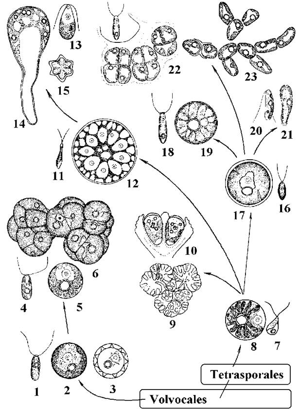
Fig. 24.23. Characteristic representatives of Chlorococcales and evolutionary trends within the order based on molecular data: 1-3 - Chlorococcum hypnosporum (1 - zoospore, 2 - vegetative cell, 3 - hypnospore); 4-6 - Tetracystis aeria (4 - zoospore, 5 - individual vegetative cell, 6 - tetrad complexes); 7, 8 - Ettlia minuta (7 - zoospore, 8 - vegetative cell); 9-10 - Botryococcus braunii (9 - colony, 10 - vegetative cells); 11-12 - Spongiochloris (11 - zoospore, 12 - vegetative cell); 13-15 - Protosiphon botryoides (13 - zoospore, 14 - vegetative cell, 15 - zygote); 16, 17 - Chlorococcum oleofaciens (16 - zoospore, 17 - vegetative cell); 18, 19 - Deasonia multinucleata (18 - zoospore, 19 - vegetative cell); 20, 21 - Chlamydopodium vacuolatum (20 - zoospore, 21 - vegetative cell); 22, 23 - Pleurastrum insigne (22 - zoospore and cells in sarcino-form state, 23 - filamentous state) (after Korshikov, 1955; Sauer, 1977; Andreeva, 1998; Ettl, Gartner, 1995; and original data).
Typical representatives of the first lineage are Chlorococcum hypnosporum, Ch. diplobionticum, and Tetracystis aeria. All of these organisms inhabit soils.
The cells of Chlorococcum hypnosporum are spherical, uninucleate, and possess a large parietal chloroplast with a pyrenoid and two contractile vacuoles. Asexual reproduction proceeds via biflagellate x-type zoospores or hemizoospores. The ULTRASTRUCTURE OF THE zoospores (particularly their cell wall architecture) resembles that of chlamydomonad vegetative cells. Under unfavorable conditions, vegetative cells develop a thick, spinose cell wall and transform into resting spores, or hypnospores. The sexual process is isogamous, and the life cycle is haplontic with zygotic meiosis. Another species of this genus, Chlorococcum diplobionticum, is a morphological sibling species to Ch. hypnosporum, yet it features a diplontic life cycle with gametic meiosis.
Tetracystis aeria possesses vegetative cells and zoospores similar to those of the preceding two species. However, the cells of this alga are capable of vegetative division, resulting in the formation of tetrads and tetrad complexes. Thus, within the first lineage, There is a clear evolutionary trend toward the transition from coccoid to multicellular sarcino-form habits while retaining features of monadic organization, specifically contractile vacuoles. This lineage is closely affiliated with volvocalean algae, notably certain species of the genus Chlamydomonas (Ch. reinhardtii, Ch. moewusii).
Within the second lineage, several evolutionary trends are evident simultaneously: the loss of zoospores accompanied by a transition to a colonial organization (the Ettlia-Botryococcus lineage), the shift from p-type to x-type zoospores (Ettlia-Chlorococcum), and transitions from unicellular to multicellular (Chlorococcum-Pleurastrum) or siphonous organization (Spongiochloris-Protosiphon; Chlorococcum-Deasonia).
Ettlia minuta features uninucleate spherical cells with a parietal, radially dissected chloroplast and a nearly central pyrenoid. Reproduction occurs primarily via naked biflagellate zoospores, and less frequently through aplanospores. It inhabits soils in tropical regions.
Botryococcus braunii is a colonial alga. Its colonies form botryoidal (cluster-like) structures embedded in a cartilaginous mucus matrix, at the periphery of which ovoid cells are located. These cells contain a parietal, deeply dissected chloroplast with a pyrenoid lacking a starch sheath. The Cytoplasm accumulates large amounts of colorless or orange-pigmented oil, and the cells exude hydrocarbon compounds to the exterior. Reproduction takes place via autospores.
This alga inhabits freshwater plankton and frequently causes water "blooms" in subtropical and tropical regions. In some countries, B. braunii has been brought into industrial cultivation as a hydrocarbon producer and a raw material for biofuel manufacturing.
A complete transitional series from the uninucleate Ettlia minuta to true siphonous forms is demonstrated by the edaphic algae Spongiochloris and Protosiphon. Both genera produce naked biflagellate zoospores (p-type, the protosiphon-type, named after one of these genera) and possess cells with reticulate METABOLISM/14.html">Chloroplasts and pyrenoids. In Spongiochloris llanoensis, cells are spherical, small (up to 25–35 µm in diameter), and uninucleate; another species, S. typica, also has spherical cells, but they are larger (up to 70 µm) and rapidly become multinucleate. In S. irregularis, cells smaller than 20 µm are spherical and uninucleate; subsequently, they become multinucleate and gradually lose their regular shape, reaching up to 120 µm.
During early and intermediate Stages of Ontogeny, Protosiphon botryoides mirrors all Developmental Stages of Spongiochloris irregularis, whereas at late stages, it develops a multinucleate rhizoidal thallus. It is differentiated into a long, colorless rhizoid (up to 3 mm long and 30–50 µm wide) and a sub-spherical apical portion containing a reticulate chloroplast, multiple pyrenoids, and several dozen nuclei; the diameter of the apical region can reach 500 µm. Under adverse conditions, thick-walled akinetes resembling adult Spongiochloris cells are formed. In the Presence of water, an isogamous sexual process takes place, yielding a stellate zygote that germinates after a dormancy period.
Chlorococcum oleofaciens belongs to the group of Chlorococcum species lacking contractile vacuoles. Vegetative cells are uninucleate, spherical, and feature a dissected parietal chloroplast. As the cells age, substantial amounts of oil accumulate in the cytoplasm. Asexual reproduction yields biflagellate x-zoospores, which nevertheless differ somewhat in ultrastructure from the zoospores of Ch. hypnosporum and chlamydomonad cells (specifically, the glycoprotein layer is absent from the cell wall). Aplanospores are also known to occur.
Molecular data, which correlate well with morphological and certain cytological traits, indicate that Chlorococcum oleofaciens is more closely related to Ettlia minuta than to Chlorococcum hypnosporum and Ch. diplobionticum. This clearly illustrates the paraphyletic nature of both the order as a whole and its individual genera.
The genus Deasonia resembles Chlorococcum oleofaciens, but features a spongy-type chloroplast. Adult cells become multinucleate, thus exhibiting a trend toward a siphonous body plan within representatives possessing x-type zoospores.
Pleurastrum insigne is a multicellular edaphic alga consisting of complexes of sarcina-like packets that occasionally form filamentous structures. Reproduction occurs via the fragmentation of aggregates, as well as through biflagellate x-type zoospores2. Under unfavorable conditions, akinete-like cells with spinose walls are formed.
Cylindrocapsales. This order unites algae characterized by a filamentous morphological body type, the Cylindrocapsa-type of mitosis, and cytokinesis mediated by a cleavage furrow. Cytologically, cylindrocapsalean algae are very close to Chlorococcales (see Table 24.3), yet they are sharply distinguished by their ability to form unbranched filaments. A typical representative is the genus Cylindrocapsa.
The thalli of cylindrocapsalean algae consist of unbranched filaments that do not dissociate into individual cells. Each cell is encased in a thick, layered wall and contains a massive central chloroplast with a pyrenoid and a single nucleus. Pores and plasmodesmata between neighboring cells are absent; therefore, the alga can be considered truly multicellular only in a conventional sense. Reproduction proceeds via naked biflagellate zoospores (p-type), which can be produced in any cell of the filament.
The sexual process is oogamous. Oogonia are formed through the proliferation of vegetative cells. Antheridia arise following a series of rapid divisions of a vegetative cell, with 1–2 spermatozoa forming within each antheridium. Following fertilization, the zygote develops a thick wall and enters a dormant state. Upon its completion, meiosis occurs, producing four daughter cells, each capable of growing into a new filament.
The Cy-type of mitosis has also been identified in filamentous algae of the genera Radiofilum, Geminella, Binuclearia (Fig. 24.24).

Fig. 24.24. Characteristic representatives of Cylindrocapsales: 1–3 — Cylindrocapsa geminella — filaments with oogonia (1) and antheridia (2), ultrastructural Cell Structure (3); 4 — Radiophylum mesomorphum; 5 — Geminellaterricola unstained (5) and stained (6) with methylene blue solution; 7 — Binuclearia tectorum (1, 2, 5, 6 — orig., 3 — after Kouwets, 1994; 4 — after Skuja, 1955; 7 — after Wichmann, 1937).
The order is considered monophyletic and is thought to have originated from chlorococcal algae with naked p-type zoospores.
Chaetophorales. This group unites true multicellular (filamentous and heterotrichous) algae possessing primary pores formed As a result of the uronema-type of mitosis. Cytokinesis proceeds with the participation of a cell plate, and the basal bodies of the flagella are shifted clockwise. All chaetophoralean algae inhabit the periphyton of inland freshwater bodies.
According to cytological, molecular-biological, and biochemical studies, Chaetophorales represents a monophyletic group. Investigations of the nuclear Gene encoding ribosomal RNA have shown that the ancestors of the order are volvocalean algae related to Chlorogonium.
Within the order, a progressive complexification of the body plan can be traced: from unbranched filamentous algae to heterotrichous representatives that are highly differentiated morphologically and functionally. This evolutionary series is clearly exemplified by the genera Uronema, Stigeoclonium, Draparnaldia (Fig. 24.25).

Fig. 24.25. Selected representatives of Chaetophorales: 1, 2 — Uronema (1 — vegetative filament, 2 — release of zoospores from the sporangium); 3–7 — Stigeoclonium (3 — fragment of the upright thallus portion, 4 — zoospore, 5–7 — stages of zoospore germination); 8, 9 — Chaetophora (8 — general appearance of the thallus, 9 — thallus fragment with setae); 10, 11 — Draparnaldia (10 — prostrate, supporting, and assimilatory filaments and setae, 11 — fragment of the upright portion with supporting and assimilatory filaments) (after Moshkova, 1986).
Uronema features a thallus in the form of a simple, unbranched filament consisting of morphologically and functionally identical cells (with the exception of the basal and apical ones). Each cell is uninucleate and contains a parietal chloroplast shaped like an open ring, enclosing one (less frequently two to four) pyrenoid. The basal cell forms a holdfast disc used by the alga to attach to underwater substrates. The apical cell has a tapered and pointed apex. Reproduction occurs via quadriflagellate zoospores or aplanospores.
Stigeoclonium possesses heterotrichous thalli consisting of branched upright and prostrate filaments. The former perform photosynthetic and reproductive Functions, while the latter serve to attach the alga to the substrate. The upright filaments are tapered at the apex and frequently terminate in single- or multicellular, thin, colorless hairs known as setae. The Abundance of branching and the number of setae depend on the nitrogen concentration in the medium: when the water contains a high level of nitrogen, branching is sparse and setae are virtually absent (in which case the alga resembles Uronema), and vice versa. Asexual reproduction occurs via quadriflagellate zoospores and aplanospores, whereas the sexual process is isogamous, mediated by biflagellate gametes. Interestingly, gametes are capable of germinating parthenogenetically, thereby also fulfilling the function of zoospores. The life cycle is haplontic, with zygotic reduction and no alternation of generations.
Close to Stigeoclonium is the genus Chaetophora. The thalli of Chaetophora are nearly identical to those of the previous genus, except that they are embedded within a massive colonial mucilage.
The thalli of Draparnaldia are differentiated into three systems of filaments: prostrate, upright supporting, and assimilators. The prostrate filaments and assimilators resemble the corresponding filaments of Stigeoclonium, performing the functions of substrate attachment, Photosynthesis, and reproduction, respectively. The supporting filaments elevate the entire mass of assimilators above the substrate. The Cells of the supporting filaments possess a reduced chloroplast, thick cell walls, and are incapable of producing zoospores, gametes, or carrying out intensive photosynthesis.
Oedogoniales. This order unites filamentous and heterotrichous algae which resemble chaetophoralean algae in cytokinesis involving a cell plate, but differ significantly in having a specialized type of mitosis (oedogonial mitosis), cell division with cap formation, stephanokont monad cells (zoospores, androspores, and spermatozoa), and a highly developed oogamous sexual process. The order comprises about 500 species of algae distributed across three genera: Oedogonium, Bulbochaete, and Oedocladium.
Cell division in Oedogoniales. At the onset of division, a fold forms near the apex of the cell from dictyosomes, which fuse to create a ring indented into the cytoplasm. Subsequently, the nucleus divides mitotically. A cell partition begins to form between the daughter nuclei via a phycoplast, and the maternal wall ruptures in the region of the ring. The ring rapidly straightens out, driving the elongation of the new upper cell. The remnants of the mother cell wall remain on the daughter cell as a cap (Fig. 24.26). The number of caps corresponds to the number of divisions that particular cell has undergone.

Fig. 24.26. Cell division (1–4), zoospore release (5), and its germination (6) in oedogonialean algae (after Vinogradova, 1977).
The monad cells of oedogonialean algae are stephanokont and can be represented by three types: (a) zoospores, formed singly within a sporangium cell; (b) androspores, from which a specialized microscopic male plant—the nannandrium—develops; androspores are formed singly, much like zoospores; (c) spermatozoa, which develop in pairs within the antheridium. It has been established that following The breakdown of their walls, the protoplasts of ordinary vegetative cells are capable of developing flagella and transforming into zoospores.
Oedogonialean algae can be either monoecious or dioecious. The life cycles in oedogonialean algae fall into two types: haplontic without alternation of generations, and haplontic with a microscopic male gametophyte stage known as the nannandrium (Fig. 24.27). Species exhibiting the first type of life cycle are termed macrandrous, while those with the second are termed nannandrous.
In macrandrous species, both antheridia and oogonia develop on ordinary vegetative filaments. Two spermatozoa are formed within each antheridium, and a single egg cell within the oogonium. The spermatozoa escape from the antheridium, swim toward the oogonium, penetrate the egg cell through a pore or a crack in the wall, and fertilize it. The zygote develops a thick wall and enters a resting stage. Upon the Conclusion of the resting period, reduction division takes place within the zygote, and each haploid nucleus yields a zoospore that subsequently germinates into a new filament.

Fig. 24.27. Characteristic representatives of Oedogoniales. 1–7 — Oedogonium: 1 — vegetative filament, 2 — filament with antheridia (macrandrous species), 3 — release of a spermatozoon from the antheridium, 4 — spermatozoon, 5 — filament with an oogonium, 6 — filament with an oogonium and two nannandria (nannandrous species), 7 — nannandrium with an apical antheridium producing spermatozoa. 8–9 — Bulbochaete: thallus fragments with setae, oogonia, and nannandria (1, 6 — orig., 2, 5, 7–9 — after Junger, 1993; 3, 4 — after Vinogradova, 1977).
In nannandrous species, oogonia and specialized sporangia develop on the filaments, within which male zoospores—androspores—are produced. An androspore swims to the oogonium, settles on it or on an adjacent cell, and germinates into a 2–4-celled nannandrium. The apical cell of the nannandrium transforms into an antheridium, inside which two spermatozoa develop. Thereafter, the developmental cycle proceeds in the same manner as in macrandrous species.
The largest genus in the order is Oedogonium (approximately 500 species), characterized by unbranched filamentous thalli. In the genus Bulbochaete (over 100 species), the filaments are branched, and the terminal branches typically bear setae that are bulbously thickened at the base. Oedogonium and Bulbochaete are widely distributed in the periphyton and benthos of freshwater bodies, frequently detaching from the substrate to form soft, non-slippery mats on the water surface. The genus Oedocladium features thalli of heterotrichous organization, differentiated into subterranean prostrate and aerial upright filaments. Species of this genus inhabit soils in tropical regions.
Scenedesmales. In classical systems, all algae of this order were assigned to Chlorococcales, and only molecular and electron microscopic studies in the 1990s revealed the evolutionary independence and phylogenetic distinctness of this group. The order comprises predominantly freshwater planktonic and terrestrial soil algae with a coccoid type of cellular structure.
Monadoid cells (zoospores and gametes) lack a cell wall (p-type) and possess a distinct flagellar orientation: the basal bodies are positioned opposite each other and are not shifted either clockwise or counter-clockwise (12-6 orientation). When viewed from the side, the basal bodies appear almost parallel to one another, which also clearly distinguishes Scenedesmales from Chlorococcales. A significant number of species within the order have lost their monadoid stages, and the placement of such taxa in Scenedesmales has been established through molecular phylogenetic studies. In terms of mitosis and cytokinesis, scenedesmalean algae do not differ from chlorococcalean algae.
Based on electron microscopic studies of the cytoskeleton, the ancestors of scenedesmalean algae are considered to be unicellular volvocalean and tetrasporalean algae with slightly unequal flagella, resembling the genera Heterochlamydomonas and Heteroteracystis, respectively.
Within the order, two main trends can be traced: transitions to multinucleate and coenobial body plans. Three evolutionary lines are currently recognized among the scenedesmales: a) a line of unicellular and coenobial zoosporaic algae showing a trend toward multinucleation; b) a line of unicellular and colonial autosporaic algae characterized by a tendency toward longitudinal division of the protoplast; and c) a line of coenobial autosporaic algae. According to modern data, all three lines within the order are considered parallel evolutionary series.
Characteristic representatives of the first line include Neochloris aquatica, Bracteacoccus, Sorastrum, Pediastrum, Hydrodictyon. All these algae possess naked biflagellate zoospores or gametes (p-type) and represent one of the pathways in the transition from unicellular forms to highly specialized coenobial ones.
The most primitive representative is Neochloris aquatica. This alga is unicellular in its adult stage. Each cell features a parietal chloroplast with a pyrenoid and several nuclei. Reproduction occurs via uninucleate biflagellate p-zoospores or aplanospores. Zoospores and aplanospores quite often do not immediately escape from the maternal cell wall, instead beginning to grow directly inside the sporangium. In this case, they join laterally and are released from the sporangium as aggregates resembling spherical coenobia. The breakdown of such coenobia into individual cells occurs only at the onset of sporogenesis, when each cell becomes multinucleate.

Fig. 24.28. Evolutionary directions in Scenedesmales based on molecular and cytological characters. 1 - Heterochlamydomonas (Volvocales), 2-5 - Heterotetracystis (Tetrasporales), 6,7 - Muriella terrestris, 8 - Oocystis, 9-11 - Scotiellopsis, 12 - Scenedesmus, 13 - Characiopodium hindakii, 14-17 - Neochloris aquatica, 18-22 - Bracteacoccus, 23 - Pediastrum, 24, 25 - Hydrodictyon, 26, 28 - Mychonastes homosphaera, 29, 30 - Monoraphidium, 31, 32 - Ankistrodesmus (phylogenetic scheme after Friedl, 1997; Lewis, 1997; Sluiman, 1997; et al. Illustrations after Oltmanns, 1904; Korshikov, 1953; Starr, 1955; Hindak, 1988; Cox, Deason, 1968, 1969; Fott, Novakova, 1969; Hindak et al., 1973; Puncocharova, Kalina, 1981; Ettl, Gartner, 1995; and original).
Algae of the genus Bracteacoccus are also multinucleate in the adult stage, are capable of forming coenobia-like aggregates, and reproduce via zoospores. Unlike Neochloris, the cells of Bracteacoccus possess numerous parietal disc-shaped chloroplasts and lack pyrenoids.
In Pediastrum and Hydrodictyon, cells are always united into coenobia characteristic of the respective genus. Both genera reproduce via zoospores and exhibit an isogamous sexual process.
In species of the genus Pediastrum, the coenobia are flattened, star-shaped, or wheel-like, consisting of 4–128 laterally united cells. Each mature cell contains a parietal chloroplast with one or several pyrenoids and several nuclei.
The coenobia of Hydrodictyon are macroscopic and consist of several thousand large cylindrical multinucleate cells connected at each pole to two or four neighboring cells. As a result, pentagonal and hexagonal meshes are formed, which together give the coenobium a net-like appearance (hence the common name of Hydrodictyon - water net).
During asexual reproduction, the protoplast of each cell of the water net breaks down into several thousand naked biflagellate zoospores that, without leaving the maternal cell, join together to form a daughter coenobium. After the rupture of the maternal cell wall, such a coenobium begins to exist as an independent Organism.
During sexual reproduction, the cell protoplast breaks down into several thousand naked biflagellate isogametes. The gametes emerge through an opening in the maternal cell wall. Subsequently, gametes of opposite mating types copulate to form a zygote that enters a resting stage. Upon its completion, meiosis occurs, and the zygote germinates into four large haploid zoospores. They are capable of swimming away from the site of formation over a considerable distance and thus perform a dispersal function. Upon cessation of movement, each zoospore germinates into a polygonal multinucleate cell, the so-called polyedr, which represents the sporophyte of the water net. The protoplast of the polyedr divides into A large number of zoospores that assemble into a new coenobium without leaving the polyedr. Thus, the life cycle of Hydrodictyon is haplophasic, with zygotic reduction and a heteromorphic alternation of generations: a coenobial gametosporophyte and a unicellular polyedr sporophyte. An analogous type of asexual and sexual reproduction is also characteristic of Pediastrum.
Examples of algae belonging to the second line of scenedesmalean algae—the line of uninucleate unicellular and colonial autosporaic algae—are the genera Mychonastes and Ankistrodesmus.
Cells of Mychonastes are solitary, small, spherical, with a parietal chloroplast lacking a pyrenoid, and a single nucleus. Reproduction occurs exclusively via autospores, which are formed in numbers of 2–8 and are released upon the rupture of the sporangial wall. Mychonastes was initially assigned to the genus Chlorella (e.g., Chlorella homosphaera), although true species of Chlorella belong to a different class, Trebouxiophyceae. Only the Application of Molecular phylogenetic Methods made it possible to determine the true position of Mychonastes within The system of Scenedesmales.
The genus Ankistrodesmus comprises colonial autosporaic algae. Colonies are formed by two to four parallel-arranged cells embedded in a common colonial mucilage. Each cell is elongated and pointed at the poles, possessing a parietal chloroplast, usually with a pyrenoid, and a single nucleus. During reproduction, the protoplast divides longitudinally, resulting in the formation of a group of parallel autospores.
An intermediate genus between the two preceding ones is Monoraphidium. The cells of this alga are similar in morphology and reproduction to Ankistrodesmus, yet they do not form colonies, thereby resembling Mychonastes.
The third line of the order is represented by uninucleate autosporaic, predominantly coenobial algae, whose cell wall contains an additional sporopollenin layer, and the wall itself is typically capable of producing various warts, spines, spinules, and bristles. Characteristic representatives include Muriella, Oocystis, Scotiellopsis, and Scenedesmus.
Algae of the genus Muriella are complete morphological "twins" of the genus Bracteacoccus: they possess spherical cells with numerous parietal
disc-shaped chloroplasts lacking pyrenoids. However, unlike Bracteacoccus, the cells of Muriella are uninucleate, do not form zoospores, and reproduce solely via autospores. Quite often, after release from the sporangium, the autospores remain laterally attached for a prolonged period, thereby resembling true coenobial algae. Older cells accumulate a large amount of secondary carotenoids and turn red.
Oocystis is characterized by ellipsoidal cells with small polar thickenings. The alga is considered colonial because the autospores germinate directly inside the sporangium, while the maternal cell wall expands but does not rupture.
Cells of species of the genus Scotiellopsis are also ellipsoidal, but solitary. The surface of the cell wall bears characteristic longitudinal Ribs that are clearly visible only under Electron Microscopy. Each cell is uninucleate, possesses a parietal chloroplast perforated by several slits, and a single pyrenoid. Reproduction occurs via 2–8 autospores that are released upon the rupture of the sporangial wall.
The genus Scenedesmus is characterized by coenobia typically formed by 4 or 8 cells laterally joined together. The cell wall features various "ornamentations" in the form of ribs, spinules, bristles, warts, and long horns. The type of such ornamentations serves as a taxonomic feature at the species level. Reproduction occurs via autospores that assemble into a new coenobium while still inside the maternal cell.
Interestingly, depending on The chemical composition of the water, the Morphology of the ornamentations can vary significantly (e.g., bristles or warts may form instead of horns). Although Scenedesmus is considered a typical example of autosporaic chlorococcalean algae, zoospores have recently been successfully obtained in culture for several species of this genus. Electron microscopic examination of these zoospores showed that their basal bodies are oriented in the 12-6 pattern, i.e., identically to those in Neochloris, Bracteacoccus, Pediastrum, Hydrodictyon.
Scenedesmal algae inhabit mainly the plankton of continental freshwater bodies and are quite frequently the dominants of this type of biocenosis (e.g., Pediastrum, Monoraphidium, Ankistrodesmus, Scenedesmus). Some representatives of the order occur in terrestrial habitats (Bracteacoccus, Muriella, Scotiellopsis).
Microsporales. This order unites algae with a filamentous type of structure, originating from scenedesmal algae. The order is represented by a single genus, Microspora, which is found in continental freshwaters.
The systematic features of the order include the presence of naked biflagellate zoospores, in which the basal bodies are not offset and are oriented according to the 12-6 scheme, and the microtubular roots correspond to the 7-1-7-1 formula. Additional microfibrillar structures are also associated with the thin microtubular roots (Fig. 24.29).

Fig. 24.29. STRUCTURE OF THE flagellar apparatus in Microspora zoospores: 1 - basal body; 2, 3 - thick (2) and thin (3) microtubular roots; 4, 5 - upper (4) and lower (5) connecting fibers; 6, 7 - additional microfibrillar roots associated with thin microtubular roots (after Lokhorst, Star, 1999).
A distinctive morphological feature that allows Microspora to be recognized quite easily is The structure of the vegetative cell wall. The wall consists of two H-shaped segments, due to which a characteristic "fork" is always present at the ends of the filaments, resembling a similar structure in yellow-green algae of the genus Tribonema (Fig. 24.30). Each cell contains a single nucleus and a parietal reticulate chloroplast, which in some species is capable of breaking down into disc-shaped segments. Pyrenoids are absent. An isogamous sexual process has been observed in a few species, but its details remain unstudied.
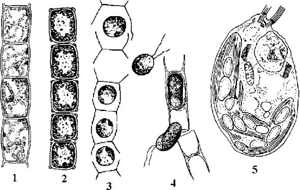
Fig. 24.30. Microsporal algae: 1-3 Microspora floccosa (1 - vegetative filament, 2 - akinetes, 3 - release of zoospores); 4 - cell walls and zoospores of Microspora willeana; 5 - ultrastructure of the zoospore of Microspora quadrata (1-4 - after Starmach, 1972; 5 - after Lokhorst, Star, 1999).
According to cytological and molecular characteristics, Microsporales are most closely related to zoospore-producing scenedesmal algae with slightly unequal flagella, resembling species of the genus Bracteacoccus.
Sphaeropleales. The order includes two genera, Sphaeroplea and Atractomorpha, and numbers about 10 species found in freshwater bodies of Africa, Australia, and South America. The order is of theoretical interest because it unites algae with a siphonocladous type of structure, flagellar root orientation of the 12-6 type, a "cylindrocapsoid" type of mitosis, and naked p-type zoospores.
Sphaeroplea appears as unbranched filaments consisting of morphologically and functionally identical cells. Each cell contains a reticulate chloroplast with pyrenoids, while two to four nuclei are located in the chloroplast-free areas, the so-called lacunae (most commonly four per cell) (Fig. 24.31).
Asexual reproduction occurs via filament fragmentation, zoospores, and special cells known as germlings (prothallia). The sexual process is oogamous. During sporulation, several large biflagellate macrozoospores are formed within the cell. Without leaving the sporangium, they germinate into multi-nucleate germlings. Afterward, the sporangium wall ruptures, and the germlings emerge into the external environment. Each germling produces several biflagellate microzoospores. Following a brief period of motility, the latter settle on the substrate and germinate once again into a germling, which gives rise to a new filament (Fig. 24.31).
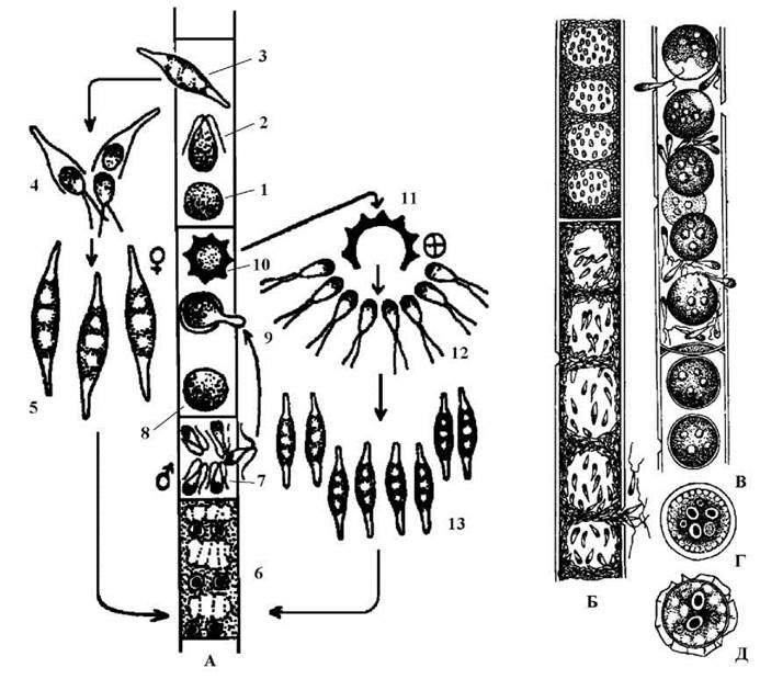
Fig. 24.31. Sphaeroplea. A - developmental cycle: 1 - zoospore protoplast, 2 - zoospore, 3 - multinucleate germling formed from a macrozoospore, 4 - germination of the germling into microzoospores, 5 - multinucleate germling formed from a microzoospore, 6 - vegetative multinucleate cell, 7 - antheridium with spermatozoa, 8 - egg cell, 9 - fertilization of the egg cell by a spermatozoon, 10 - zygote, 11 - germination of the zygote, 12 - zoospores formed from the zygote, 13 - multinucleate germlings formed from zygotic zoospores; B - filament with a vegetative multinucleate cell (top) and an antheridium with numerous spermatozoa (bottom); C - egg cells and the sexual process; D - zygote with three unfertilized haploid egg nuclei and one zygotic diploid nucleus; E - mature zygote with a spinous wall (after Starmach, 1972).
During sexual reproduction, ordinary vegetative cells transform into oogonia and antheridia. Both monoecious and dioecious species are known in Sphaeroplea. Typically, 4 egg cells are formed in a single oogonium (corresponding to the number of lacunae), each with four nuclei. Numerous biflagellate spermatozoa develop within the antheridia. A spermatozoon fertilizes one of the four egg nuclei. The other three nuclei degenerate, after which a zygote with a spinous wall is formed. Following a resting period, a reductional division occurs in the zygote, and the zygote germinates either into zoospores that rapidly transform into germlings, or directly into germlings. The germlings give rise to new filaments.
Distribution and Ecology
Chlorophyte algae inhabit mainly Two Types of ecotopes: continental freshwater bodies and soils, with only a few volvocine algae occurring in hypersaline lakes.
In water bodies, unicellular and coenobial species are represented mainly by planktonic algae. Some genera occur almost universally and frequently rank among dominants. These are primarily Monoraphidium, Ankistrodesmus, Scenedesmus, Pediastrum. In ephemeral water bodies, characteristic dominants of planktonic communities include Chlamydomonas, Chlorogonium, Pandorina, Eudorina.
In the periphyton, Chlorophyceae are most frequently represented by filamentous and heterotrichous algae from Chaetophorales, Oedogoniales, Microsporales—specifically the genera Uronema, Stigeoclonium, Oedogonium, Microspora, and less commonly by tetrasporalean and certain chlorococcalean and scenedesmal algae—Chaetochloris, Chlamydopodium, Characiopodium. Characteristic benthos representatives include Dicranochaete, Bulbochaete, Draparnaldia.
Chlorophyte algae are capable of causing water "blooms" and forming macroscopic growths known as water silk or algal mats. For instance, the "bloom" of brine in salt lakes is usually caused by Dunaliella salina. The water acquires a red coloration and a violet odor. The color is caused by the accumulation of a large amount of carotene in the cells, and the odor by secondary volatile compounds produced during its decomposition. In ephemeral freshwaters, a red "bloom" can be caused by Haematococcus pluvialis. Interestingly, this very alga is most frequently the coloring component of "Blood" rains and "blood" snow. In water bodies polluted with organic matter, green "blooms" are sometimes caused by various chlorococcalean and scenedesmal algae.
Algal mats (water silk) are most commonly formed by Oedogonium and Microspora. Both algae undergo initial Stages of development in the periphyton, then detach from the substrate and, continuing to reproduce, accumulate in cotton-like, soft, and non-slippery green masses on the water surface.
The genus Nautococcus (Tetrasporales) is a neustonic alga. It is most often responsible for the formation of a green, shiny surface film in ephemeral water bodies, especially those polluted with organic matter.
Soil chlorophyte algae constitute a numerous group. In arid steppe and desert soils, chlorococcalean algae of the genus Ettlia and scenedesmal algae of the genus Scotiellopsis develop massively. Upon Aging, they generally accumulate large amounts of carotenoids. Thus, Ettlia carotinosa is the main CAUSATIVE AGENT OF the red soil "bloom" in arid steppes and deserts.
In moderately moistened forest soils, common chlorophytes include volvocine (Chlamydomonas), chlorococcalean (Chlorococcum, Spongiochloris, Tetracystis), and scenedesmal (Bracteacoccus) algae. They rarely cause green soil "blooms". Typical inhabitants of waterlogged soils are Protosiphon, Pleurastrum, Cylindrocapsa.
Some chlorophytes, notably Chloromonas nivalis (Volvocales), represent an ecological group of cryophilic algae. This species grows On the surface of mountain snow and glaciers, causing their green and red "blooms".
Chlorophyte algae are virtually absent in marine and aerophytic habitats.
1 In modern taxonomic keys, the zoospores of Pleurastrum insigne are described as naked; however, studies by T. Friedl (1996) have shown that a wall is indeed present in the zoospores of this species, and electron micrographs reveal that it possesses an electron-dense second layer, resembling the zoospore walls of Chlorococcum oleofaciens.
Last update: 07/08/2026
Editorial and Educational Adaptation: This material has been compiled based on the primary/original source text. The project team performed an editorial review, corrected technical inaccuracies, structured sections, and adapted the content for an educational format.
What was processed:
- elimination of formatting defects (OCR errors, structural breaks, corrupted characters);
- editorial organization of content;
- standardization of terminology in accordance with academic sources;
- verification of factual statements against the original source text.
All mentions of the author, publication year, and origin of the primary text have been preserved in accordance with the source.