BIOLOGY Volume 2 - A Guide to General Biology - 2004
17. ANIMAL COORDINATION AND REGULATION
17.5. Structure and Function of Receptors
17.5.3. The Eye
The eye is a sensory organ that detects electromagnetic radiation of specific wavelengths (light) emitted by or reflected from objects within a certain area of the surroundings—the field of vision—and converts these light rays into electrical impulses. These impulses travel via the Optic nerve to the Brain, where they are processed to form a perceived image.
The human eye is sensitive exclusively to radiation within the visible spectrum, spanning wavelengths from 380 to 760 nm. Light energy is emitted and absorbed in discrete packets called quanta or photons. Each quantum of visible light is capable of triggering a photochemical reaction in the light-sensitive Cells of the eye, known as photoreceptors.
The operation of our human eye relies on the same fundamental principles as that of a camera, namely: 1) it controls The amount of light entering the eye; 2) it focuses the images of external objects using a system of lenses; 3) it registers the image on a sensitive surface; and 4) it translates the raw optical image into an internal perception of the visual world.
Structure AND Functions of the Human Eye
The Eyeball is roughly spherical in shape, measuring about 24 mm in diameter and weighing 6–8 g. It is situated within a bony depression of the Skull known as the Orbit, and is held in place by four rectus and two oblique Muscles that govern its movements. The bulk of the eye consists of accessory structures that transmit light to the photoreceptor cells, which form the innermost layer of the sphere—the retina. The retina is surrounded by the choroid, which continues anteriorly into the ciliary body and the iris with its central pupil. The outermost layer of the eyeball, the fibrous tunic, is divided into the cornea and the sclera (Fig. 17.33). Positioned immediately behind the pupil is the lens. The interior of the sphere is filled with the vitreous body and the aqueous humor, which maintain an internal intraocular pressure of approximately 3.3 kPa.
Class="center">

Fig. 17.33. STRUCTURE OF THE human eye: A — diagram of a vertical longitudinal section; B — histological preparation upon which the diagram is based.
The structure of the human eye is illustrated in Fig. 17.33, and the functions of its various components are summarized below.
Sclera — a tough, Collagen-rich white outer layer that protects the eye from damage and helps maintain its shape.
Cornea — the transparent anterior portion of the sclera; owing to its curved surface, it acts as the primary refractive structure, bending light rays to focus them onto the retina.
Conjunctiva — a thin, transparent mucous membrane consisting of epithelial cells that protects the cornea and is continuous with the epithelium lining the inner surface of the eyelids.
Eyelid — a fold of Skin that uses reflex movements to protect the front surface of the eyeball from mechanical and chemical injury, and shields the retina from excessively bright light.
Choroid — a vascular layer rich in Blood Vessels that nourish the retina, lined on the inside with a black pigmented epithelium that prevents internal light reflection within the eye.
Ciliary body — the region where the sclera and cornea meet; it contains epithelial cells, blood vessels, and the ciliary Muscle.
Ciliary muscle — a ring of smooth muscle fibers (circular and radial) that alters the curvature of the lens during accommodation.
Suspensory ligaments (zonule of Zinn) — fibers that connect the lens to the ciliary body.
Lens — a transparent, elastic, biconvex structure that provides fine-tuning and focusing of light rays on the retina by changing its curvature, while separating the chambers filled with aqueous humor and the vitreous body.
Aqueous humor — a transparent saline fluid secreted by the ciliary body that fills the anterior and posterior Chambers of the eye between the cornea and the lens; it drains into the bloodstream via the canal of Schlemm.
Iris — a pigmented ring-shaped Diaphragm that determines eye color; it divides the fluid-filled space into the anterior and posterior chambers and regulates the amount of light entering the eye.
Pupil — the central aperture of the iris that allows light to enter the interior of the eye.
Vitreous body — a transparent, gel-like mass enclosed within a membrane that fills the interior of the eyeball and helps maintain its shape.
Fovea centralis — the region of the retina with the highest resolving power (visual acuity), measuring less than 0.5 mm in diameter and containing exclusively cones. The majority of light rays are focused here.
Optic nerve — a bundle of nerve fibers (axons) that transmits visual impulses from the retina to the brain.
Blind spot — the region where the optic nerve exits the retina (approximately 1.7 mm in diameter); it lacks both rods and cones and is therefore completely insensitive to light.
17.6. List in order the structures through which light passes before reaching the retina.
Accommodation
Accommodation is the reflex mechanism by which light rays coming from an object are sharply focused on the retina. It involves two distinct processes, each of which will be examined separately.
PUPILLARY Reflexes. In bright light, the circular muscle of the iris (the sphincter pupillae) contracts, while the radial muscle (the dilator pupillae) relaxes. As a result, the pupil constricts, reducing the amount of light entering THE EYE AND thereby preventing retinal damage (Fig. 17.34). Conversely, in dim light, the radial muscles contract and the circular muscles relax, causing the pupil to dilate. An additional benefit of pupillary constriction is a purely optical phenomenon known as increased depth of field: the smaller the aperture through which light passes, the sharper the objects appear.
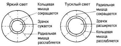
Fig. 17.34. Responses of the iris (and consequently the pupil) to changes in illumination.
REFRACTION OF LIGHT. Light rays from distant objects (more than 6 m away) enter the eye practically parallel, whereas those from closer objects diverge. In both cases, to form a sharp image on the retina, these rays must be refracted and focused onto it, with closer objects requiring a stronger degree of refraction. A normal human eye can accurately focus light from objects situated anywhere from about 25 cm to infinity. Light refraction occurs when it passes from one medium into another with a different refractive index, specifically at the air-cornea interface and across the surfaces of the lens.
Since the curvature of the cornea is fixed, corneal refraction depends solely on the angle of incidence of incoming light, which in turn varies with the object's distance. The cornea provides the majority of the eye's refractive power, while the lens serves to fine-tune the focus. The shape of the lens is regulated by the ciliary muscle: the degree of its contraction determines the tension on the suspensory ligaments (zonules of Zinn) that hold the lens in place. These ligaments exert tension on the elastic lens, altering its shape (radius of curvature) and, consequently, its refractive angle. When the radius decreases, the ciliary body moves closer to the lens, relaxing the suspensory ligaments, and the lens becomes more convex due to its inherent elasticity. This increases light refraction, allowing the eye to "accommodate" for viewing near objects. Relaxation of the ciliary muscle causes the ciliary body to pull away elastically from the lens, placing tension on the suspensory ligaments and stretching the lens. It flattens out, reducing refraction and establishing a new focal length adjusted for distant objects. These changes are summarized in Table 17.7 and Fig. 17.35.
Table 17.7. Relationship between the structures involved in changing lens shape and the degree of light refraction
Ciliary muscle |
Suspensory ligaments (Zonules) |
Lens curvature |
Light refraction |
Contracted (object is near) |
Slack / relaxed |
Increased (lens is more convex) |
Enhanced |
Relaxed (object is far) |
Taut / under tension |
Decreased |
Reduced |

Fig. 17.35. Accommodation for viewing objects at various distances from the eye. A. Side view of the eye. B. Front view of the eye.
The image formed on the retina is inverted and reversed According to the laws of optics; however, we navigate our surroundings effortlessly because the brain (visual cortex) processes this sensory information, allowing us to perceive the visual world the right way up.
Structure of the retina
The photoreceptor cells of the retina (rods and cones) have their light-sensitive portions directed toward the choroid, while on the vitreous side they are overlapped by The Cell bodies and processes of Neurons that connect them to the brain (Fig. 17.36).

Fig. 17.36. Diagram of retinal structure, including ultrastructural details of rods and cones. The connections between sensory cells and optic nerve neurons are shown. Light rays must pass through the layers of ganglion, amacrine, and bipolar cells before reaching the rods and cones.
The retina consists of three distinct cellular layers. The outermost photoreceptor layer is composed of light-sensitive cells—rods and cones—which are partially embedded in the pigmented epithelium of the choroid. Next is the intermediate layer containing bipolar neurons, which form synaptic connections between the photoreceptors and the cells of the third layer, as well as horizontal and amacrine cells responsible for lateral inhibition (see below). The third layer, the inner surface layer, is formed by ganglion cells, whose dendrites synapse with bipolar neurons and whose axons converge to form the optic nerve.
Structure and function of rods and cones
Rods and cones share a very similar structure (Fig. 17.36). In both cell types, photosensitive (visual) pigments are located on the outer surface of stacked membranes within the so-called outer segment; both consist of four distinct regions, whose structure and function are briefly outlined below.
OUTER SEGMENT. This is the photosensitive region where light energy is transduced into a generator potential. The entire segment consists of a stack of flattened membranous discs containing visual pigments. In a rod, these comprise 600–1000 such discs enclosed by The Plasma Membrane, whereas in a cone, they are formed by folds of the plasma membrane itself (fewer in number). The outer segment is cylindrical in rods and conical in cones.
CONSTRICTION. Here, the outer segment is almost completely separated from the inner segment by an invagination of the outer membrane. The two segments are connected via the Cytoplasm and a pair of cilia passing from one segment into the other. Each cilium consists of only nine peripheral microtubule triplets (the central pair typical of such Organelles is absent) and performs no motile function.
INNER SEGMENT. This is a region of high metabolic activity; it is packed with Mitochondria, which supply energy for visual processes, and polyribosomes for the synthesis of Proteins involved in The formation of membrane vesicles and visual pigments. The Nucleus is also located in this region.
SYNAPTIC REGION. Here, photoreceptor cells form synapses with bipolar cells. Some bipolar cells synapse with multiple rods simultaneously, which provides stimulus convergence—a mechanism that, as mentioned earlier, increases the eye's light sensitivity while decreasing visual acuity (Sec. 17.4.2). Other bipolar cells connect a single cone to a single ganglion cell; consequently, the acuity of "cone vision" is higher, whereas its sensitivity is lower. Horizontal and amacrine cells interconnect several rods and cones. Through these cells, visual information undergoes preliminary Processing even before leaving the retina, notably participating in lateral inhibition (see below).
Differences between rods and cones
The retina contains far more rods than cones (approximately 120 million and 6 million, respectively). These photoreceptors are also distributed unevenly across the retina. Rods are uniformly distributed throughout the retina except in the fovea centralis, which is dominated by cones. Because cones are densely packed in the fovea (50,000 per 1 mm), this region exhibits exceptionally high visual acuity (see below).
Rods (collectively) are much more sensitive to light than cones, reacting even to minute amounts of it. Containing only a single visual pigment, they are incapable of color discrimination and serve us primarily in dim light. Each cone contains one of three visual pigments, the interaction of which provides Color Vision. However, owing to their low sensitivity, these photoreceptors are used mainly during daylight.
Rod vision has lower acuity because rods are not packed as tightly as cones in the fovea, and their signals undergo convergence; however, this exact arrangement provides the high sensitivity required for night vision (see below).
Sensitivity and visual acuity
Visual acuity is the resolving power of vision, i.e., The ability to distinguish fine details of an object. For example, if one retinal region can resolve two closely spaced points as two separate entities, whereas another region perceives them as a single blurred point, visual acuity is higher in the first case. It reaches its maximum in the fovea centralis, which typically corresponds to the central part of our visual field, and decreases toward the periphery. Therefore, when examining an object closely, we shift our gaze from one part to another, bringing each in turn before the fovea. This area contains only cones, accounting for about 90% of their total number. Many of these cones synapse with just a single bipolar neuron (Fig. 17.36), which in turn connects to a single ganglion cell. This 1:1 ratio ensures maximum theoretical visual acuity, because each part of the image is processed by a dedicated cell, meaning the transmitted information is neither pooled nor "blurred" by a flood of other visual signals. Visual acuity increases with the number of cones per unit area (much like dots in a newspaper photograph: the more dots, the sharper the image). Cones located away from the fovea (as well as some within it) synapse with two or three bipolar neurons, which is why visual acuity declines toward the retinal periphery.
Against roughly 120 million rods and 6 million cones in each human eye, there are only about 1.2 million ganglion cells; consequently, signal convergence occurs (Sec. 17.4.2), averaging about 105 photoreceptors per ganglion cell. For rods, the degree of convergence is much higher than for cones, resulting in much lower visual acuity but higher sensitivity (see Question 17.7 below). Under dim lighting, the fovea centralis—devoid of rods—performs the worst, whereas the retinal periphery—dominated by rods—performs the best. This can be verified on a clear night sky: we perceive far more stars at the edges of our visual field than at its center. When light levels are low, only rods operate, causing visual acuity to drop (the perceived image becomes blurred) and color discrimination to vanish.
17.7. Explain why synaptic convergence increases visual sensitivity.
17.8. Explain why at night we can distinguish objects better if we look slightly to the side of them rather than directly at them.
Rods contain the light-sensitive pigment rhodopsin (visual purple), located on the outer surface of the membrane discs. Rhodopsin is a complex molecule formed by the protein scotopsin (an opsin) reversibly bound to a small light-absorbing carotenoid molecule, retinal. The latter is a vitamin A derivative that exists in two isomeric forms (cis and trans) that interconvert depending on illumination conditions (Fig. 17.37).

Fig. 17.37. A. Conversion of cis-retinal to trans-retinal under METABOLISM/18.html">The Influence of light. B. Bleaching and regeneration of rhodopsin.
It is known that the absorption of a single photon of Light triggers the Conversion of the cis-retinal molecule into its trans-form. This is accompanied by the dissociation of retinal from scotopsin (a process known as pigment bleaching):

In the dark, rhodopsin is resynthesized immediately: first, trans-retinal is converted back into the cis-form, after which the latter recombines with scotopsin (Fig. 17.37, B). This process forms the chemical basis of dark adaptation and takes about 30 minutes in total darkness across all rods, thereby restoring the eye's maximum photosensitivity.
How exactly does the light-induced breakdown of rhodopsin lead to the generation of an Action Potential? This is linked to Changes in the membranes of the outer and inner segments of the rods (Fig. 17.38). The inner segment houses a sodium pump that continuously extrudes sodium ions from the cell. In the dark, the outer segment membrane is permeable to these ions, and they diffuse back inward, thereby maintaining a steady resting Membrane Potential (around —40 mV rather than —70 mV, as seen in most cells). In the light, however, this permeability decreases while the sodium pump continues to operate, leading to an buildup of negative charge inside the rod (Fig. 17.38). Ultimately, this results in membrane hyperpolarization. This situation is the exact opposite of what typically occurs in other receptor cells, where the signal-induced generator potential is caused instead by membrane depolarization. Rod hyperpolarization slows down the release of the excitatory synaptic neurotransmitter, meaning that transmitter release is actually highest in the dark. Consequently, photoreceptor-linked bipolar neurons undergo hyperpolarization, yet the ganglion cells communicating with these neurons respond by generating action potentials.
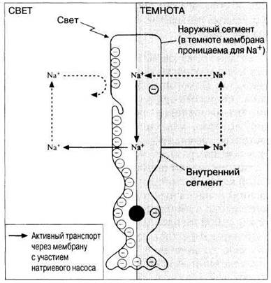
Fig. 17.38. Diagram of rod structure illustrating the hypothesized changes in outer segment permeability to sodium ions upon light stimulation. Negative charges (circles with a minus sign inside) on the right side of the rod correspond to the Resting Potential, and on the left side, to hyperpolarization.
Role of horizontal and amacrine cells
Horizontal cells, each stimulated by several rods and synapsing with multiple bipolar neurons (Fig. 17.36), mediate The phenomenon of lateral inhibition, which enhances both visual sensitivity and acuity. Simply put, if these cells simultaneously receive signals of equal intensity from two adjacent rods, they "mutually cancel" them out, thereby inhibiting further propagation. As a result, only signals from unequally stimulated receptors remain effective, which increases image contrast by sharpening the difference between strongly and weakly illuminated areas of the visual field, thereby facilitating the perception of object contours, for instance. Amacrine cells, each stimulated by several bipolar neurons and synapsing with multiple ganglion cells, transmit information regarding changes in illumination levels.
Color vision
The human eye absorbs light of all wavelengths within the visible spectrum, perceiving them as six distinct colors, each roughly corresponding to a specific region of the spectrum (Table 17.8). Colors as such do not exist in nature; they are a "creation" of the brain. Color differentiation is enabled by Three types of cones containing different pigments that absorb light within their respective wavelength ranges. Consequently, based on their maximum spectral sensitivity, these are classified as "red", "green", and "blue" cones. Experiments have shown that any color shade can be produced by combining these three primary colors.
17.9. Using Table 17.8, deduce what color an object will appear if you view it with one eye covered by a green filter (mean wavelength 530 nm) and the other by a red filter (mean wavelength 620 nm).
Table 17.8. Colors of the visible spectrum and approximate corresponding wavelength ranges
Color |
Wavelength, nm |
Red |
Above 620 |
Orange |
590-620 |
Yellow |
570-590 |
Green |
500-570 |
Blue |
440-500 |
Violet |
Below 440 |
Color vision is explained by the widely accepted trichromatic theory, according to which the perceived color depends on the degree of stimulation of each cone type. For instance, equal stimulation of all cones produces the sensation of white. Fig. 17.39 illustrates the sensitivity of the three cone types to various wavelengths. It is evident that although they are referred to as "red", "blue", and "green" cones, each photoreceptor responds not only to its "own" color but also to others to a lesser extent, with the sensitivity spectra of different types overlapping partially. Therefore, the perceived color is simply determined by the varying degree of stimulation of each cone type. For example, orange rays stimulate green and red cones to approximately the same extent, blue rays strongly stimulate blue receptors and weakly stimulate green ones, while green rays activate all three cone types.

Fig. 17.39. Sensitivity of the three cone types to light of different wavelengths (spectral colors).
While the trichromatic theory accounts for most experimental data on color vision, certain facts remain unexplained. Although discussing them is beyond The Scope of this book, they highlight The Need for further theoretical development in this field.
Initial color differentiation occurs in the retina, but the final Formation of the perceived hue requires interpretation of sensory signals by the brain.
Color Blindness. The complete absence or deficiency of a specific cone type leads to Various Forms of color blindness or color vision deficiencies, i.e., the inability to distinguish certain colors. For instance, individuals lacking "red" or "green" cones cannot differentiate between red and green, whereas those with an insufficient number of either type struggle to perceive unsaturated shades of these colors. Special test charts (such as Ishihara plates) composed of multicolored dots are used to diagnose color vision defects. Some of these plates contain numbers formed by the dots. A person with normal color vision easily reads these numbers, whereas individuals with impaired color perception see a different number or none at all. Color blindness is inherited as an X-linked recessive trait. Because it stems from Gene defects on the X chromosome, it is much more common in males: about 2% cannot see red (protanopes) and 6% cannot see green (deuteranopes), whereas both types of anomalies affect only 0.4% of females.
Binocular and Stereoscopic Vision
Binocular vision is the perception of an object using both eyes simultaneously. In this process, their visual fields overlap, and the image is focused concurrently on the two central foveae of the retinas. Because each eye views the object from a slightly different angle, the two images differ marginally, yet the visual cortex interprets them as a single unified image. Binocular vision forms The basis of stereoscopic, or three-dimensional, vision; this enables us to distinguish the relative distance of various visible details of an object—in other words, we perceive spatial depth.
The closer together the eyes are positioned, the greater their visual fields overlap and the wider the zone of stereoscopic vision. For example, the total combined field of view in humans spans 180°, with stereoscopic vision covering 140°. In horses, the eyes are set on the sides of the HEAD, and depth perception is limited to distant objects within a narrow strip directly ahead. To examine a close-range object, a horse turns its head and relies on monocular vision, yielding a flat visual image akin to a movie screen. Effective stereoscopic vision requires forward-facing eyes with foveae located centrally within their fields of view, which ensures high visual acuity. Under these conditions, stereoscopic vision provides a more accurate assessment of an object's size, shape, and distance. This type of vision is characteristic primarily of predators, for whom it is vital when capturing prey through sudden ambushes or high-speed dives, as seen in felines, hawks, and eagles. Conversely, in animals whose survival strategy depends on fleeing from predators, the eyes are typically positioned laterally—broadening the total field of view while narrowing the binocular zone (in rabbits, for instance, these are 360° and 26°, respectively). For them, spotting danger as early as possible and bolting indiscriminately in the opposite direction is far more critical.
The analysis of retinal images in stereoscopic vision is performed within two symmetrical regions that constitute the visual cortex.
The Role of the Brain
Nerve impulses originating in the retina travel via approximately one million nerve fibers of the optic nerve to the primary visual area of the visual cortex, located in the occipital lobe of the cerebral hemispheres (Fig. 17.26). Here, each retinal region—likely encompassing only a few rods and/or cones—is mapped to a specific processing area so that we "see". However, what we see acquires meaning only after signal exchange with other cortical areas, primarily the temporal lobes, where prior visual information is stored and utilized to analyze and interpret ongoing visual inputs (Section 17.2.4). In humans, axons originating from the left side of the retina in both eyes (which, according to optical principles, perceives the right visual field) project to the left hemisphere, while those from the right half (left visual field) project to the right hemisphere. Axons emerging from the medial (nasal) halves of the retinas cross over; this crossing point is clearly visible on the ventral surface of the brain as an X-shaped structure formed by the converging optic nerves, known as the optic chiasm (Fig. 17.40). Approximately 20% of optic nerve fibers bypass the Cerebral Cortex AND terminate in the Midbrain, where they participate in reflexes associated with pupillary constriction and Eye Movements.
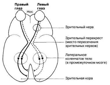
Fig. 17.40. Diagram of human visual pathways. Ventral view of the brain.
Last update: 06/08/2026
Editorial and Educational Adaptation: This material has been compiled based on the primary/original source text. The project team performed an editorial review, corrected technical inaccuracies, structured sections, and adapted the content for an educational format.
What was processed:
- elimination of formatting defects (OCR errors, structural breaks, corrupted characters);
- editorial organization of content;
- standardization of terminology in accordance with academic sources;
- verification of factual statements against the original source text.
All mentions of the author, publication year, and origin of the primary text have been preserved in accordance with the source.