BIOLOGY Volume 1 - A Guide to General Biology - 2004
3. CHEMICAL COMPONENTS OF LIVING ORGANISMS
3.5. Proteins
3.5.3. Protein Structure
Each protein possesses a distinct and specific geometric shape, or conformation. To describe the three-dimensional Structure of Proteins, four Levels of Organization are typically considered, which we will outline here.
Primary structure refers to The sequence of Amino Acids in a polypeptide chain. The first researcher to determine the Amino Acid Sequence of a protein molecule was Fred Sanger, working at the Cavendish Laboratory of the University of Cambridge—the very same place where Watson and Crick determined The structure of DNA. Sanger worked with the hormone Insulin, the smallest protein he could find. The work took 10 years, and the results were published in 1953 (Fig. 3.28). Another prominent molecular biologist from the Cavendish, Max Perutz, recalled: "This discovery was a sensation, for it was shown for the first time that amino acids in protein polypeptide chains are arranged in a completely definite manner." In 1958, Sanger was awarded the Nobel Prize for this work (he received his second Nobel Prize for studying the Structure of Nucleic Acids). The insulin molecule contains 51 amino acids. It consists of two polypeptide chains held together by disulfide bridges.
Class="center">
Fig. 3.28. Primary structure (amino acid sequence) of insulin. The molecule consists of two polypeptide chains held together by two disulfide bridges.
Currently, most amino acid sequencing work is automated, and primary structures are now known for well over a hundred thousand proteins. Figure 3.29 illustrates the Introduction/19.html">Primary structure of another protein, the enzyme Lysozyme. Table 3.6 provides An Overview of the number of amino acid residues in the molecules of various proteins.
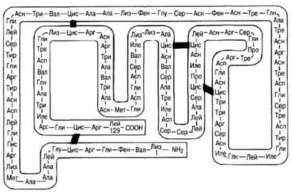
Fig. 3.29. Primary structure of lysozyme. Lysozyme is an enzyme found in many Tissues and secretions of The Human Body, as well as in plants and egg white. This enzyme catalyzes The breakdown of bacterial Cell walls. The lysozyme molecule consists of a single polypeptide chain containing 129 amino acid residues, with four intrachain disulfide bridges.
The human body contains thousands of different proteins, all built from the same 20 standard amino acids. A protein's amino acid sequence determines its biological function. In turn, this amino acid sequence is determined by The nucleotide sequence of DNA (Ch. 23). The substitution of a single amino acid in a given protein molecule can drastically alter its function, as seen, for example, in Sickle-Cell Anemia (Ch. 25). Interesting insights can be gained by analyzing the Amino acid sequences of Homologous proteins from different biological species; such data allow us to infer the potential taxonomic relationships among these species. This topic will be discussed in Chapter 26.
3.12. a) Write out The amino acid sequences of all tripeptides that can be constructed from two different amino acids, A and B.
b) Based on your results, formulate an equation to determine the number of different tripeptides that can be formed from two different amino acids.
c) How many Polypeptides of 100 amino acid residues in length can be constructed from two different amino acids?
d) How many polypeptides of 100 amino acid residues in length (which is a relatively small protein) can be constructed from all 20 standard amino acids?
e) How many Peptides or polypeptides (of a given length) can be constructed from all 20 standard amino acids?
In addition to primary structure, every protein is characterized by a specific secondary structure. Typically, a protein molecule resembles an elongated spring. This is the so-called α-Helix, stabilized by numerous Hydrogen Bonds formed between closely positioned CO and NH groups. The hydrogen atom of the NH group of one amino acid forms such a bond with the oxygen atom of the CO group of another amino acid located four residues away (Fig. 3.30). Thus, amino acid 1 is linked to amino acid 5, amino acid 2 to amino acid 6, and so on. X-Ray Diffraction Analysis shows that there are 3.6 amino acid residues per turn of the helix.

Fig. 3.30. STRUCTURE OF THE α-helix. A. Only α-carbon atoms are shown; the line connecting them traces the α-helix. B. Ball-and-stick model of the α-helix. C. A portion of the α-helix in an extended view, stabilized by hydrogen bonds. D. Ribbon representation of the α-helix.
The protein keratin has a fully α-helical conformation and, consequently, a fibrillar structure. It is a structural protein found in Hair, wool, Nails, beaks, feathers, and horns, and is also a component of vertebrate Skin. The hardness and extensibility of keratin vary depending on the number of disulfide bridges between adjacent polypeptide chains (the degree of cross-linking).
Theoretically, all CO and NH groups can participate in hydrogen bonding, making the α-helix a very stable and therefore widespread conformation. Regions of the α-helix resemble rigid rods within the molecule. Nevertheless, most proteins exist in a globular form, which also contains regions of β-sheets (see below) and non-regular structures. This is because Hydrogen bond formation is hindered by several factors: the presence of Certain amino acid residues in the polypeptide chain, disulfide bridges between different PARTS OF THE same chain, and, finally, the fact that the amino acid Proline is entirely incapable of forming hydrogen bonds.
The β-sheet, or pleated sheet, is another type of secondary structure. The silk protein Fibroin, produced by the silk-glands of silkworm caterpillars during cocoon spinning, exists entirely in this form. Fibroin consists of a series of polypeptide chains that are more extended than those with an α-helical conformation. These chains run parallel to one another, but adjacent chains have opposite directions (antiparallel; Fig. 3.31). They are linked by hydrogen bonds formed between the C=O and NH groups of neighboring chains. In this case as well, all NH and C=O groups participate in hydrogen bonding, meaning this structure is also highly stable. This polypeptide conformation is referred to as the β-conformation, and the overall structure as a pleated sheet. Fibroin has high tensile strength and is inextensible, yet this arrangement of polypeptide chains makes silk highly flexible. In Globular proteins, the polypeptide chain can fold back on itself, creating regions with a pleated-sheet structure at the turns of the globule.
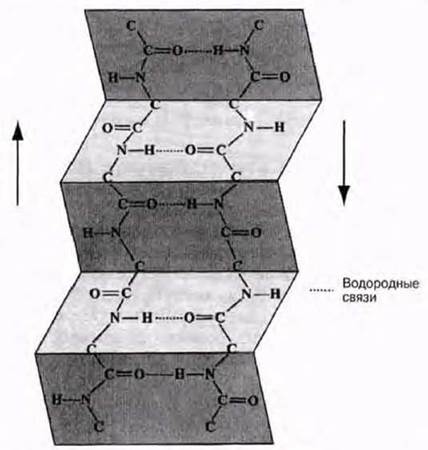
Fig. 3.31. β-pleated sheet. The chains are arranged parallel to one another and held in position by hydrogen bonds between the C=O and NH groups of adjacent chains. Side groups (R) are not shown; they project above and below the plane of the sheet.
Another way in which polypeptide chains are organized is found in the fibrous protein Collagen. This is another structural protein that, like keratin and fibroin, has a high tensile strength. In collagen, three polypeptide chains are wound together like the strands of a rope to form a triple helix. Each polypeptide chain of this complex helix, known as tropocollagen, contains about 1000 amino acid residues. The individual polypeptide chain is a loosely coiled helix (though not an α-helix; Fig. 3.32). The three chains are held together by hydrogen bonds. Many triple helices, lying parallel to one another and linked by covalent bonds between adjacent chains, form fibrils, which in turn combine to form fibers. Thus, the structure of collagen is formed stepwise—at several levels—much like the structure of Cellulose. Collagen is also virtually inextensible, a property essential for its function in tendons, bones, and Other types of Connective Tissue. Proteins that exist only in a fully helical form, such as keratin and collagen, are exceptions among other proteins.

Fig. 3.32. Triple-helical structure of collagen.
Tertiary structure
In most proteins, The polypeptide chains are folded in a specific way into a compact globule. The manner in which the polypeptide chains of globular proteins are folded is called their tertiary structure. Tertiary structure is maintained by the Three types of bonds already discussed—ionic, hydrogen, and Disulfide Bonds—as well as by hydrophobic interactions (Fig. 3.33). Quantitatively, hydrophobic interactions are the most important; the protein folds in such a way that its hydrophobic side chains are tucked away inside the molecule, while the hydrophilic ones are exposed on the outside.
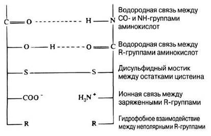
Fig. 3.33. Bonds stabilizing the secondary and tertiary structures of proteins. Hydrophobic interactions (the association of non-polar molecules or parts of molecules) are particularly important for maintaining the structure, as they exclude Water molecules—a crucial factor, for example, in membranes, given the aqueous cellular environment.
X-ray crystallography can be used to determine the Tertiary Structure of proteins. Following several years of research, John Kendrew and Max Perutz determined the secondary and tertiary structure of Myoglobin by this method by early 1959 and proposed a model of its molecule (Fig. 3.34). For this work, they were awarded the Nobel Prize in 1962. The following features of myoglobin were now known:
primary structure — the molecule consists of a single polypeptide chain made up of 153 amino acid residues (their sequence was established in the early 1960s);
secondary structure — about 75% of the chain has an α-helical conformation (eight helical regions);
tertiary structure — the α-helix is irregularly folded into a compact globule;
prosthetic group — a heme group (containing iron).
Myoglobin is synthesized in Muscles, where it serves to store oxygen. As in Hemoglobin, oxygen in the myoglobin molecule binds to the heme group, which is responsible for the red color of muscles. We will discuss the Functions of myoglobin in more detail in Chapter 14. Determining the tertiary structure of proteins remains a very laborious process. Recently, molecular biologists have been putting increasing effort into using computers and other technology to predict a protein's tertiary structure based on its already known Primary and secondary structure. This could open up possibilities for designing proteins with specific structures for specific functions, which could play a vital role in both industry and medicine.
Fig. 3.34 shows several ways of representing the tertiary structure of a protein. Another method is illustrated in Fig. 3.35. In proteins with a tertiary structure, function is intimately dependent on the precise shape of the molecule. This is particularly easy to see when studying Enzymes (Section 4.1.2).
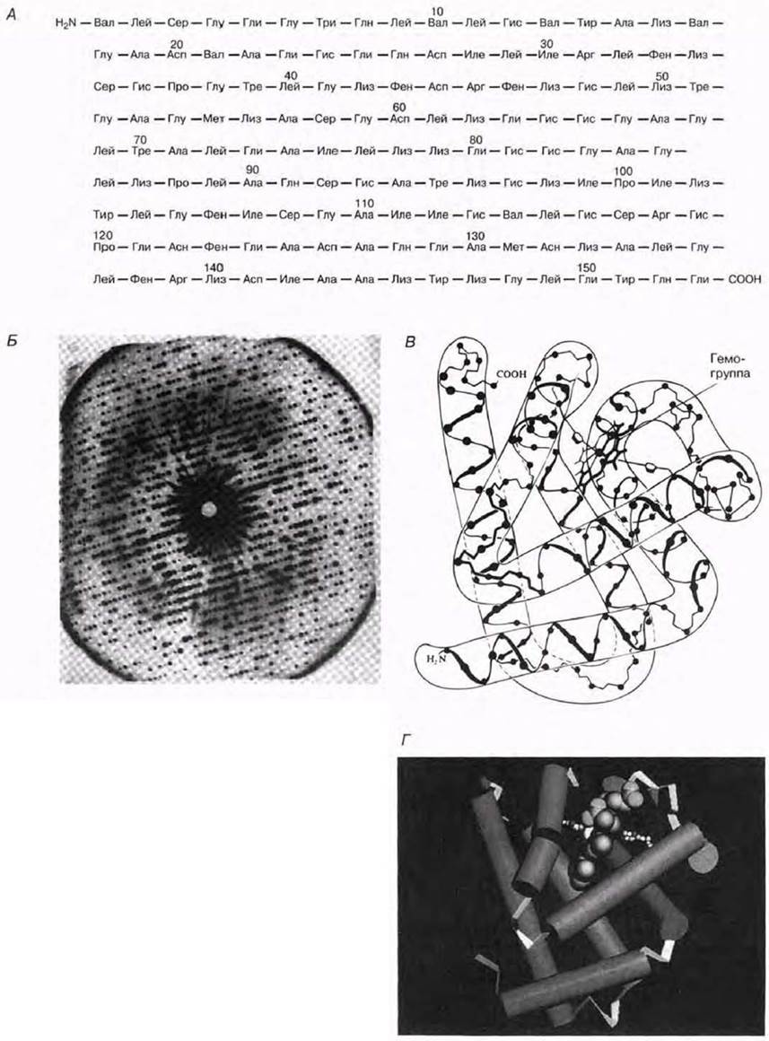
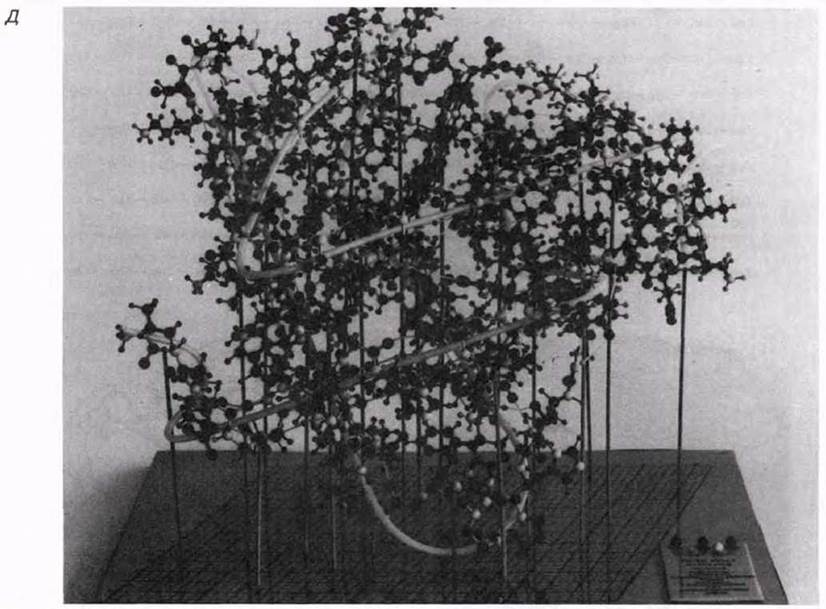
Fig. 3.34. A. Primary structure of myoglobin. B. X-ray diffraction pattern of myoglobin (from sperm whale Muscle). The regular arrangement of spots results from the scattering (diffraction) of incident X-rays interacting with myoglobin atoms in the crystal. The photograph is a two-dimensional section of the three-dimensional diffraction pattern. The positions of individual atoms in the molecule are determined from the distribution and intensity of the diffraction spots. (After J. G. Kendrew, Scientific American, December, 1961.) C. Conformation of myoglobin determined by high-resolution X-ray analysis. Eight α-helical regions surround the flat, disk-shaped heme group. D. Another way of representing the three-dimensional structure of proteins. In this computer-generated diagram, the α-helical regions in the myoglobin molecule are depicted as cylinders (eight such regions are visible). The model shows the heme group held in place by Two amino acids (small white spheres). E. Ball-and-stick model of myoglobin.

Fig. 3.35. Tertiary structure of lysozyme. Arrows indicate regions with a β-sheet structure. Regular coils represent regions with an α-helical structure. The rest of the molecule is shown as a winding ribbon, and the four disulfide bridges are depicted as four zig-zag lines (see Fig. 3.29).
Quaternary structure
Many highly complex proteins consist of several polypeptide chains held together within the molecule by hydrophobic interactions, as well as hydrogen and ionic bonds. The manner in which these polypeptide chains are packed and assembled together is called the protein's quaternary structure. Quaternary structure is found, for example, in hemoglobin—the red pigment contained in vertebrate erythrocytes that binds and transports oxygen. The hemoglobin molecule consists of four separate polypeptide chains of two different types: two α-chains and two β-chains. Structurally, these chains resemble the polypeptide chain of myoglobin. The two α-chains each contain 141 amino acid residues, and the two β-chains each contain 146 residues. The complete structure of hemoglobin was determined by Kendrew and Perutz and is shown schematically in Fig. 3.36.

Fig. 3.36. Structure of hemoglobin. The molecule consists of four polypeptide chains: two α-chains and two β-chains. Associated with each chain is a heme group to which an oxygen molecule binds. Hemoglobin is an example of a protein consisting of separate subunits, i.e., possessing a quaternary structure.
Like other globular proteins, the hydrophobic side chains of hemoglobin are buried within the interior of the molecule, while the hydrophilic ones are exposed On the surface, making hemoglobin water-soluble. A mutation that replaces one of the hydrophilic amino acids with a hydrophobic one, thereby reducing hemoglobin solubility, is responsible for the condition known as sickle-cell anemia (Chap. 25).
Some Viruses, such as tobacco mosaic virus, have a protein coat composed of many polypeptide chains packed in a highly ordered arrangement (see Fig. 2.18).
Last update: 06/08/2026
Editorial and Educational Adaptation: This material has been compiled based on the primary/original source text. The project team performed an editorial review, corrected technical inaccuracies, structured sections, and adapted the content for an educational format.
What was processed:
- elimination of formatting defects (OCR errors, structural breaks, corrupted characters);
- editorial organization of content;
- standardization of terminology in accordance with academic sources;
- verification of factual statements against the original source text.
All mentions of the author, publication year, and origin of the primary text have been preserved in accordance with the source.