Human Biochemistry Volume 2 - Murray R. 1993
Special Topics
Cancer, Oncogenes, Growth Factors
Oncogenic Viruses - Oncogenes
Oncogenes are regions of DNA (genes) whose functioning leads to the neoplastic transformation of Cells. The discovery of oncogenes was of great importance for investigating the fundamental mechanisms of carcinogenesis. Oncogenes were first discovered in tumor-causing Viruses and identified as factors responsible for the transformation process (Viral Oncogenes).
Oncogenes of the Rous Sarcoma Virus
Currently, The Structure of the Rous sarcoma virus oncogene has been investigated and the product of this oncogene characterized. The Genome of this retrovirus consists of four genes: gag, pol, env, and src. Schematically, it can be represented as follows:
Class="center">
The gag Gene encodes the synthesis of viral group-specific Antigens, the pol gene encodes retrovirus-specific Reverse Transcriptase, and the env gene encodes the synthesis of certain viral envelope Glycoproteins. The product of the src gene, which is responsible for transformation (The Development of sarcoma), is Tyrosine protein kinase. These findings turned out to be extremely important. They demonstrated the existence of a specific biochemical mechanism that can, at least in part, explain the pleiotropic effect of Retroviruses during transformation. Cellular Proteins whose altered phosphorylation potentially leads to transformation remain to be identified. One of these is likely vinculin, a protein found in adhesion plaques (structures involved in Cell-Cell Adhesion). The rounding of cells upon transformation, as well as the decrease in cell adhesion to Glass and other cells, is presumably explained by Changes in the phosphorylation of focal adhesion vinculin (Table 57.5). The targets of the tyrosine protein kinase encoded by the src gene appear to be certain glycolytic Enzymes. This is consistent with the elevated level of Glycolysis frequently observed in transformed cells. The src gene product may also catalyze the phosphorylation of phosphatidylinositol, resulting in The formation of phosphatidylinositol mono- and bisphosphate. The Hydrolysis of phosphatidylinositol-4,5-bisphosphate by phospholipase C yields Inositol trisphosphate and diacylglycerol (see Chapter 44). The former compound acts as a mediator in the release of Ca2+ ions from intracellular compartments (e.g., The Endoplasmic reticulum). Diacylglycerol stimulates The activity of Plasma Membrane-bound protein kinase C; this enzyme, in turn, phosphorylates A number of other proteins, likely including protein components of ion pumps. It has been suggested that a slight increase in intracellular pH, caused by the activation of the Na+/H+ antiporter system, stimulates the mitotic process (see Chapter 42). Thus, the src gene product can influence a wide range of cellular processes by phosphorylating various protein targets and enzymes, as well as by stimulating polyphosphoinositide synthesis pathways.
Table 57.5. Changes in cell culture indicative of neoplastic transformation (e.g., following infection with tumor-causing viruses). A decisive sign of malignancy is the ability of cells to form tumors upon Introduction into animals
|
Morphological changes: transformed cells have a more rounded shape than control cells Increase in cell density (loss of contact Inhibition of Growth): transformed cells frequently form multi-layered structures, whereas control cells grow as a monolayer Loss of anchorage dependence: transformed cells can grow without attaching to The surface of the culture vessel and often grow in Agar Loss of contact inhibition of movement: transformed cells can grow and migrate over other cells, whereas normal cells cease movement upon contact with other cells Various biochemical changes, including an increased rate of glycolysis, altered cell surface composition (e.g., changes in glycoprotein or glycosphingolipid composition), and the secretion of several proteases Alterations in cytoskeletal structures, such as Actin microfilaments Decreased growth factor requirements of tumor cells; the secretion of several factors into the surrounding medium is frequently elevated |
Tyrosine Protein Kinase in Normal and Tumor Cells
The Discovery of the Rous sarcoma virus tyrosine protein kinase spurred intensive research into the phosphorylation of tyrosine residues in proteins. It is now known that tyrosine protein kinase is present in many normal cells. In most such cells, The amount of phosphotyrosine is low, but upon transformation by Oncogenic Viruses carrying protein kinase, it typically
increases, although the absolute amount remains low (1% of all phosphorus-containing Amino Acids). A number of receptors (e.g., for epidermal growth factor, Insulin, and platelet-derived growth factor) in both normal and tumor cells possess tyrosine protein kinase activity, which is enhanced upon receptor-Ligand interaction (see "Growth Factors" below). Thus, tyrosine protein kinase plays a vital role in both normal and transformed cells.
Oncogenes of Other Retroviruses
In addition to the Rous sarcoma virus oncogenes, about 20 oncogenes from other retroviruses have been discovered. The products of roughly half of these are protein Kinases (predominantly of the tyrosine type). Some viral oncogenes and their products are listed in Table 57.6. While some of them encode protein kinases, a number of other oncogenes determine various other biologically active proteins. The product of the erb-B gene of the avian erythroblastosis virus is a truncated form of the epidermal growth factor receptor, and the product of the sis oncogene of the simian sarcoma virus is a truncated form of the B-chain of the platelet-derived growth factor molecule. The fms oncogene of a virus isolated from feline sarcoma cells encodes the synthesis of macrophage colony-stimulating factor. The product of the myc gene, first discovered in avian myelocytomatosis viruses, is a DNA-binding protein that can regulate cell mitosis. The product of the ras oncogene of the murine sarcoma virus binds GTP and exhibits GTPase activity. It is likely related to proteins that regulate the activity of one of the key membrane enzymes, adenylate cyclase (Chapter 44).
Table 57.6. Some retroviral oncogenes. (Modified and reproduced, with permission, from Franks L. M., Teich N. M. (editors). Introduction to the CELLULAR AND MOLECULAR Biology of Cancer, Oxford Univ. Press, 1986.)
|
Oncogene |
Retrovirus |
Origin |
Oncogene product |
Subcellular localization |
|
abl |
Abelson virus (murine leukemia) |
Mice |
Tyrosine protein kinase |
Plasma membrane |
|
erb-B |
Avian erythroblastosis virus |
Chickens |
Truncated EGF receptor |
Plasma membrane |
|
fes |
Feline sarcoma virus |
Cats |
Tyrosine protein kinase |
Plasma membrane |
|
fos |
Murine sarcoma virus |
Mice |
? |
|
|
myc |
Myelocytomatosis virus 29 |
Chickens |
DNA-binding protein |
Nucleus |
|
sis |
Simian sarcoma virus |
Monkeys |
Truncated platelet-derived growth factor (B-chain) |
Membranes secreted (?) |
|
src |
Rous sarcoma virus |
Chickens |
Tyrosine protein kinase |
Plasma membrane |
The central question that has occupied researchers since the discovery of viral oncogenes is their origin. Nucleic acid Hybridization experiments (see Chapter 36) have shown that normal cells contain DNA sequences similar (and possibly identical) to viral oncogenes. Apparently, during intracellular development, viruses incorporate cellular genes into their genome. Their presence in the viral genome presumably confers certain selective advantages related, for instance, to altered growth patterns of transformed cells.
Cellular DNA sequences homologous to viral oncogenes have been found in many Eukaryotic cells, meaning they represent essential COMPONENTS OF NORMAL cells. Furthermore, corresponding mRNAs and the proteins they encode can be detected at various Selection/3.html">Stages of development or the Cell Cycle. Such normal cellular genes have been termed proto-oncogenes. Proto-oncogene products play a crucial role in normal differentiation and other cellular processes.
Tumor Cell Oncogenes
Experiments with DNA isolated from tumor cells also provide Evidence for the existence of oncogenes. The method for detecting cellular oncogenes is known as "gene transfer" or "transfection." It is based on the principle that certain genes present in tumor cells can induce the transformation of "normal" cells in culture. DNA is extracted from tumor cells, precipitated with calcium phosphate, and added to recipient cells (typically the NIH/3T3 mouse fibroblast cell line). After 1–2 weeks, transformation foci are observed under the Microscope. The cells forming a focus alter their Morphology, changing from flat to rounded. DNA is extracted from the transformed cells, and the experiment is repeated. This cycle is performed several times, which reduces the amount of DNA not involved in transferring the transformation trait and thereby facilitates the identification of specific genes (using Southern blot hybridization; see Chapter 36). Using this method, about 20 cellular oncogenes have been identified; some of these are similar to the ras gene of the murine sarcoma virus. These cellular oncogenes either do not differ at all from normal genes or possess minor structural features (see below). In the former case, tumor transformation may alter The regulation of their expression.
Abbreviations of Cellular and Viral Oncogenes
An oncogene contained within tumor cells is typically designated in literature as "c-onc" (short for cellular oncogene; e.g., c-ras). Their counterparts in normal cells—proto-oncogenes—are correspondingly termed c-onc proto-oncogenes (e.g., c-ras proto-oncogene). A viral oncogene is called v-onc (e.g., v-ras; from viral oncogene), and the corresponding proto-oncogene is termed v-onc proto-oncogene (e.g., v-ras proto-oncogene).
Mechanism of Conversion of Proto-oncogenes into Oncogenes
We will discuss five mechanisms by which the expression or structure of proto-oncogenes is altered, resulting in their conversion into oncogenes. The process whereby gene METABOLISM/31.html">Transcription is upregulated (from a zero or low level) is termed activation. Understanding the mechanisms of activation is fundamental to comprehending the Current state of the cancer problem.
A. Promoter insertion. Some retroviruses (such as avian leukosis viruses) do not contain oncogenes, yet they are capable of causing cancer. In such cases, tumors appear after a longer latency period (months rather than days, as with oncogene-containing viruses). When cells are infected with these and other retroviruses, a DNA copy (cDNA) is synthesized from the viral RNA genome via reverse transcriptase and integrates into the host cell genome. The integrated double-stranded cDNA is called a provirus. Retroviral cDNA copies, much like certain Transposons (jumping genes) found in plants and Bacteria, contain sequences known as long terminal repeats at both ends. These sequences play a critical role in the proviral integration mechanism and act as transcription promoters (see Chapter 39). When chicken B lymphocytes are infected with certain avian leukosis viruses, their proviruses integrate near the myc gene. This gene is activated by the upstream Long Terminal Repeat functioning as a promoter. This results in the transcription and subsequent Translation of the corresponding myc mRNA (Fig. 57.3). In turn, this leads to the development of a B-cell tumor, although the specific role of myc gene products in this process remains unclear. Similar processes occur when various cells are infected by other retroviruses.
B. Enhancer insertion. Sometimes a provirus integrates downstream or upstream of the myc gene but in the reverse orientation; nevertheless, the myc gene is activated (Fig. 57.4). In this case, activation cannot be explained by promoter insertion, since a promoter must be located upstream of the gene whose transcription is enhanced, and the sequence must be in the correct orientation (5' to 3'). Consequently, activation is presumably explained by the presence of enhancers within the long terminal repeats of retroviruses (see Chapters 39 and 41).

Fig. 57.3. Schematic representation of proto-oncogene activation via promoter insertion. A. A normal chicken chromosome contains the inactive myc gene. B. Avian leukosis virus is integrated into the chromosome as a provirus in the region adjacent to the myc gene. The right long terminal repeat (LTR) of the provirus, containing a strong promoter, is located upstream of the myc gene, activates it, and subsequently initiates myc mRNA transcription. For simplicity, only a single DNA strand is shown.
The two mechanisms described above (promoter and enhancer insertion) can be considered typical of viral carcinogenesis.
C. Chromosomal translocations. As noted earlier, chromosomal abnormalities can be observed in many tumor cells. One such type of abnormality is translocation, which occurs when a fragment of one chromosome breaks off and attaches to another chromosome. If this latter chromosome, in turn, yields a corresponding fragment to the first chromosome, a so-called "reciprocal translocation" takes place. Characteristic translocations have been found in A wide variety of tumor cells. For example, the Philadelphia chromosome can be detected in the cells of patients with chronic granulocytic leukemia, involving Chromosomes 9 and 22.
Reciprocal translocation has been identified in some patients with Burkitt's lymphoma, a rapidly growing human B-cell tumor (Fig. 57.5). This translocation illustrates The Mechanism of activation of potential cellular oncogenes, involving chromosomes 8 and 14. The fragment of chromosome 8 that attaches to chromosome 14 contains the myc gene. As shown in Figure 57.6, As a result of this rearrangement (transposition), the inactive gene falls under the control of an enhancer that stimulates the transcription of genes encoding immunoglobulin heavy chains, thereby activating the myc gene. The synthesis of large amounts of the DNA-binding protein encoded by the myc gene apparently induces cell malignancy, possibly by interfering with mitotic regulation. This mechanism is similar to enhancer insertion; however, in this case, a chromosomal translocation (rather than proviral integration) places the proto-oncogene (myc in this instance) under the control of an enhancer.

Fig. 57.4. Schematic representation of proto-oncogene activation via enhancer insertion. A. A normal chicken chromosome contains the inactive myc gene. B. Avian leukosis virus integrates into the chromosome as a provirus in a region adjacent to the myc gene. In this case, however, the integration site is located downstream of the myc gene and cannot function as a promoter (Fig. 57.6). A specific sequence of the provirus acts as an enhancer, leading to the activation of the myc gene and its transcription. For simplicity, only a single DNA strand is shown.
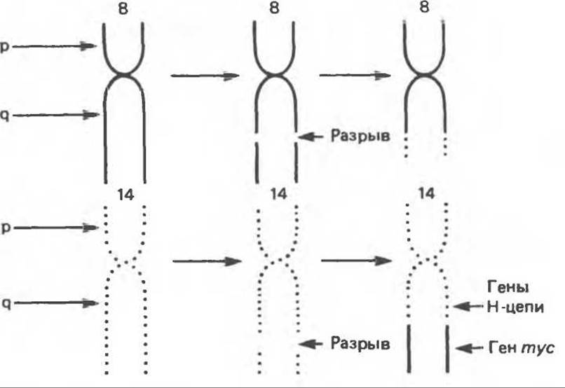
Fig. 57.5. Schematic representation of the reciprocal translocation found in Burkitt's lymphoma cells, involving chromosomes 8 and 14. The end of the q-arm of chromosome 8 is translocated to chromosome 14, and the homologous fragment of chromosome 14 is translocated to chromosome 8. The myc gene is located within the segment of chromosome 8 that is transferred to chromosome 14; it integrates downstream of the genes encoding immunoglobulin heavy chain molecules and becomes activated.
D. Gene Amplification. The amplification of certain genes (see Chapter 38) has been detected in the cells of various tumors. As it turns out, this can be induced by administration of the antitumor drug methotrexate, a Dihydrofolate Reductase inhibitor. As a result, tumor cells develop resistance to methotrexate. This resistance is caused by the amplification of the dihydrofolate reductase gene, which increases enzyme activity by approximately 400-fold. Amplified genes, with a total length of 1000 kb or even more, appear as homogeneously staining regions on the respective chromosomes. Amplified genes can also be detected in double minute chromosomes lacking centromeres. The relationship between homogeneously staining regions and double minute chromosomes is currently under investigation. The process of amplification—and consequently activation—can also affect certain cellular oncogenes. Evidence suggests that an increased copy number of certain oncogenes (e.g., c-ras) resulting from amplification may play a role in enhancing tumor malignancy (see "Tumor Progression" below).
E. Point Mutations. The c-ras oncogene was first discovered in certain rodent retroviruses (e.g., rats and mice). The product of this gene—a protein with a Molecular Weight of 21,000 (p21)—is structurally related to G-proteins that regulate adenylate cyclase activity, and thus plays a key role in cellular responses to Hormones and drugs. A sequence comparison of the c-ras proto-oncogene from normal human cells and the c-ras oncogene from bladder tumor cells revealed that they differ by only a single base and, consequently, a single amino acid (at position 12 of the p21 protein). This interesting finding was confirmed by analysis of c-ras genes from other human tumors. In all cases, the results were identical: the gene isolated from tumor cells contained only a single point mutation compared to the c-ras proto-oncogene from normal cells. Since THE POSITION OF such a mutation varies, the Location OF THE amino acid substitution varies as well. Mutations in p21 alter protein conformation and decrease its GTPase activity. This reduction in enzyme activity leads to The stimulation of adenylate cyclase activity, which is normally inhibited during The conversion of GTP to GDP (see Chapter 44). The stimulation of adenylate cyclase increases cAMP levels, which in turn affects the activity of various cAMP-dependent protein kinases, thereby shifting the balance of cellular metabolism toward transformation or its stabilization.
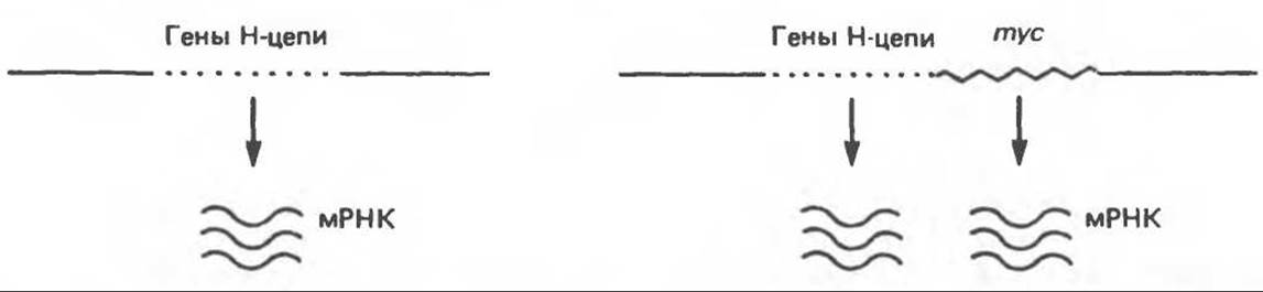
Fig. 57.6. Schematic representation of myc proto-oncogene activation via translocation in Burkitt's lymphoma cells. A small fragment of chromosome 14 before translocation, containing genes encoding portions of immunoglobulin heavy chains. Following translocation, the initially inactive myc gene falls under the control of an enhancer located within the heavy chain gene region, resulting in the activation of the myc gene. Only a single DNA strand is shown.
General Remarks on oncogene activation
Of the five oncogene activation mechanisms considered, the first four (promoter insertion, enhancer insertion, chromosomal translocations, and gene amplification) involve enhanced transcription and a consequent increase in the amount of the oncogene product, while its Primary Structure remains unchanged. An increase in product concentration alone may be sufficient to trigger cell malignant transformation. The fifth activation mechanism (point mutations) involves an alteration in the STRUCTURE OF THE oncogene product, while its quantity may remain unchanged. Thus, the presence of a structurally abnormal regulatory protein can be sufficient to drive the transformation process.

Fig. 57.7. Schematic diagram of the mechanisms by which oncogene products can affect cellular metabolism and thereby stimulate growth. cAMP influences cellular processes by activating cAMP-dependent protein kinases. Tyrosine protein kinase and protein kinase C can activate a wide range of target proteins. Cellular responses are also modulated by Ca2+ ions, Prostaglandins, and Leukotrienes derived from arachidonic acid. P — receptor; G — G-protein; AC — adenylate cyclase; PI — phosphatidylinositol; PKC — protein kinase C; TPK — tyrosine protein kinase; IP3 — inositol trisphosphate; DAG — diacylglycerol; ER — endoplasmic reticulum.
When discussing The Role of oncogenes in oncogenesis, it should be noted that oncogenes have been isolated from only about 15% of human tumors. In some cases, oncogene activation may be a consequence of transformation rather than its cause. The involvement of oncogenes in the development of experimental tumors induced by chemical carcinogens is only beginning to be explored. For instance, it was recently demonstrated that rat mammary gland tumorigenesis induced by N-nitrosomethylurea involves activation of the c-ras gene, specifically through a G→A transition mutation. This finding indicates that oncogenes are indeed involved in chemical carcinogenesis. Because the carcinogen was administered as a single dose (without a promoter) in these experiments, the detected mutation likely represents a component of the initiation stage of chemical carcinogenesis. Further research is required to elucidate the potential role of oncogenes in tumor initiation, promotion, progression, and metastasis.
Mechanisms of oncogene action
There are at least three mechanisms by which oncogenes stimulate cell growth (Fig. 57.7). 1. Oncogenes can act on key intracellular processes involved in cell growth control, rendering external stimuli unnecessary. Examples include the src gene product, which is a tyrosine protein
kinase; the ras gene product, which (indirectly) stimulates adenylate cyclase activity; or the myc gene product, which is a DNA-binding protein. In each of these instances, the oncogene can affect mitosis (with the first two oncogene products acting via the phosphorylation of key regulatory proteins). A significant gap in our understanding of cell growth regulation is that very little is known about the molecular aspects of mitotic control, even in normal cells. 2. Oncogene products can mimic the action of Polypeptide Growth Factors. 3. Oncogene products can mimic the situation arising from the interaction between a receptor and its corresponding growth factor.
Last update: 06/08/2026
Editorial and Educational Adaptation: This material has been compiled based on the primary/original source text. The project team performed an editorial review, corrected technical inaccuracies, structured sections, and adapted the content for an educational format.
What was processed:
- elimination of formatting defects (OCR errors, structural breaks, corrupted characters);
- editorial organization of content;
- standardization of terminology in accordance with academic sources;
- verification of factual statements against the original source text.
All mentions of the author, publication year, and origin of the primary text have been preserved in accordance with the source.