LEHNINGER PRINCIPLES OF BIOCHEMISTRY - VOL. 1. THE FOUNDATIONS OF BIOCHEMISTRY: STRUCTURE AND CATALYSIS - 2011
Class="right">The Cell is an atom in biology... To understand what life is, one must study the cell and determine its Structure so as to comprehend the laws of vital activity of any cell, to find those differences that account for its specific Functions.
François Jacob, The Logic of Life: A History of Heredity, 1970
1. THE FOUNDATIONS OF BIOCHEMISTRY
Some fifteen to twenty billion years ago, the universe originated in a cataclysmic explosion that hurled high-energy, incandescent subatomic particles in all directions. The simplest elements (hydrogen and helium) formed within seconds. As the universe expanded and cooled, matter condensed under the pull of gravity to form stars. Some stars became gigantic and exploded as supernovae, releasing the energy required to fuse simple atomic nuclei and form more complex chemical elements. Thus, several billion years ago, the Earth came into being with the chemical elements that exist on it today. Life arose about four billion years ago: simple microorganisms appeared, capable of deriving energy from chemical compounds, and later from sunlight, using it to synthesize a vast array of complex Biomolecules from the simple elements and compounds found on the Earth's surface.
Biochemistry aims to explain how the remarkable properties of living organisms arise from the thousands of different inanimate molecules. When isolated and studied individually, these molecules obey all the Physical and Chemical laws that describe The behavior of inanimate matter; this holds true for all processes occurring in living organisms. Biochemical research helps us understand how inanimate molecules within living organisms interact with one another to preserve and continuously maintain life, with these interactions occurring strictly in accordance with the physical and chemical laws governing inanimate matter.
Nevertheless, living organisms possess distinctive characteristics that set them apart from other material objects. What are these distinguishing features?
High degree of chemical complexity and microscopic Organization. Thousands of different molecules make up the intricate Internal Structure of the cell (Fig. 1-1, a). Among them are extremely long polymers (each macromolecule containing a characteristic set of subunits and possessing a unique three-dimensional structure), as well as the capacity for highly specific, i.e., selective, binding to other molecules within the cell.
Systems for extracting, transforming, and utilizing energy from the environment (Fig. 1-1, b) enable the Organism to build and maintain its internal structures and perform mechanical, chemical, osmotic, and electrical work. This counteracts the tendency of all inanimate matter toward decay and transition into a disordered state, ultimately achieving a dynamic steady state with the environment.
Fig. 1-1. Some characteristics of living matter. (a) Electron micrograph of a stained thin section of vertebrate Muscle tissue illustrating The complexity of the microscopic organization of living tissue; (b) a saker falcon obtains the nutrients it needs by preying on smaller birds; (c) biological reproduction is characterized by astonishing fidelity.
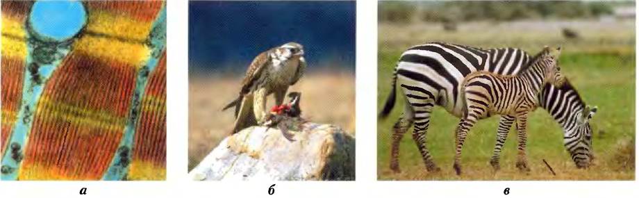
Capacity for precise self-Replication (Fig. 1-1, c). A single bacterial cell placed in a sterile nutrient medium can give rise to a billion identical "daughter" Cells within 24 hours. Each cell contains thousands of different molecules, some of extremely complex structure, yet every bacterium is an exact copy of the original, and its structure is completely determined by the information contained in the genetic material of the parent cell.
Mechanisms for sensing Changes in the surroundings continuously adjust the organism to environmental conditions by adapting its internal chemical structure or altering its position in the environment.
Defined functions for each component of a living organism and regulation of their interactions. This is a hallmark not only of macro-structures such as stem and leaf or Heart and lung, but also of microscopic intracellular structures and individual chemical compounds. The interplay of chemical components in a living organism is dynamic; changes in one compound trigger coordinated or compensatory changes in others, and the ensemble as a whole acquires traits distinct from those of its individual parts. The collection of molecules executes a program whose ultimate result is the replication of that program and of the molecular assemblage itself—that is, life.
Capacity to change over time by gradual evolution. Organisms change their inherited survival strategies very gradually in response to changing environmental conditions. The result of endless evolution is the vast diversity of life forms, outwardly quite different (Fig. 1-2), but fundamentally linked by common descent. This communality of All living organisms at THE MOLECULAR LEVEL is manifested in similarities of Gene sequences and protein structures.
Fig. 1-2. Diverse living organisms share the same chemical makeup. Birds, animals, plants, soil microorganisms, and humans possess the same basic structural units (cells) and macromolecules (DNA, RNA, Proteins) built from the same types of monomeric units (NUCLEOTIDES, Amino Acids). Living organisms employ the same metabolic pathways to synthesize cellular components, share a common Genetic Code, and descend from the same evolutionary ancestors. Shown is a detail from the painting "The Garden of Eden" by Jan van Kessel the Younger (1626–1679).

Despite these shared properties and the fundamental unity of life, it is difficult to make generalizations that apply to all living organisms. The Diversity of life on Earth is enormous. The range of habitats from hot springs to arctic tundra, and from animal intestines to college dormitories, corresponds to a wide spectrum of specific biochemical adaptations achieved within a common chemical framework. For the sake of clarity, in this book we occasionally use generalizations that may be imperfect yet useful. Furthermore, we frequently draw attention to exceptions that can be supported by Examples.
Biochemistry describes at the molecular level the structures, mechanisms, and chemical processes shared by all organisms and provides organizing principles that underlie all life forms—principles that can be termed the molecular logic of life. Although biochemistry contributes significantly to basic and applied medicine, agriculture, Nutrition, and industry, its primary objective is The Study of life itself.
In this introductory chapter, we will briefly review the cellular, chemical, physical (thermodynamic), and Genetic foundations of biochemistry, as well as the overarching principle of evolution—the alteration of living cell properties across generations. As you read through the book, returning to this chapter may prove helpful for refreshing the fundamental material presented here.
1.1. Cellular Foundations
The unity and diversity of organisms become apparent already at THE CELLULAR LEVEL. The smallest organisms consist of a single cell and are microscopic in size. Larger Multicellular Organisms contain many different cell types, differing in size, shape, and specific functions. Despite these obvious differences, all cells, from the simplest to the most complex organisms, share fundamental properties that can be investigated at the biochemical level.
Cells are the Structural and functional units of all living organisms
Cells of all types share common structural features (Fig. 1-3). The Plasma Membrane defines the periphery of the cell, separating its contents from the external environment. The membrane consists of lipid and protein molecules that form a thin, tough, pliable, hydrophobic barrier around the cell. The membrane prevents the free passage of inorganic ions and most other charged or polar compounds. Transport proteins embedded in The Plasma Membrane facilitate the passage of specific ions and molecules; receptor proteins transmit signals into the cell; and membrane Enzymes participate in certain metabolic reactions. Because individual lipid and protein molecules in the plasma membrane are not covalently linked, the entire structure is remarkably flexible, allowing the cell to change its shape and size. As the cell grows, newly synthesized lipid and protein molecules are incorporated into the plasma membrane. When each cell divides, it yields two new cells, each surrounded by its own membrane. Cell Growth and Division (Cleavage) take place without loss of membrane integrity.
Fig. 1-3. Universal features of living cells. All cells have a Nucleus or nucleoid, a plasma membrane, and Cytoplasm. The Cytosol refers to that portion of the cytoplasm that remains in the supernatant after centrifugation of a cell extract at 150,000 × g for 1 h.
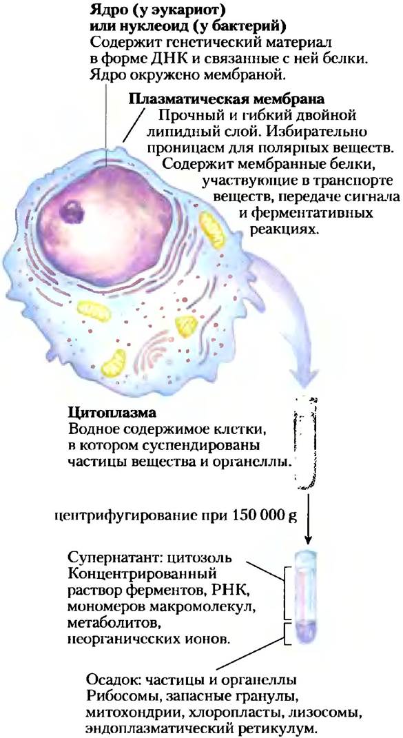
The internal space enclosed by the plasma membrane is filled with cytoplasm (Fig. 1-3), which is an aqueous medium (cytosol) containing a myriad of particles performing diverse functions. The cytosol is a concentrated solution containing enzymes and the RNA molecules that encode them, the building blocks for these macromolecules (Amino Acids and nucleotides), hundreds of small organic molecules (metabolites, which are intermediates in Biosynthesis AND DEGRADATION), Coenzymes for numerous enzymatic reactions, inorganic ions, and supramolecular structures such as Ribosomes, where Protein Synthesis takes place, and proteasomes, where proteins no longer needed by the cell are degraded.
All cells, at least during a certain period of their life cycle, have a nucleus or nucleoid in which the genome—the complete set of genes consisting of DNA—is stored and replicated. In Bacteria and archaea, the nucleoid is not separated from the cytoplasm by a membrane. In the large group of eukaryotes (from the Greek eu, meaning true, and karyon, meaning nucleus), The Nucleus consists of nuclear material enclosed within a double-layered membrane, the nuclear envelope. Cells possessing a nuclear envelope are called eukaryotic. All microorganisms lacking a nuclear envelope were formerly referred to as prokaryotes (from the Greek pro, meaning before, and karyon, meaning nucleus); however, they are now classified separately into the domain Archaea and the domain Bacteria (see below).
Cell size is limited by oxygen diffusion
Most cells are microscopic and invisible to the naked eye. Animal and plant cells typically range from 5 to 100 µm in diameter, whereas the length of most single-celled microorganisms usually does not exceed 1–2 µm. What limits cell size? The lower limit is presumably determined by the minimum number of different biomolecules required by the cell. The smallest bacterial cells, Mycoplasmas, have a diameter of 300 nm and a volume of about 10-14 mL. A bacterial ribosome measures about 20 nm in its largest dimension, meaning that just a few ribosomes occupy a significant portion of a mycoplasma cell's volume.
The upper limit of cell size is likely determined by The rate of diffusion of dissolved substances in an aqueous medium. For example, a bacterial cell that relies on energy-yielding reactions involving oxygen consumption obtains molecular oxygen by diffusion from the surrounding environment across the plasma membrane. The cell is so small, and its surface-to-volume ratio is so large, that the diffusing oxygen easily reaches any part of the cytoplasm. However, as cell size increases, the surface-to-volume ratio decreases, and metabolic oxygen consumption rises faster than the rate at which oxygen enters the cell via diffusion. Thus, above a certain cell size, METABOLISM utilizing O2 becomes impossible, which establishes the theoretical upper limit for cell size.
There are three domains of living organisms
Every living organism can be assigned to one of three major groups (domains)—the three branches of evolution stemming from a common ancestor (Fig. 1-4). Two Major Groups of unicellular microorganisms differ in their biochemical properties: bacteria and archaea. Bacteria inhabit soil, surface waters, and the Tissues of living or decaying organisms. Most of the best-studied bacteria, including Escherichia coli, belong to eubacteria. Archaea were designated as a separate domain by Carl Woese in the 1980s. Many of these organisms thrive in extreme natural environments, such as salt lakes, hot springs, highly acidic bogs, and the deep ocean. Available data suggest that archaea and bacteria diverged into separate branches early in evolution. All eukaryotic organisms constitute the third domain, eukaryotes, which originates from the same evolutionary branch that gave rise to archaea. Thus, archaea are more closely related to eukaryotes than to bacteria.
Fig. 1-4. Phylogeny of the three domains of living organisms. Phylogenetic relationships are often illustrated using a "family tree" of this type. The construction of such a tree is based on the sequence similarity of ribosomal RNA within each group; the greater the sequence similarity, the closer the branches are positioned, so that the distance between any two branches reflects the degree of divergence between the two sequences. Phylogenetic trees can also be based on Amino Acid Sequence similarities in a single protein. For example, the tree shown in Fig. 3-32 was constructed by comparing sequences of the GroEL protein (a bacterial protein involved in protein folding). Fig. 3-33 presents a "consensus" tree, constructed by incorporating multiple comparison parameters—such as those listed above—to achieve a more accurate representation of the evolutionary affinities among groups of organisms.

Members of the domains Archaea and Bacteria are subdivided based on their environmental conditions. Aerobic organisms, inhabiting oxygen-rich environments, derive energy by transferring electrons from fuel molecules to oxygen. Microorganisms adapted to anaerobic conditions, where oxygen is virtually absent, obtain energy by transferring electrons to nitrate (yielding N2), sulfate (yielding H2S), or CO2 (yielding CH4). Many organisms that evolved under anaerobic conditions are obligate anaerobes, meaning they are killed by the presence of oxygen. Others are facultative anaerobes, capable of living both in the absence and presence of oxygen.
Organisms can be classified According to the way they obtain the energy and carbon required for synthesizing cellular material (Fig. 1-5). Based on their energy source, organisms are divided into two broad groups: phototrophs (from the Greek trophe, meaning nourishment) use sunlight, whereas chemotrophs obtain energy by oxidizing fuel molecules. A subset of chemotrophic organisms, known as lithotrophs, can oxidize inorganic substances: HS- to elemental sulfur S0, S0 to SO42-, NO2- to NO3-, and Fe2+ to Fe3+. Organotrophs oxidize various Organic compounds found in their environment. Phototrophs and chemotrophs can also be subdivided according to whether they use CO2 as a carbon source (autotrophs) or must obtain carbon from organic nutrients (heterotrophs).
Fig. 1-5. Organisms can be classified according to their sources of energy (sunlight or The oxidation of chemical compounds) and carbon used for the synthesis of cellular constituents.

The bacterium Escherichia coli is the most studied bacterium
Bacterial cell architecture shares common structural features as well as certain group-specific traits (Fig. 1-6). Under normal conditions, E. coli is a harmless resident of the human intestine. Its cell is about 2 µm long and slightly less than 1 µm in diameter. The cell is protected by an outer membrane, while the inner plasma membrane encloses the space containing the cytoplasm and nucleoid. The space between the outer and inner membranes is occupied by a thin yet rigid layer of polymers called peptidoglycans, which impart mechanical strength and a definite shape to the cell. The plasma membrane, together with the layers external to it, constitutes the cell envelope. The cells of archaea derive their mechanical strength from a different type of polymer, pseudopeptidoglycans. The bacterial plasma membrane consists of a thin lipid bilayer with embedded proteins. Archaeal Membranes have a similar architecture, yet their Membrane Lipids are strikingly different from those of bacteria (see Fig. 10-12).
Fig. 1-6. Common Structural elements of bacterial cells. Due to differences in Cell wall structure, some eubacteria (gram-positive bacteria) retain the stain in the Gram Staining Procedure, whereas others do not (gram-negative bacteria). E. coli is a gram-negative bacterium. Cyanobacteria are eubacteria characterized by a specialized internal Membrane Structure that houses Photosynthetic Pigments. The cell walls of archaea and gram-positive eubacteria appear similar under an Electron microscope, but the chemical structures of their membrane lipids and Polysaccharides differ markedly (see Fig. 10-12).

The cytoplasm of E. coli contains about 15,000 ribosomes, ten to a thousand copies of each of approximately 1,000 different enzymes, and about 1,000 organic compounds with molecular weights below 1,000 (metabolites and Cofactors). The nucleoid houses a single circular DNA molecule, while the cytoplasm, like that of many other bacteria, contains one or more smaller circular DNA molecules called Plasmids. In nature, certain plasmids confer resistance to various environmental toxins and Antibiotics. In laboratory settings, these DNA molecules are exceptionally convenient for experimental manipulation and are widely utilized by molecular geneticists.
Most bacteria (including E. coli) exist as individual cells, but certain species (such as myxobacteria) exhibit primitive "social behavior" by forming multicellular aggregates.
Eukaryotic cells contain various membrane-bound Organelles that can be isolated and studied
Typical eukaryotic cells (Fig. 1-7) are many times larger than bacterial cells, typically ranging from 5 to 100 µm in diameter, with volumes thousands to millions of times greater. A hallmark of eukaryotes is the presence of a nucleus and numerous membrane-bound organelles with specialized functions: Mitochondria, The Endoplasmic reticulum, the Golgi apparatus, Peroxisomes, and Lysosomes. Plant cells additionally contain vacuoles and Chloroplasts (Fig. 1-7). The cytoplasm of many cells also contains granules or droplets storing nutrient reserves, such as starch and fats.
Fig. 1-7. Structure of a Introduction/5.html">Eukaryotic Cell. Schematically illustrating the two main cell types: top, an animal cell; bottom, a plant cell. Plant cells typically range from 10 to 100 µm in diameter, whereas animal cells range from 5 to 30 µm. Structures highlighted in red are exclusive to either animal or plant cells. Cells of eukaryotic microorganisms (such as protists and Fungi) possess structures similar to those of PLANT AND ANIMAL cells, and many also contain specialized organelles not depicted here.

A significant contribution to The Development of biochemistry was made by the work of Albert Claude, Christian de Duve, and George Palade on Methods for separating cellular organelles—a crucial step in isolating and studying the functions of biomolecules and larger cellular structures. In a standard fractionation procedure (Fig. 1-8), cells or tissues in solution are gently homogenized. This disrupts the plasma membrane while leaving most cellular organelles intact. The homogenate is then centrifuged. Nuclei, mitochondria, and lysosomes differ in size and consequently sediment at different rates. They also differ in buoyant density and are thus separated into distinct fractions during density gradient centrifugation.
Fig. 1-8. Fractionation of subcellular tissue structures. To isolate intracellular organelles, tissues such as Liver are first mechanically homogenized to rupture the cells and disperse their contents into a buffer. A sucrose solution provides an osmotic pressure nearly identical to that inside the cellular organelles, preventing Water from entering, Swelling, and destroying them. a) Large and small particles in suspension can be separated by differential centrifugation at varying speeds. b) Particles of different densities can be separated by isopycnic centrifugation. In this method, centrifuge tubes are filled with a solution whose density increases from top to bottom. This density gradient is created, for example, by preparing solutions with varying sucrose concentrations. The organelle mixture is then layered on top of the tube and centrifuged at high speed. As a result, the organelles sediment through the gradient until they reach a position whose density exactly matches their own. After centrifugation, each layer can be collected separately from the tube.

Differential centrifugation results in a crude fractionation of the cytoplasmic contents. These fractions can then be further separated by isopycnic* centrifugation. In this procedure, organelles with different buoyant densities (resulting from different protein-to-lipid ratios) are separated by centrifugation through a solvent with a density gradient. By carefully isolating the contents of each gradient fraction and examining them under a microscope, a biochemist can determine THE POSITION OF each organelle fraction within the centrifuge tube and isolate a purified fraction for further study. Using this method, it was established, for example, that lysosomes contain hydrolytic enzymes, mitochondria contain oxidative enzymes, and chloroplasts contain photosynthetic pigments. The isolation of an organelle containing a specific enzyme is frequently the first step in the purification of that enzyme.
* Isopycnic — having the same density.
The Cytoplasm Possesses a Cytoskeleton and Dynamic Properties
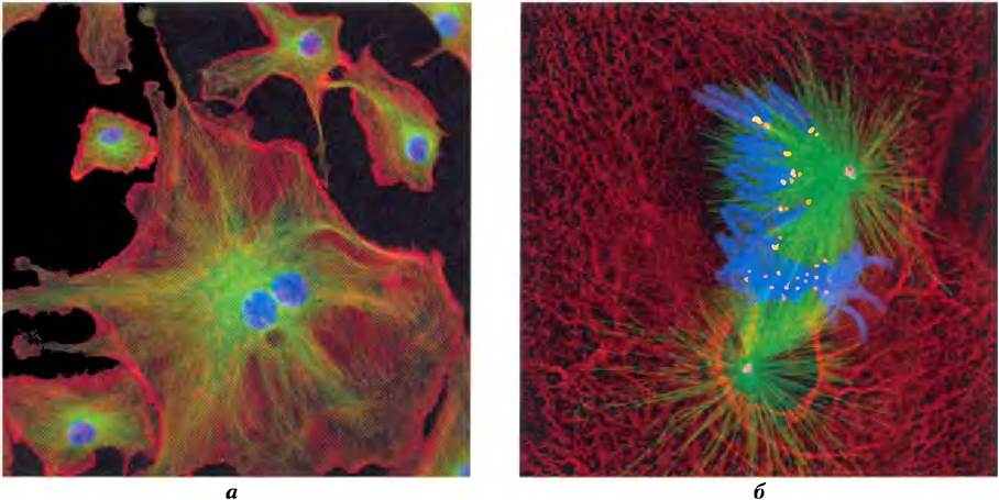
Fluorescence Microscopy reveals several types of protein filaments that crisscross The Eukaryotic Cell, forming a three-dimensional network known as the cytoskeleton. There are three principal types of cytoplasmic filaments—Actin filaments, microtubules, and Intermediate filaments (Fig. 1-9); they differ in thickness (ranging from 6 to 22 nm), structure, and specific functions. All of these filaments serve to organize and structure the cytoplasm and impart shape to the cell. Actin filaments and microtubules also facilitate the movement of organelles and entire cells.
All cytoskeletal filaments, regardless of type, are built from simple protein molecules that associate noncovalently to form strands of defined thickness. These filaments are not static structures; they continuously dissociate into their constituent subunits and reassemble. Their intracellular positioning is likewise not strictly fixed, but undergoes significant changes during mitosis, cytokinesis, amoeboid movement, or alterations in cell shape. The disassembly, assembly, and localization of all filament types are regulated by accessory proteins required for cross-linking the filaments or for the translocation of cytoplasmic organelles along them.
Fig. 1-9. The Three types of cytoskeletal fibers: actin filaments, microtubules, and intermediate filaments. Cellular structures can be visualized using Antibodies (which recognize specific proteins) covalently linked to fluorescent tags. When cells treated in this manner are viewed under a fluorescence microscope, the corresponding structures become visible. a) Bovine pulmonary artery endothelial cells. Bundles of actin filaments, known as stress fibers, are stained red; microtubules radiating from the cell center are stained green; and Chromosomes within the nucleus are stained blue. b) Newt lung cells in mitosis. Microtubules (green) attached to kinetochores (yellow) on condensed chromosomes (blue) pull the chromosomes toward opposite poles of the cell (centrosomes; magenta). Intermediate filaments composed of keratin (red) serve to maintain cellular structure.
The structure described above corresponds to the condition observed in eukaryotic cells, featuring a reticular network of filaments and a complex Organization of Membrane-bounded compartments (Fig. 1-7). Filaments disassemble and reassemble elsewhere in the cell. Membrane-enclosed vesicles bud off from one organelle and fuse with another. Organelles move through the cytoplasm along protein filaments, with motor proteins providing the energy for these movements. Internal membranes compartmentalize distinct metabolic processes and provide surfaces on which specific enzymatic reactions take place. The transport mechanisms of Endocytosis and Exocytosis, which mediate The entry of substances into the cell and their release from it, respectively, involve the fusion and fission of membranes. These processes facilitate exchange between the cytoplasm and the extracellular environment, allowing the secretion of cellular products and the uptake of extracellular Materials.
This cytoplasmic organization is complex, yet far from chaotic. The movement and localization of organelles and cytoskeletal elements are under strict control. At specific Stages of the eukaryotic Cell Cycle, major, precisely orchestrated rearrangements occur, such as mitosis. Interactions between the cytoskeleton and organelles are noncovalent and reversible; they are regulated in response to various intracellular and extracellular signals.
Supramolecular Structures Exist Within Cells
Macromolecules and their monomeric units vary greatly in size (Fig. 1-10). An Alanine molecule is less than 0.5 nm long. The oxygen-carrying Hemoglobin molecule in erythrocytes consists of approximately 600 amino acid residues organized into four long chains folded into globular structures, with native hemoglobin reaching a diameter of 5.5 nm. Proteins, in turn, are much smaller than ribosomes (the latter being about 20 nm in diameter), which are themselves vastly smaller than organelles such as mitochondria (which have a diameter of about 1000 nm). There is a colossal scale difference between simple biomolecules and the cellular structures visible under a Light Microscope. The hierarchy of cellular structural organization is illustrated in Fig. 1-11.
Fig. 1-10. Organic molecules that constitute the bulk of cellular matter: the biochemical alphabet. (a) Six of the twenty amino acids that make up all proteins (side chains are highlighted in pink); (b) five nitrogenous bases, two five-carbon sugars, and phosphoric acid, which comprise all Nucleic Acids; (c) five components of membrane lipids; (d) D-glucose, the monomeric unit of most CARBOHYDRATES. Note that phosphoric acid is a constituent of both Nucleic Acids and membrane lipids.
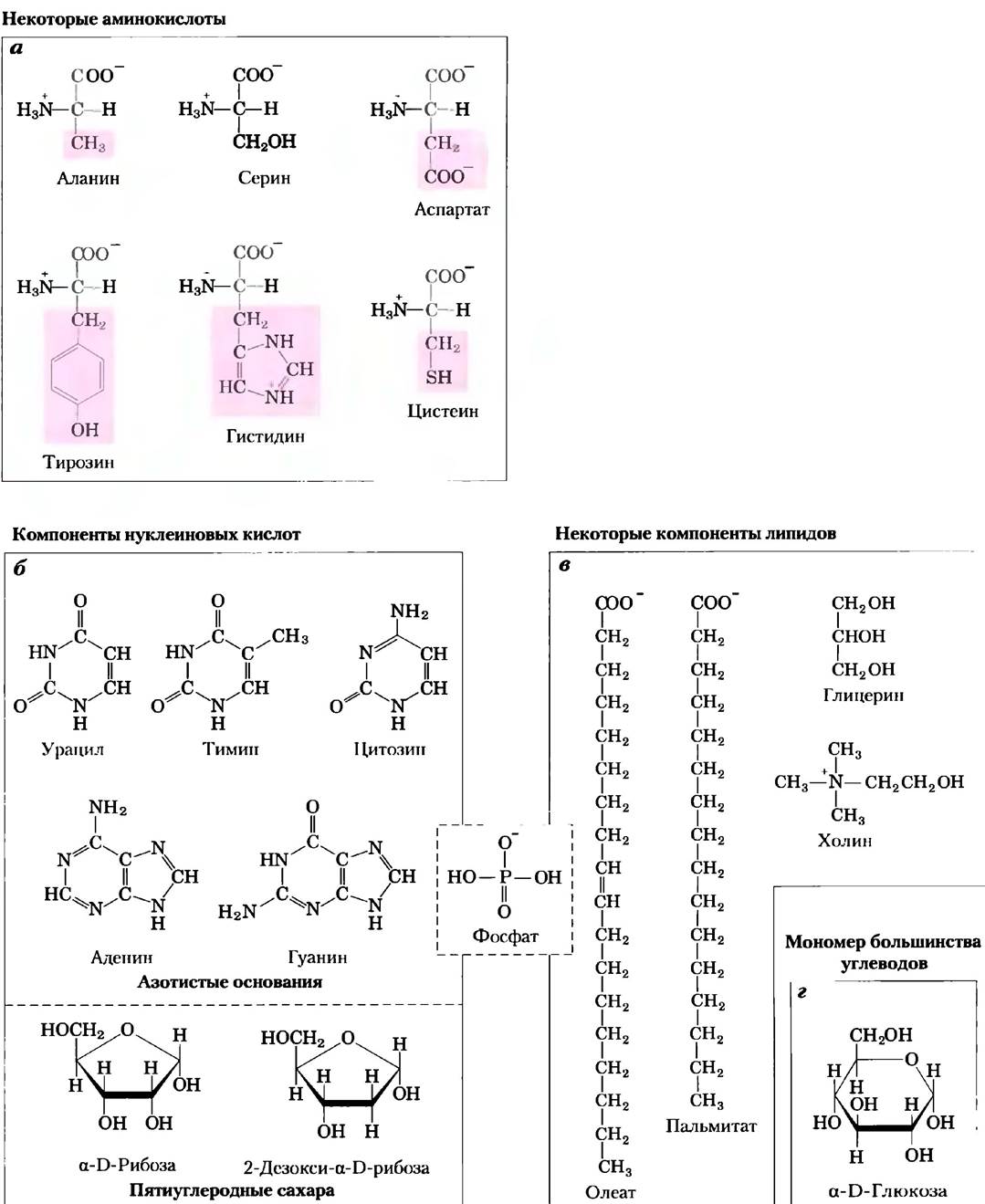
Fig. 1-11. Structural hierarchy in the Molecular organization of a cell. In this plant cell, the nucleus contains several types of supramolecular complexes, including chromosomes. Chromosomes consist of DNA macromolecules and a variety of proteins. Each type of macromolecule is built from simple monomeric units; for example, DNA is composed of nucleotides (deoxyribonucleotides).

The monomeric units of proteins, nucleic acids, and polysaccharides are linked by covalent bonds. In supramolecular complexes, molecules are held together by much weaker noncovalent interactions, prominent among which are Hydrogen Bonds (between polar groups), ionic interactions (between charged groups), hydrophobic interactions (between nonpolar groups in aqueous environments), and Van der Waals interactions—all of which possess considerably less energy than a covalent bond.
The Nature of these noncovalent interactions is discussed in Chapter 2. Macromolecules within supramolecular structures are held together by a vast number of weak interactions that confer a unique and stable architecture upon these complexes.
In vitro studies may fail to reveal crucial interactions between molecules
One approach to investigating biological processes is to study purified molecules in vitro ("in Glass") in the absence of other "extraneous" molecules present within the intact (i.e., in vivo, "in the living organism") cell. While this reductionist approach is immensely useful, one must not forget that the interior of a cell differs profoundly from the contents of a test tube. The "extraneous" components removed during purification can play a vital role in the biological action or Regulation of the isolated molecule. For example, studies of pure enzymes in vitro are typically performed at very low enzyme concentrations in thoroughly stirred aqueous solutions. Within cells, however, enzymes are dissolved or suspended in a gel-like cytosol alongside thousands of other proteins, some of which bind to the enzyme in question and modulate its activity. Many enzymes are integrated into multienzyme complexes, wherein reaction intermediates are directly "channeled" from one enzyme to the next without ever escaping into the surrounding medium. Because diffusion is restricted in the gel-like cytosol, its composition can vary significantly from one region of the cell to another. In short, a molecule under investigation may behave quite differently in a test tube than it does inside a cell. A primary objective of biochemistry is to evaluate how Cellular Organization and macromolecular complexes influence the function of individual enzymes and other biological molecules, thereby bridging our understanding of their behavior both in vivo and in vitro.
Summary of Section 1.1. Principles of Cellular Organization
■ All cells are bounded by a plasma membrane, contain a cytosol enriched with dissolved metabolites, coenzymes, inorganic ions, and enzymes, and harbor a set of genes enclosed either in a nucleoid (in bacteria and archaea) or a nucleus (in eukaryotes).
■ Phototrophs harness sunlight for their energy needs, whereas chemotrophs oxidize fuel molecules, transferring electrons to electron acceptors such as inorganic substances, organic compounds, or molecular oxygen.
■ Bacterial and archaeal cells feature a cytosol, nucleoid, and plasmids. Eukaryotic cells contain a true nucleus and are subdivided into numerous compartments where specific processes take place within distinct organelles; these organelles can be isolated and studied independently.
■ Cytoskeletal proteins are assembled into long filaments that give the cell mechanical strength, determine its shape, and serve as tracks for the movement of cellular organelles.
■ Supramolecular complexes are held together by non-covalent interactions and feature a hierarchical structure; some of these complexes can be observed under a light microscope. When individual molecules are isolated from these complexes for in vitro studies, certain crucial patterns of cellular activity may be overlooked.
Last update: 06/08/2026
Editorial and Educational Adaptation: This material has been compiled based on the primary/original source text. The project team performed an editorial review, corrected technical inaccuracies, structured sections, and adapted the content for an educational format.
What was processed:
- elimination of formatting defects (OCR errors, structural breaks, corrupted characters);
- editorial organization of content;
- standardization of terminology in accordance with academic sources;
- verification of factual statements against the original source text.
All mentions of the author, publication year, and origin of the primary text have been preserved in accordance with the source.