BIOCHEMISTRY: A TEXTBOOK FOR HIGHER EDUCATION - E. S. Severin - 2004
CHAPTER 11. HORMONAL REGULATION OF METABOLISM AND BODY FUNCTIONS
V. Changes in Hormonal Status and Metabolism in Diabetes Mellitus
Diabetes Mellitus is a condition caused by an absolute or relative Insulin deficiency.
A. Main Clinical forms of Diabetes Mellitus
According to the World Health Organization, diabetes mellitus is classified into two main forms based on genetic and clinical differences: Type I diabetes, or insulin-dependent diabetes mellitus (IDDM), and Type II diabetes, or non-insulin-dependent diabetes mellitus (NIDDM).
1. Insulin-Dependent Diabetes Mellitus
Insulin-dependent diabetes mellitus is a disease caused by the destruction of β-Cells in the pancreatic islets of Langerhans.
The destruction of β-cells results from autoimmune reactions, involving lymphocytes and macrophages (monocytes). These cells produce cytokines that either directly damage β-cells or mediate cellular responses against them.
Type I diabetes can be triggered by viral infections that destroy β-cells. Such Viruses, known as β-cytotropic viruses, include smallpox, rubella, measles, cytomegalovirus, mumps, Coxsackie, and Adenoviruses. Some β-cytotropic viruses cause lysis of β-cells.
Certain toxic substances are also known—such as nitrosourea derivatives and other nitro- or amino-containing compounds—that selectively target β-cells and trigger an autoimmune reaction. Additionally, IDDM may result from a partial, genetically determined defect in the immune surveillance system and often coexists with other autoimmune disorders.
IDDM accounts for approximately 25–30% of all cases of diabetes mellitus. Typically, the destruction of β-cells occurs slowly, and the onset of the disease is not accompanied by metabolic disturbances. Once 80–95% of the cells are destroyed, an absolute insulin deficiency occurs, leading to severe Metabolic Disorders. IDDM most commonly affects children, adolescents, and young adults, but it can manifest at any age (starting from one year old).
2. Non-Insulin-Dependent Diabetes Mellitus
Non-insulin-dependent diabetes mellitus is an umbrella term for several conditions that develop As a result of a relative insulin deficiency. This deficiency arises from impaired insulin secretion, defective conversion of proinsulin to insulin, an increased rate of insulin Catabolism, or disrupted insulin signaling mechanisms in target cells (e.g., Insulin Receptor defects or impaired intracellular insulin signaling mediators). NIDDM typically affects individuals over the age of 40. Type II diabetes is characterized by a high frequency of familial forms; the risk of NIDDM in close relatives of a patient reaches 50%, whereas for IDDM it does not exceed 10%. The disease predominantly affects residents of developed countries, especially urban populations.
Potential causes of NIDDM include The formation of Antibodies against insulin receptors, Genetic Defects in the post-receptor apparatus of insulin-dependent Tissues, and dysregulation of insulin secretion. Key Factors Determining the development and clinical course of the disease include obesity, poor diet, a sedentary lifestyle, and stress.
Mutations in genes controlling insulin secretion, METABOLISM/26.html">Energy Metabolism in β-cells, and Glucose metabolism in insulin target cells lead to several forms of NIDDM with an Autosomal dominant inheritance pattern.
Obesity serves as the primary triggering factor for non-insulin-dependent diabetes. This type of diabetes is frequently associated with hyperinsulinemia, which further promotes obesity. Thus, obesity acts both as a major risk factor and as one of the early manifestations of diabetes mellitus.
B. Metabolic Changes in Diabetes Mellitus
In diabetes mellitus, the insulin/Glucagon ratio is typically decreased. As a result, The stimulation of Glycogen and fat storage processes is weakened, while the mobilization of energy reserves is intensified. Even after a meal, the Liver, Muscles, and adipose tissue continue to function in a postabsorptive state.
1. Symptoms of Diabetes Mellitus
All forms of diabetes are characterized by an elevated Blood glucose concentration—hyperglycemia. Following a meal, glucose levels can reach 300–500 mg/dL and remain persistently high during the postabsorptive period, indicating impaired glucose tolerance. Impaired glucose tolerance is observed in latent (subclinical) forms of diabetes mellitus. In such cases, individuals lack the Complaints and clinical symptoms typical of diabetes, and their fasting blood glucose levels are within the normal range. However, provocative tests (such as a glucose tolerance test) reveal decreased glucose tolerance (Fig. 11-30).
Class="center">Fig. 11-30. Changes in glucose tolerance in patients with latent diabetes mellitus. Glucose tolerance testing is used to diagnose diabetes mellitus. The subject ingests a glucose solution at a dosage of 1 g per 1 kg of body weight (glucose load). Blood glucose concentrations are measured over 2–3 hours at 30-minute intervals. 1 — healthy individual; 2 — diabetes mellitus patient.

The elevated Blood Plasma glucose concentration is caused by a reduced rate of glucose utilization by tissues, resulting from insulin deficiency or diminished biological activity of insulin in target tissues.
Insulin deficiency leads to a decrease in the number of glucose transporter Proteins (GLUT-4) on the membranes of insulin-dependent cells (adipose tissue and Muscle). In muscles and the liver, glucose is not stored as glycogen, and in adipose tissue, The rate of fat synthesis and storage declines. Furthermore, a lowered insulin-glucagon index activates Gluconeogenesis from Amino Acids, glycerol, and lactate. The elevated blood glucose concentration in diabetes exceeds the renal threshold, causing glucose to be excreted in the urine (glucosuria). Under normal conditions, the proximal renal tubules reabsorb all glucose filtered by the glomeruli, provided its level does not exceed 8.9 mmol/L (160 mg/dL).
A characteristic sign of diabetes mellitus is also an elevation of ketone body concentration in the blood, known as ketonemia. At a low insulin-to-glucagon ratio, fats are not stored but instead undergo accelerated catabolism because hormone-sensitive lipase in Adipose tissue is in its phosphorylated, active form. The concentration of non-esterified Fatty acids in the blood rises. The liver takes up these Fatty Acids and oxidizes them to acetyl-CoA, which is subsequently converted into β-hydroxybutyric and acetoacetic acids. In the tissues, acetoacetate is partially decarboxylated to acetone, the distinct smell of which can be detected on the breath of diabetes patients, sometimes from a distance. An increased concentration of Ketone Bodies in the blood (above 20 mg/dL, and occasionally reaching 100 mg/dL) leads to ketonuria. The accumulation of ketone bodies reduces the blood's buffer capacity and causes acidosis.
Another hallmark of diabetes mellitus is an elevated blood level of Lipoproteins (mainly VLDL), termed hyperlipoproteinemia. Dietary fats are not stored in adipose tissue due to impaired storage mechanisms; instead, they enter the liver, where they are partially converted into triacylglycerols and transported out of the liver as part of VLDL.
In diabetes mellitus, insulin deficiency leads to a decreased rate of Protein Synthesis in the body and enhanced protein breakdown, resulting in elevated blood amino acid levels. These Amino acids are transported to The Liver and undergo deamination. The carbon skeletons of glucogenic amino acids enter gluconeogenesis, which further exacerbates hyperglycemia. The ammonia produced in this process enters the Ornithine cycle, leading to an increased concentration of urea in the blood and, consequently, in the urine—a condition known as azotemia and azouria.
High concentrations of glucose, ketone bodies, and urea require their enhanced excretion from the body. Because the Kidneys have a limited concentrating capacity, the elimination of large volumes of Water increases dramatically, which can result in severe dehydration. Urine output in patients increases several-fold, occasionally reaching 8–9 L per day, though most commonly ranging between 3–4 L, a condition known as polyuria. This fluid loss triggers constant thirst, or polydipsia.
2. Acute Complications of Diabetes Mellitus. Mechanisms of Diabetic Coma Development
Disorders of carbohydrate, lipid, and Protein metabolism in diabetes mellitus can lead to comatose states (acute complications). Diabetic coma manifests as a drastic impairment of all body Functions accompanied by loss of consciousness. The primary precursors of diabetic coma are acidosis and tissue dehydration (Fig. 11-31).
Fig. 11-31. Metabolic changes in diabetes mellitus and the causes of diabetic coma.
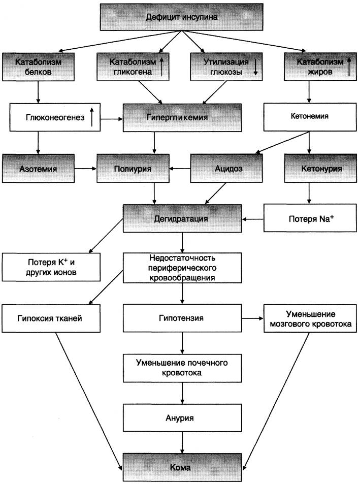
Parallel to ketoacidosis, decompensated diabetes involves a disruption in water-electrolyte balance, driven fundamentally by hyperglycemia accompanied by increased intravascular osmotic pressure. To preserve osmolarity, a compensatory fluid shift begins from cells and the extracellular space into the vascular bed. This leads to a loss of water and electrolytes from tissues—primarily Na+, K+, Cl-, and HCO3- ions. As a result, severe cellular dehydration develops along with a deficiency of intracellular ions (especially K+), which is followed by generalized dehydration. This reduces peripheral Circulation, diminishes cerebral and renal blood flow, and causes Hypoxia. Diabetic coma develops slowly, over the course of several days, but can occasionally onset within a few hours. The earliest signs may include nausea, vomiting, and lethargy. Patients exhibit decreased blood pressure.
Comatose states in diabetes mellitus can manifest in three main forms: ketoacidotic, hyperosmolar, and lactic acidotic. Ketoacidotic coma is characterized by pronounced insulin deficiency, ketoacidosis, polyuria, and polydipsia. Hyperglycemia (20–30 mmol/L), caused by insulin insufficiency, is accompanied by massive fluid and electrolyte losses, dehydration, and plasma hyperosmolality. The total concentration of ketone bodies reaches 100 mg/dL or higher.
In hyperosmolar coma, extremely high plasma glucose levels, polyuria, and polydipsia are observed, consistently accompanied by severe dehydration. It is believed that in most patients, hyperglycemia is caused by concurrent Renal Dysfunction. Ketone bodies are typically undetectable in blood serum.
In lactic acidotic coma, hypotension, reduced peripheral circulation, and tissue hypoxia predominate, shifting metabolism toward anaerobic Glycolysis and thereby raising blood lactic acid concentration (lactic acidosis).
Pure forms of specific diabetic coma variants are rarely encountered in clinical practice. Their onset can be triggered by various factors, such as infectious diseases, trauma, surgical interventions, or toxic compounds.
3. Late Complications of Diabetes Mellitus
The primary cause of late complications in diabetes mellitus is hyperglycemia. Hyperglycemia damages Blood Vessels and impairs the functions of various tissues and Organs.
One of the MAIN MECHANISMS OF tissue damage in diabetes mellitus is protein glycosylation, which leads to altered conformation and function. Although some proteins normally contain carbohydrate components—where such Glycoproteins are formed enzymatically (e.g., adenohypophyseal glycoprotein Hormones)—glucose can also non-enzymatically interact with free amino groups of proteins in The Human Body, a process known as non-enzymatic protein glycosylation. In healthy individuals, this reaction proceeds slowly. During hyperglycemia, however, the glycosylation rate accelerates. The degree of protein glycosylation depends on their turnover rate; slowly turning over proteins accumulate more modifications. One of the earliest signs of diabetes mellitus is a 2- to 3-fold increase in glycosylated Hemoglobin (normal HbA1C is 5.8–7.2%). Another example of slowly turning over proteins is crystallins, the Proteins of the lens. Upon glycosylation, crystallins form multimolecular aggregates that increase the refractive index of the lens. Lens transparency decreases, leading to opacification, or a cataract.
Proteins of the Extracellular matrix and basement membranes also belong to the slowly turning over protein group. The thickening of basement membranes, one of the characteristic complications of diabetes mellitus, leads to The Development of diabetic angiopathies.
Another cause of many late complications in diabetes mellitus is the increased rate of glucose conversion to sorbitol (see Section 7).
✵ The reaction converting glucose into the six-carbon alcohol (sorbitol) is catalyzed by the enzyme aldose reductase. Sorbitol is not utilized in other metabolic pathways, and its rate of cellular diffusion is low. In diabetic patients, sorbitol accumulates in the retina, lens, renal glomerular cells, Schwann cells, and vascular endothelium.
✵ High concentrations of sorbitol are toxic to cells. Its accumulation in Neurons leads to increased osmotic pressure, Cell Swelling, and tissue edema. For instance, lens opacification can develop as a result of sorbitol-induced lens swelling and disruption of the orderly crystallin Structure.
Diabetic angiopathies. Diabetic angiopathies are primarily caused by damage to vascular basement membranes. When plasma glucose concentration is high, Proteoglycans, collagens, and glycoproteins become glycosylated, impairing the metabolism and ratio of basement membrane components and disrupting their structural organization.
Macroangiopathies manifest as lesions in large and medium-sized Vessels of the Heart, Brain, and lower extremities. Pathological Changes in the tunica intima and damage to the middle and outer layers of the arterial wall are consequences of the glycosylation of basement membranes and extracellular matrix proteins (Collagen and Elastin), leading to reduced arterial elasticity. Combined with hyperlipidemia, this can promote the development of atherosclerosis. In diabetes mellitus, atherosclerosis occurs more frequently, develops at an earlier age, and progresses significantly faster than in non-diabetic individuals.
Microangiopathies are the result of damage to capillaries and small vessels, manifesting as nephropathy, neuropathy, and retinopathy.
Nephropathy develops in approximately one-third of diabetes patients. Electron-microscopic changes in the glomerular basement membrane can be detected as early as the first year following Diagnosis. However, in most patients, clinical signs of diabetic nephropathy appear 10–15 years after the onset of diabetes. An indicator of early-stage nephropathy is microalbuminuria (ranging from 30–300 mg/day), which subsequently progresses to classic Nephrotic Syndrome characterized by heavy proteinuria, hypoalbuminemia, and edema.
Retinopathy, the most serious complication of diabetes mellitus and the leading cause of blindness, develops in 60–80% of diabetic patients. In the early stages, Background retinopathy develops, manifesting as retinal hemorrhages, dilation of retinal vessels, and edema. If the changes spare the macula, Vision loss typically does not occur. Subsequently, proliferative retinopathy may develop, characterized by the formation of new vessels in the retina and vitreous body. The fragility and high permeability of these newly formed vessels lead to frequent hemorrhages into the retina or vitreous. Fibrosis develops at the sites of thrombi, resulting in retinal detachment and vision loss.
V. Diagnosis of Diabetes Mellitus
The diagnosis of diabetes mellitus can typically be established based on its classic symptoms — hyperglycemia, polyuria, polydipsia, polyphagia, and dry Mouth. The key biochemical markers for IDDM are identified through:
✵ the glucose tolerance test (see Fig. 11-30). A plasma glucose level exceeding 10 mmol/L at 2 hours post-glucose load indicates diabetes mellitus;
✵ measurement of glycated hemoglobin. In diabetes mellitus, the level of HbA1C, which normally accounts for about 5% of total hemoglobin, increases by 2 to 3 times;
✵ the absence or low levels of insulin and C-peptide in the blood and urine. Normally, insulin and C-peptide are secreted in equimolar concentrations. Because the liver retains approximately 2/3 of insulin, the normal insulin-to-C-peptide ratio in the portal vein and peripheral vessels is 1/3. Determining serum or urinary C-peptide levels provides a quite accurate assessment of functional β-cell reserve;
✵ albuminuria. In diabetes mellitus, daily urinary albumin excretion is approximately 30 — 300 mg, a condition known as microalbuminuria (compared to a normal baseline of about 8 mg).
Since NIDDM develops much more gradually, classic clinical symptoms, hyperglycemia, and insulin deficiency are diagnosed later, frequently in conjunction with the manifestations of long-term diabetic complications.
VI. Management Approaches for Diabetes Mellitus
The Treatment of diabetes mellitus depends on its type (I or II), is multifaceted, and includes medical Nutrition therapy, oral antidiabetic agents, insulin therapy, as well as the Prevention and management of complications.
Modern antidiabetic medications are divided into two main categories: sulfonylureas and biguanides. Agents that stimulate insulin secretion include sulfonylureas (e.g., glybenclamide/maninil). The MECHANISM OF ACTION of sulfonylureas is attributed to their effect on ATP-sensitive K+ channels. The increase in intracellular K+ concentration leads to membrane depolarization and accelerates the influx of Calcium Ions into The Cell, thereby stimulating insulin secretion.
Biguanides represent the other major class of antidiabetic drugs. According to certain studies, biguanides increase the number of GLUT-4 glucose transporters on The Plasma Membrane surface of adipose tissue and muscle cells.
Promising experimental approaches to treating diabetes mellitus include pancreatic islet or isolated β-cell transplantation, transplantation of genetically engineered cells, and stimulation of pancreatic islet regeneration.
Medical nutrition therapy is of paramount importance in both types of diabetes mellitus. A well-balanced diet is recommended: CARBOHYDRATES should account for 50 — 60% of total caloric intake (excluding easily digestible carbohydrates, beer, alcoholic beverages, syrups, pastries, etc.); proteins should comprise 15 — 20%; and total fats should not exceed 25 — 30%. Meals should be portioned into 5 — 6 intakes throughout the day.
Last update: 06/08/2026
Editorial and Educational Adaptation: This material has been compiled based on the primary/original source text. The project team performed an editorial review, corrected technical inaccuracies, structured sections, and adapted the content for an educational format.
What was processed:
- elimination of formatting defects (OCR errors, structural breaks, corrupted characters);
- editorial organization of content;
- standardization of terminology in accordance with academic sources;
- verification of factual statements against the original source text.
All mentions of the author, publication year, and origin of the primary text have been preserved in accordance with the source.