BIOCHEMISTRY: A TEXTBOOK FOR MEDICAL UNIVERSITIES - E. S. Severin - 2004
SECTION 11. HORMONAL REGULATION OF METABOLISM AND BODY FUNCTIONS
IV. Regulation of Major Energy Substrate Metabolism
Major dietary nutrients (CARBOHYDRATES, fats, and Proteins) are oxidized in the body to release Free energy, which is utilized in anabolic processes and to support physiological Functions. The energy content of major dietary components is expressed in kilocalories and is as follows: carbohydrates — 4 kcal/g, fats — 9 kcal/g, proteins — 4 kcal/g. A healthy adult requires 2,000 – 3,000 kcal (8,000 – 12,000 kJ) of energy per day.
Under a typical eating pattern, the intervals between meals are 4 – 5 hours, with an 8 – 12 hour overnight fast. During Digestion and the absorptive period (2 – 4 hours), the primary energy substrates utilized by Tissues (glucose, Fatty acids, Amino Acids) can come directly from the digestive tract. In the postabsorptive period and during starvation, energy substrates are produced through the Catabolism of stored energy reserves.
Changes in energy substrate intake and Energy Expenditure are coordinated through the precise Regulation of Metabolic processes across various Organs and systems, ensuring energy Homeostasis.
The primary role in maintaining energy homeostasis is played by the Hormones Insulin and Glucagon, as well as other counter-regulatory hormones — adrenaline, cortisol, iodothyronines, and somatotropin. Insulin and glucagon play The Central Role in regulating METABOLISM during the transition between absorptive and postabsorptive periods and during fasting.
A. Absorptive Period
The absorptive period is characterized by a temporary increase in the concentration of glucose, amino acids, and fats in Blood Plasma. Pancreatic Cells respond to this increase by enhancing insulin secretion and decreasing glucagon secretion. An elevated insulin/glucagon ratio accelerates the utilization of metabolites for energy storage, leading to the synthesis of Glycogen, fats, and proteins. The storage mode is triggered following a meal and is replaced by the mobilization mode once digestion is complete. The type of metabolites consumed, stored, and exported depends on the tissue type. The primary organs involved in shifting the metabolic flux between mobilization and storage modes are the Liver, adipose tissue, and Muscles (Fig. 11-28).
Class="center">Fig. 11-28. Pathways of major energy substrate utilization during the absorptive period. 1 — hepatic glycogen Biosynthesis; 2 — Glycolysis; 3 — hepatic TAG biosynthesis; 4 — TAG biosynthesis in adipose tissue; 5 — Muscle glycogen biosynthesis; 6 — Protein Biosynthesis in various tissues, including the liver.

1. Metabolic Changes in the Liver During the Absorptive Period
Following a meal, the liver becomes the primary consumer of glucose derived from the digestive tract. Nearly 60 out of every 100 g of glucose transported by the portal system is retained in the liver. The increased hepatic uptake of glucose is not a result of accelerated transport into cells (Glucose Transport into hepatocytes is not stimulated by insulin), but rather a consequence of accelerated metabolic pathways that convert glucose into stored energy forms: glycogen and fats.
As the glucose concentration rises within hepatocytes, glucokinase is activated, converting glucose into glucose-6-phosphate. Glucokinase has a high Km for glucose, which ensures a high phosphorylation rate at elevated glucose concentrations. Furthermore, glucokinase is not inhibited by glucose-6-phosphate (see Section 7). Insulin induces the synthesis of glucokinase mRNA. The elevated concentration of glucose-6-phosphate in hepatocytes accelerates glycogen synthesis. This process is facilitated by the simultaneous inactivation of Glycogen phosphorylase and the activation of glycogen synthase. Under The Influence of insulin, glycolysis is accelerated in hepatocytes due to an increase in the activity and amount of Key Enzymes: glucokinase, Phosphofructokinase, and Pyruvate kinase. At the same time, Gluconeogenesis is inhibited As a result of the inactivation of fructose-1,6-bisphosphatase and a decrease in the levels of phosphoenolpyruvate carboxykinase, which are Key Enzymes of gluconeogenesis. The increased concentration of glucose-6-phosphate in hepatocytes during the absorptive period is coupled with the active utilization of NADPH for fatty acid synthesis, thereby stimulating the Pentose Phosphate Pathway.
The acceleration of fatty acid synthesis is supported by the availability of substrates (acetyl-CoA and NADPH) generated through glucose metabolism, as well as the activation and induction of key enzymes involved in fatty acid synthesis (see Section 8 and Table 11-7).
Protein Synthesis is accelerated in the liver during the absorptive period. However, The amount of amino acids arriving at the liver from the digestive tract exceeds the capacity for their utilization in the synthesis of proteins and other nitrogen-containing compounds. Excess Amino acids are either released into the blood and transported to other tissues or deaminated, with their carbon skeletons subsequently entering the common catabolic pathway (see Section 9).
2. Metabolic Changes in Adipocytes
The primary function of Adipose tissue is to store energy in the form of triacylglycerols (TAGs). Under the influence of insulin, glucose transport into adipocytes is accelerated. The rise in intracellular glucose concentration and the activation of key glycolytic enzymes ensure The production of acetyl-CoA and glycerol-3-phosphate, which are required for TAG synthesis. Stimulation of The pentose phosphate pathway provides the NADPH needed for fatty acid synthesis. However, de novo FATTY ACID BIOSYNTHESIS in human adipose tissue proceeds at a high rate only following prior starvation. Under a normal dietary rhythm, TAG synthesis relies mainly on fatty acids delivered from chylomicrons (CM) and very-low-density Lipoproteins (VLDL) via lipoprotein lipase (see Section 8). Meanwhile, when the insulin/glucagon ratio increases, hormone-sensitive TAG lipase remains in its dephosphorylated, inactive form, thereby suppressing lipolysis.
3. Metabolic Changes in Muscles During the Absorptive Period
During the absorptive period, insulin accelerates glucose transport into muscle cells. Glucose is phosphorylated and oxidized to provide energy for The Cell, and is also used for glycogen synthesis. Fatty acids derived from CM and VLDL play a minor role in muscle Energy Metabolism during this period. The influx of amino acids into muscles and protein synthesis are also enhanced by insulin, particularly following a protein-rich meal.
B. Postabsorptive Period
The postabsorptive state refers to the period following the completion of digestion up until the next meal. If food is withheld for 24 hours or longer, this state is defined as starvation. The state following a 12-hour overnight fast is typically considered the standard postabsorptive period. At the beginning of the postabsorptive period, blood glucose levels decline, leading to a decrease in insulin secretion and an increase in glucagon concentration. As the insulin/glucagon index drops, the mobilization of stored energy reserves is accelerated (Fig. 11-29).
Fig. 11-29. Changes in major energy substrate metabolism during the transition from the absorptive to the postabsorptive state. KT — Ketone Bodies; ЖК (FA) — fatty acids.
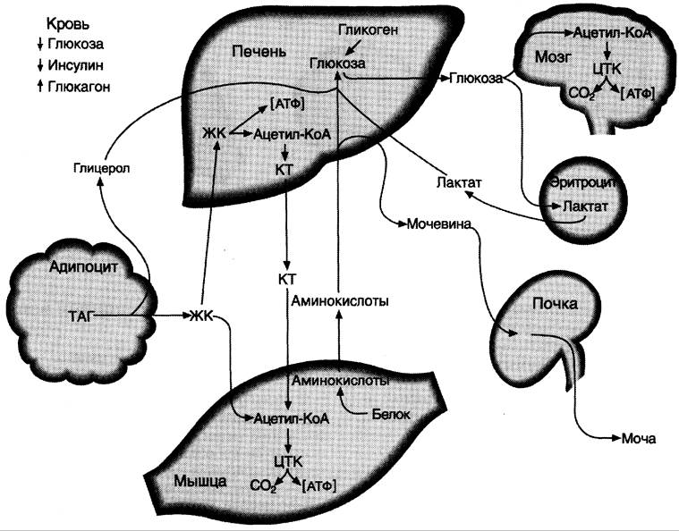
In the postabsorptive period, metabolic adjustments are aimed primarily at maintaining blood glucose concentrations, which serves as the primary energy substrate for the Brain and the sole energy source for erythrocytes. The major metabolic shifts during this period occur in The Liver and adipose tissue.
1. Metabolic changes in the liver
Glycogen mobilization in the liver is accelerated first (see Section 7). However, hepatic glycogen stores are depleted within 18–24 hours of fasting. As glycogen reserves become exhausted, gluconeogenesis emerges as the primary source of glucose, beginning to accelerate 4–6 hours after the last meal. Glycerol, amino acids, and lactate serve as substrates for glucose synthesis. When glucagon concentrations are high, The rate of fatty acid synthesis decreases due to the phosphorylation and inactivation of acetyl-CoA carboxylase, whereas the rate of β-oxidation increases. Concurrently, the liver receives an increased supply of fatty acids transported from adipose tissue depots. Acetyl-CoA generated from Fatty acid oxidation is utilized by the liver for Ketone Body Synthesis.
2. Metabolic changes in adipose tissue
In adipose tissue, elevated glucagon levels lead to a decreased rate of TAG synthesis and stimulated lipolysis. This stimulation of lipolysis results from the activation of adipocyte hormone-sensitive TAG lipase under the influence of glucagon. Fatty acids become crucial Energy Sources for the liver, muscles, and adipose tissue.
Thus, during the postabsorptive state, blood glucose concentration is maintained at 80–100 mg/dL, while the levels of Fatty Acids and ketone bodies rise.
C. Changes in hormonal status and metabolism during fasting
Fasting can be short-term, lasting up to a day (Phase I), extend for a week (Phase II), or persist for several weeks (Phase III).
In the absence of food, blood levels of glucose, amino acids, and triacylglycerols decrease. The insulin-glucagon ratio drops, and the concentration of counter-regulatory hormones, primarily cortisol, increases. Under these conditions, a state develops that is characterized by the predominance of fat, glycogen, and Protein Catabolism against the backdrop of a generally reduced metabolic rate. Driven by counter-regulatory hormones during this period, substrates are exchanged among the liver, adipose tissue, muscles, and brain. This exchange serves two main purposes: 1) maintaining blood glucose levels to supply glucose-dependent tissues (brain, erythrocytes); 2) mobilizing alternative energy sources, primarily fats, to meet the energy demands of all other tissues. Due to this metabolic shift toward energy mobilization, blood glucose concentration remains at least 60 mg/dL even after 5–6 weeks of fasting.
Because glycogen mobilization only Supports short-term fasting, gluconeogenesis becomes the main source of glucose during prolonged fasting, with amino acids, lactate, and glycerol acting as the primary substrates. Under low insulin concentrations, glucose is utilized exclusively by insulin-independent tissues, primarily the brain and erythrocytes. Other tissues meet their Energy Requirements through fatty acids and ketone bodies.
Fatty acids released during fat mobilization from adipose depots become the primary energy sources for most organs during the initial period of fasting. In Phase II, fat mobilization continues, and blood fatty acid concentrations increase 3- to 4-fold compared to the postabsorptive state. Ketone body synthesis begins within the first few days of fasting. During Phase II of fasting, the rate of ketone body synthesis increases significantly. Blood ketone body concentrations during this period can reach 20–30 mg/dL (compared to a normal 1–3 mg/dL). Ketone bodies are utilized predominantly by the muscles. During this phase of fasting, a portion of the brain's energy requirements is met by ketone bodies, while the rate of ketone body oxidation in muscles decreases.
During the first few days of fasting, Muscle Proteins—the primary source of substrates for gluconeogenesis—are rapidly degraded. When fasting exceeds 3 weeks, the Rate of protein catabolism stabilizes at approximately 20 g per day. During this period, brain consumption of ketone bodies increases, while the rate of gluconeogenesis declines. The reduction in the rate of gluconeogenesis helps conserve body proteins. Ketone bodies also become a significant energy source for the brain during this time. However, The oxidation of ketone bodies requires oxaloacetate and other TCA cycle intermediates. Normally, these are synthesized from glucose and amino acids, but during
fasting, they are derived exclusively from amino acids. If fasting continues for more than 4 weeks, atrophy develops, resulting in the loss of substantial amounts of protein. In a 70 kg human body, total protein mass is about 15 kg. The loss of 1/3 to 1/2 of body protein leads to death.
Last update: 06/08/2026
Editorial and Educational Adaptation: This material has been compiled based on the primary/original source text. The project team performed an editorial review, corrected technical inaccuracies, structured sections, and adapted the content for an educational format.
What was processed:
- elimination of formatting defects (OCR errors, structural breaks, corrupted characters);
- editorial organization of content;
- standardization of terminology in accordance with academic sources;
- verification of factual statements against the original source text.
All mentions of the author, publication year, and origin of the primary text have been preserved in accordance with the source.