Molecular Biology of the Cell - Volume 1 - Alberts B., Bray D., Lewis J., Raff M., Roberts K., Watson J. 1994
Molecular Organization of Cells
Basic Genetic Mechanisms
Viruses, Plasmids, and Transposons
In exploring the Cell/27.html">Basic Genetic Mechanisms, we have so far focused on the selective advantages they provide to The Cell. We have seen that for a cell to survive, it is absolutely essential to preserve Genetic information through DNA Repair, and for cell reproduction, it is equally necessary to replicate it rapidly and accurately. The survival of a species as a whole is known to depend on The Emergence of new, more fit genetic variants, which is greatly facilitated by Gene rearrangement and the random redistribution of DNA sequences through genetic recombination. Now we are to examine a group of genetic elements that behave as parasites, exploiting the cell's genetic machinery for their own purposes.
Under certain conditions, specific DNA nucleotide sequences can replicate independently of the rest of The Genome. These sequences vary significantly in their degree of independence from the host cell. Viral Chromosomes are the most independent, as Viruses possess protein coats that allow them to move freely from Cell to Cell. Somewhat related to viruses (though more dependent on the cell) are nucleotide sequences called Plasmids and Transposons; they lack a protein coat and therefore can replicate only within a single cell or its progeny. Even more primitive are certain DNA sequences that might tentatively be considered Mobile Genetic Elements because they occur in multiple copies within host chromosomes; however, these sequences move or replicate so rarely that it is difficult to determine whether they constitute distinct genetic elements at all.
Class="center">
Fig. 5-68. Cytology/cytology/93.html">ELECTRON MICROGRAPHS OF Viral Particles (negative staining). All viruses are photographed at the same magnification; the scale bar is shown in photo B. A. Bacteriophage T4 is a large DNA virus that infects the bacterium E. coli. The DNA is located in the HEAD of the bacteriophage and is injected into the bacterial cell through its cylindrical tail. (Courtesy of James Paulson.) B. Potato virus X. The filamentous particles of this plant virus contain an RNA genome. (Courtesy of Graham Hills.) C. Adenovirus is a DNA virus that infects various human Cells. Externally, the viral particles are surrounded by a protein coat, the capsid. (Courtesy of Mei Lie Wong.) D. Influenza virus is a large RNA animal virus. In addition to the protein capsid, it has a membrane envelope—a lipid bilayer with protruding viral glycoprotein spikes. (Courtesy of R. C. Williams, H. W. Fisher.)
All such pseudo-independent elements can replicate only by fully exploiting the host cell's METABOLISM, making them useful tools for studying normal cellular mechanisms. We will begin our Structure/133.html">Discussion with viruses, as they are the most thoroughly studied of all mobile genetic elements. We will then examine The properties of plasmids and transposons, which are sometimes remarkably similar to viruses and may be their ancestors.
5-39
5.5.1. Viruses are mobile genetic elements [43]
Viruses were first described as disease-causing agents that replicate only inside cells and are so small that they can pass through ultra-fine filters that retain even the smallest Bacteria. Before the advent of the Electron microscope, their nature remained unclear, although it was already suggested that they might simply be genes that had acquired The ability to move from cell to cell. In the 1930s, The Use of the ultracentrifuge made it possible to separate viruses from host cell components. As a result, by the early 1940s, it became clear that all viruses contain Nucleic Acids. This reinforced the idea among researchers that viruses and genetic material perform similar Functions. Confirmation of this view came from the STUDY OF BACTERIAL viruses (Bacteriophages). In 1952, it was shown that only the bacteriophage DNA (without its protein) enters the host bacterial cell, and that it is this DNA that initiates Replication, ultimately leading to the appearance of several hundred progeny viral particles in the infected cell. Thus, viruses can be viewed as genetic elements enclosed in a protective coat and capable of moving from one cell to another. Viral replication itself is often lethal to the host cell. Many viruses destroy the infected cell (causing its lysis), allowing the viral progeny to spread to neighboring cells. The Clinical symptoms of a viral infection often reflect this cytolytic capacity of the virus. For example, cold sores from a herpes simplex infection or smallpox pustules reflect the death of epithelial cells in localized areas of the Skin. The structure of the viral coat, the type of nucleic acid, the mode of entry, and The Mechanism of replication within the cell vary widely among different viruses. The electron micrographs in Fig. 5-68 illustrate some of these structural differences.
5.5.2. A virus is enclosed in a protein capsid or a membrane envelope [44]
It was originally assumed that the outer coat of viruses was composed of a single type of protein molecule. Viral infections were thought to begin with the disassembly of the viral chromosome (i.e., nucleic acid) from its protein coat inside the host cell. This was followed by replication of the chromosome to produce A large number of copies, and the synthesis of virus-specific coat Proteins. Progeny viral particles were then formed by the spontaneous assembly of the protein coat around the daughter viral chromosomes (Fig. 5-69).
We now realize that these early descriptions, while generally correct, presented a simplified view of the highly diverse life cycles of viruses. First, the protein coat (or capsid) of almost all viruses consists of more than one type of polypeptide chain, and these chains are often arranged in multiple layers. Second, in many viruses, the protein capsid is further surrounded by a membrane containing both proteins and Lipids. For many of these viruses, the assembly of the outer envelope occurs at the host cell's Plasma Membrane, and progeny viral particles are released by budding from this plasma membrane (Fig. 5-70). Budding allows the viral progeny to leave the cell without disrupting its plasma membrane, thereby not killing the cell. The Lipid Components of the viral envelope are identical to the lipids of the host cell's plasma membrane, whereas the proteins present in The Lipid Bilayer are virus-specific. We will discuss the assembly of the viral membrane at the host cell's plasma membrane in Chapter 8; the assembly of the viral protein capsid is illustrated in Fig. 3-43.
5-45
5.5.3. Viral genomes exist in diverse forms, and their genetic material can be either DNA or RNA [45]
Once the structure of DNA was deciphered, it was natural to conclude that all genetic information is stored in this double-helical form, as its advantages in maintaining DNA stability and enabling repair seemed completely indisputable. Indeed, accidental damage to one of the polynucleotide strands can always be corrected using the complementary strand. However, this advantage seems minor when it comes to tiny viral chromosomes of only a few thousand NUCLEOTIDES—the probability of their accidental damage is very low compared to the risk faced by a cellular genome containing millions of nucleotides.
For this reason, viral genetic information can be stored in a variety of unusual forms, particularly as RNA.

Fig. 5-69. The simplest viral life cycle. The hypothetical virus shown here contains a small double-stranded DNA molecule encoding a single viral protein that makes up the viral capsid. No viruses with such a simple structure are known to exist.

Fig. 5-70. Electron micrograph of a thin section of an animal cell from which several enveloped viral particles (Semliki Forest virus) are budding. The genome of this virus consists of single-stranded RNA. (Courtesy of M. Olsen, G. Griffiths.)

Fig. 5-71. Schematic representation (not to scale) of various TYPES OF VIRAL genomes. In the smallest viruses, the genome consists of only a few genes, and the genetic material can be either DNA or RNA; in the largest viruses, the genome is always double-stranded DNA containing hundreds of genes. Chromosomal features such as circular DNA molecules or specialized structures at the ends of linear molecules allow viruses to avoid the difficulties associated with replicating the last few nucleotides at the end of a DNA strand.
The viral chromosome can be single-stranded RNA (tobacco mosaic virus), double-stranded RNA (reovirus), circular single-stranded DNA (bacteriophages M13 and phiX174), or linear single-stranded DNA (Parvoviruses). Although simple linear double-stranded DNAs were the first viral chromosomes to be studied, circular double-stranded DNAs or more complex forms of linear double-stranded DNAs were later found to be just as common. In some viruses, a protein is covalently attached to the 5' ends of the DNA strands, while in the very large Poxviruses, the two complementary DNA strands are joined at their ends by phosphodiester bonds (Fig. 5-71). Each type of viral genome has its own unique enzymology of replication, and therefore must encode not only the proteins that form the viral coat but also one or more Enzymes required for the replication of the viral nucleic acid.
5-40
5.5.4. The viral chromosome contains information for the synthesis of enzymes involved in the replication of viral nucleic acid [46]
The Amount of Information a virus introduces into an infected cell to ensure its replication varies considerably among different viruses. For example, the DNA of the relatively large bacteriophage T4 encodes at least 30 different enzymes that ensure the selective and rapid replication of the T4 chromosome at the expense of host E. coli DNA replication (Fig. 5-72). These proteins participate in continuous cycles of T4 DNA Replication and direct the selective incorporation of 5-hydroxymethylcytosine, which replaces cytosine in T4 DNA. The bacteriophage T4 genome also encodes Nucleases that selectively destroy E. coli DNA (the bacteriophage's own genome is resistant to these nucleases due to its unusual base composition). In addition, it encodes proteins that modify bacterial RNA polymerase molecules so that they transcribe different sets of bacteriophage genes at different stages of infection.
Smaller DNA viruses, such as the simian virus SV40 or the tiny bacteriophage ΦX174, carry much less genetic information and are far more dependent on host cell enzymes for both their protein and DNA Synthesis. They hijack and exploit host cell enzymes involved in DNA replication, including DNA polymerase, for their own purposes.
Yet, even the genomes of the smallest DNA viruses encode enzymes that selectively initiate their own DNA synthesis by recognizing a specific nucleotide sequence on the viral chromosome—THE ORIGIN OF replication. This is crucial because a virus can replicate successfully only if it bypasses host regulatory signals that would otherwise prevent viral DNA from replicating more than once per Cell Cycle. We still do not fully understand how Eukaryotic cells regulate their own DNA synthesis, and it is hoped that studying the mechanisms by which viruses evade this regulation (which are, of course, much easier to study) will provide key insights into host cell regulatory mechanisms.
RNA virus replication poses unique challenges, as reproducing their genome requires copying RNA molecules, which involves polymerizing nucleoside triphosphates on an RNA template. Since host cells typically lack the enzymes to carry out this reaction, even the smallest RNA viruses must encode their own RNA-dependent nucleic acid polymerases.
Let us now examine the replication mechanisms of Different types of viruses in greater detail.
5-42
5.5.5. RNA and DNA viruses replicate by forming complementary strands [47]
Just like DNA replication, the replication of RNA virus genomes involves The formation of complementary polynucleotide strands. In most RNA viruses, this process is catalyzed by RNA-dependent RNA polymerases (replicases) encoded by the viral RNA chromosome. These enzymes are often packaged into progeny viral particles, making them immediately available upon infection to initiate viral RNA replication. In negative-strand RNA viruses, such as influenza and vesicular stomatitis viruses, the replicase is always packaged within the capsid. This group of viruses is so named because their infecting strand does not encode any proteins; only the complementary strand contains the necessary coding sequences. Thus, the infecting strand cannot initiate viral replication without a pre-packaged replicase. The situation is different for positive-strand RNA viruses, such as poliovirus, where the viral RNA can function directly as mRNA, making their "naked" genome infectious.
Viral RNA Synthesis always begins at the 3' end of the RNA template (i.e., at the 5' end of the new RNA molecule) and proceeds until the 5' end of the template is reached. There are no proofreading mechanisms to correct viral RNA synthesis, and the error rate is roughly the same as in Introduction/24.html">DNA Transcription (averaging one error per 104 nucleotides). However, the lack of proofreading mechanisms does not severely impact replication due to The small size of the viral RNA chromosome. The genomes of all RNA viruses are small compared to those of large DNA viruses, which is a direct consequence of their more primitive replication mechanism.
In all DNA viruses, replication is initiated at an origin of replication, where specific initiator proteins bind and recruit the host cell's replication machinery (see Section 5.3.9). There are many different pathways for viral genome replication. Their complexity arises from the need to solve The problem of replicating the ends of a simple linear DNA molecule, given that DNA replicase cannot initiate synthesis without a primer. DNA viruses solve this problem in various ways: some have circular chromosomes with no ends; in some linear-genome viruses, terminal nucleotide sequences are repeated or chromosome ends form loops; finally, some viruses carry specific terminal proteins in their genome that can directly prime DNA polymerase.

Fig. 5-72. The chromosome of bacteriophage T4, showing more than 30 genes involved in its DNA replication. The bacteriophage T4 genome consists of over 160,000 nucleotide pairs encoding more than 200 different proteins, including those involved in DNA replication (some of which are indicated here). Many of the remaining proteins are involved in the assembly of the head and tail (see Fig. 5-68, A).
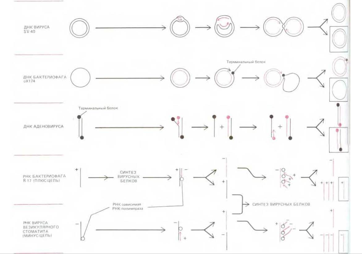
Fig. 5-73. Examples illustrating various strategies of viral genome replication. In two of the cases shown, terminal proteins are covalently attached to the ends of the DNA strands; these proteins play a crucial role in the respective replication processes. Note the key difference between positive- and negative-strand RNA viruses: negative-strand viruses must synthesize a positive strand before they can produce viral proteins. For this purpose, the capsid of a negative-strand RNA virus must package one or more molecules of viral RNA-dependent RNA polymerase (replicase). The boxed diagram on the right shows the final RNA or DNA product for each case, which is identical to the infecting viral genome shown on the left.
Figure 5-73 illustrates some of the genome replication strategies found in viruses.
5-43
5.5.6. Viral chromosomes can integrate into host cell chromosomes [48]
The entry of a viral chromosome into a cell does not always lead to immediate replication of viral particles. Many viruses can exist in a latent state: although their genomes are present in the cell, they remain inactive, and no progeny

Fig. 5-74. The life cycle of bacteriophage λ. The bacteriophage genome consists of approximately 50,000 nucleotide pairs and encodes about 50 proteins. Its double-stranded DNA can exist in both linear and circular forms. Bacteriophage development can proceed via either the lytic or lysogenic pathway, as shown here. Damage to the DNA of a lysogenic cell triggers the integrated phage DNA (prophage) to excise from the host chromosome and enter the lytic cycle. The integration of phage DNA into the host chromosome and its excision are mediated by site-specific genetic recombination catalyzed by a specific bacteriophage λ protein called integrase.
particles are produced. The ability of viruses to exist in a latent state became known when it was discovered that many seemingly uninfected bacteria produce bacteriophages when exposed to ultraviolet light. Subsequent experiments demonstrated that these lysogenic bacteria contain a complete viral chromosome integrated into their own chromosome. These integrated viral chromosomes became known as proviruses (or prophages).
Bacteriophages capable of integrating into bacterial chromosomes are called temperate (or lysogenic) bacteriophages. The most thoroughly studied of these is bacteriophage lambda (λ), whose enzyme, lambda integrase, has already been discussed. When bacteriophage λ infects a suitable E. coli cell, it typically replicates inside, producing several hundred progeny phage particles that are released upon cell lysis; this is known as the Lytic Pathway of infection. Less frequently, the linear infecting DNA molecules circularize and integrate into the circular chromosome of the host bacterium via Site-Specific Recombination (see Section 5.4.7). Once integrated, the resulting lysogenic bacterium, carrying the bacteriophage λ chromosome as a prophage, reproduces normally until triggered by a damaging environmental factor, such as ultraviolet light or ionizing radiation. This exposure induces the integrated prophage to excise from the host chromosome and initiate a standard lytic replication cycle. The integrated prophage is thus not doomed to perish with the damaged host cell; it has a chance to escape and infect a neighboring, undamaged E. coli cell (Fig. 5-74).
5.5.7. Continuous synthesis of viral proteins can transform normal cells into Cancer cells [49]
In animal cells, as in bacteria, there is an alternative to the lytic pathway for viral replication. Animal cells in which DNA viruses replicate lytically, leading to cell death, are called permissive cells. Cells in which viral replication is blocked are termed nonpermissive; in these cells, the viral chromosome either integrates into the host genome and replicates along with it, or forms a plasmid—a circular DNA molecule whose replication is regulated and does not kill the cell. Occasionally, this induces a genetic change in nonpermissive cells that leads to uncontrolled growth, transforming normal cells into cancer cells. In such cases, the DNA virus is called a DNA tumor virus, and this conversion is known as viral neoplastic transformation. Among DNA tumor viruses, two members of the papovavirus family, namely SV40 and polyomavirus, have been studied most extensively. Their transforming ability has been shown to depend on several viral proteins whose cooperative action drives resting cells from the G0 phase into the S phase (see Section 3.3). In permissive cells, this transition into the S phase makes all the host cell replication enzymes required for viral DNA synthesis available to the virus. In a nonpermissive cell, the synthesis of these viral proteins by the provirus overrides some of the normal regulatory mechanisms of both the cell and its progeny.
5.5.8. RNA tumor viruses belong to the class of Retroviruses [50]
RNA tumor viruses behave in a unique manner: their entry into a permissive cell often leads simultaneously to the non-lethal release of progeny viral particles (budding from the cell surface) and to a stable genetic alteration in the infected cell, transforming it into a cancer cell. How viral infection could cause a stable genetic change remained a mystery until the Discovery of the enzyme Reverse Transcriptase, which transcribes the infecting viral RNA strands into complementary DNA strands. RNA tumor viruses, which include the first well-studied tumor virus, Rous Sarcoma virus, belong to a large class of viruses known as retroviruses. This name reflects the fact that a portion of their life cycle involves a process that is the reverse of normal transcription (i.e., the Transcription of DNA into RNA). The AIDS (Acquired Immunodeficiency Syndrome) virus also belongs to the retrovirus family.
Figure 5-75 illustrates the Life Cycle of a retrovirus. The enzyme reverse transcriptase is an unusual form of DNA polymerase capable of using both RNA and DNA AS A template; this enzyme is encoded in the retroviral RNA and is always packaged into the capsid during the assembly of progeny viral particles. When the single-stranded retroviral RNA enters a cell, reverse transcriptase first synthesizes a DNA copy of this RNA strand, resulting in a hybrid DNA-RNA helix, which the same enzyme then uses to produce a double helix consisting of two DNA strands. This DNA copy of the viral RNA genome is integrated into the host cell chromosome. Integration is facilitated by a virus-encoded enzyme that performs site-specific recombination; it recognizes a specific nucleotide sequence in the viral DNA and catalyzes the Integration of the viral DNA into almost any site on the host cell chromosome (see Fig. 5-67, B). The next stage of the infectious process is the Transcription of the integrated viral DNA by the host cell RNA polymerase, resulting in a large number of viral RNA molecules identical to the original infecting genome. The process concludes with the Translation of these RNA molecules to produce capsid and Membrane Proteins, as well as reverse transcriptase; finally, new enveloped viral particles are assembled and bud off from The cell membrane (see Fig. 5-75).
Both RNA and DNA tumor viruses cause neoplastic cell transformation because the presence of viral DNA in the cell induces the synthesis of novel proteins that disrupt the Regulation of Cell division. The genes encoding these proteins are called oncogenes. In DNA tumor viruses, oncogenes typically encode normal viral proteins required for viral replication. The situation is different for RNA tumor viruses: the oncogenes they carry are modified forms of normal host cell genes and are not required for viral replication. Since only a limited amount of RNA can fit into a retroviral capsid, the required oncogenic nucleotide sequences often replace an essential part of the retroviral genome, rendering the virus defective. We will discuss later (see Section 13.4.2 and Section 21.2.1) why The Study of Viral Oncogenes provided the key to understanding the causes and nature of cancer, as well as the mechanisms that normally regulate Cell Growth and Division in Multicellular Organisms.
5.5.9. Some transposons are very similar to retroviruses [51]
Since many viruses can integrate into and excise from host cell chromosomes, it is reasonable to assume that all large genomes contain a certain number of different proviruses. It is highly likely that these genomes also contain a variety of mobile DNA sequences that do not form viral particles and cannot leave the cell; these are called transposons. Transposons (ranging in size from a few hundred to tens of thousands of nucleotide pairs) are typically present in the cell in multiple copies. They act as tiny parasites lurking within the chromosomes. From time to time, any such transposon becomes activated and, under The Influence of its own enzyme that catalyzes site-specific recombination, moves to another DNA site within the same cell. This process is called transposition. The enzymes that catalyze transposition—known as transposases—are mostly encoded within the transposon's own DNA. Most transposons move from place to place extremely rarely (in bacteria, approximately once per 105 generations), making them very difficult to distinguish from stationary Regions of the chromosome. We also do not know what triggers this process.

Fig. 5-75. The life cycle of a retrovirus. The retroviral genome is an RNA molecule of about 8500 nucleotides; two such molecules are packaged in each viral particle. The enzyme reverse transcriptase is a DNA polymerase that first makes a DNA copy of the viral RNA molecule and then a second DNA strand, resulting in a DNA copy of the RNA genome. The integration of this double-stranded DNA into the host cell chromosome, catalyzed by a viral protein, is required for the synthesis of new viral RNA molecules by the host cell RNA polymerase.
The mechanisms of transposition can vary. In one large family of transposons, the mechanism used is identical to a portion of the retrovirus life cycle. These elements—known as retrotransposons—are found in organisms as diverse as Yeast, fruit flies, and mammals. One of the most thoroughly studied retrotransposons is the Ty1 element found in yeast. In the first step of its transposition, the entire element is transcribed to produce an RNA copy of more than 5000 nucleotides. This RNA transcript encodes a reverse transcriptase that synthesizes a circular double-stranded DNA copy of the RNA molecule via a hybrid RNA-DNA double helix intermediate, just as occurs in a retrovirus-infected cell (see Fig. 5-75). The analogy continues as the circular DNA integrates into a randomly selected site on the chromosome. As with a retrovirus, integration occurs via site-specific recombination, as shown in Fig. 5-67, B; this likely utilizes a transposase also encoded in the aforementioned long RNA transcript. Despite its striking similarity to a retrovirus, the Ty1 element differs in that, lacking a functional protein coat, it can only move within a single cell or be passed on to its progeny.
5-44
5.5.10. In another group of transposons, transposition occurs directly from one genomic site to another [52]
Many transposons differ from retrotransposons in that they apparently never exist outside the host cell chromosome; the transposases that catalyze their movement act on the transposon DNA while it remains integrated in the host genome. The transposase is thought to bind to short, inverted repeat nucleotide sequences at both ends of the transposon, thereby bringing these ends together for the subsequent recombination it catalyzes (Fig. 5-76). In some transposons, the transposition mechanism is limited to DNA Cleavage and rejoining; the two ends of the transposon are joined to the chromosome ends at another site where a staggered cut has occurred (see Fig. 5-67, B). Such transposons move directly from one chromosomal site to another without accompanying DNA replication. However, when the chromosome is repaired at the donor site from which the transposon was excised, its nucleotide sequence is often disrupted, resulting in a mutation at that chromosomal locus.
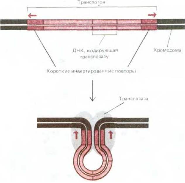
Fig. 5-76. Structure of a transposon that moves directly from one chromosomal site to another. Transposons of this type are recognized by the presence of two inverted repeat DNA sequences at their ends. Experiments have shown that these repeat sequences (sometimes as short as 20 nucleotides) are required for the intervening DNA to be transposed to a new site by the specialized enzyme transposase. The diagram shows the protein complex formed During the first step of the transposition process. Subsequent events include DNA Cleavage at the ends of the inverted repeats and several other steps that can be viewed as a Modification of the scheme shown in Fig. 5-67, B (see also Fig. 5-77).
There are also transposons that replicate during transposition. The best-studied example is one in which site-specific recombination 'triggers' local DNA synthesis; one copy of the replicated transposon is integrated into a new, randomly selected chromosomal site, while the other remains at the original site (Fig. 5-77). The mechanism of this process is very similar to the non-replicative pathway described above. Some transposons are known to be capable of using either pathway.
As they move, transposons of all types sometimes carry along adjacent host genomic nucleotide sequences or cause rearrangements within them. This often results in deletions in neighboring genomic regions or the transposition of adjacent sequences to a new site. The presence of transposons in chromosomes makes their nucleotide sequences less stable than previously thought; these elements may well be responsible for many genomic changes crucial to evolution.
Can we attribute another important evolutionary role to transposons, namely, can they be considered the ancient ancestors of viruses? As for retroviruses, they indeed appear to have evolved from retrotransposons. However, we must not overlook that all currently existing transposons are intimately dependent on DNA metabolism, whereas the genomes of the earliest cells are thought to have consisted of RNA rather than DNA. Therefore, the path to the very origin of viruses should likely be sought in RNA metabolism.

Fig. 5-77. Diagram illustrating the transposition of one type of transposon. This type of transposon replicates during transposition, so that while a new copy appears at a new chromosomal site, one copy also remains at the original site. The two inverted repeat DNA sequences at the ends of the transposon are shown here as red rectangles. At THE START OF transposition, transposase cleaves one of the two DNA strands at both ends of the transposon to initiate DNA synthesis, for which the transposon serves as a template. Synthesis proceeds by adding nucleotides to the 3' ends of the chromosomal DNA sequences.
Subsequent steps of this process are also known, but the entire mechanism is too complex to be detailed here.
5.5.11. Most viruses probably evolved from plasmids [53]
Even the largest viruses are incapable of carrying out biosynthetic processes on their own; they depend entirely on host cells in this regard. None of the known viruses possess their own Ribosomes or the ability to synthesize the ATP they require. It is clear, therefore, that cells must have evolved before viruses. The ancestors of viruses were likely small nucleic acid fragments that acquired the ability to replicate independently of their host cells' chromosomes. Such independently replicating elements—called plasmids—replicate autonomously. Plasmids exist in both DNA and RNA forms and, like viruses, contain a specific nucleotide sequence that serves as an origin of replication. Unlike viruses, however, they cannot synthesize proteins to form a protective coat and, lacking such a coat, cannot freely move from cell to cell. Many of them are also unable to integrate into the host cell chromosome.
It is possible that the earliest RNA plasmids resembled the Viroids found in some plant cells. These small, circular RNA molecules (no more than 300–400 nucleotides) replicate even though they do not encode any proteins (see Fig. 10-61). Lacking a capsid, viroids exist only as naked RNA molecules and are transmitted from plant to plant only when both the donor and recipient cells are damaged—that is, when there is no membrane barrier that the viroid cannot cross. Under natural Selection, such autonomously replicating elements could presumably acquire host cell nucleotide sequences that facilitated their independent replication, including some protein-coding sequences. Indeed, some known plasmids are quite complex, encoding proteins and RNA molecules that regulate their replication, as well as proteins that control their partitioning between daughter cells. The largest known plasmids are circular double-stranded DNA molecules of over 100,000 nucleotide pairs.
The first virus probably arose when a plasmid acquired a gene encoding a capsid protein. However, a capsid can accommodate only a limited amount of nucleic acid, meaning the number of genes a virus can carry is limited by the size of its capsid. Forced to make optimal use of their small genomes, some small viruses (such as the bacteriophage φX174) evolved overlapping genes, in which part of The nucleotide sequence encoding one protein is used (either in the same or a different reading frame) to encode a second protein. Other viruses evolved larger capsids, which must have allowed them to acquire new beneficial genes.
By possessing The unique ability to transfer DNA across species barriers, viruses have almost certainly played a major role in the Evolution of the organisms they infect. Many viruses frequently recombine both with one another and with host cell chromosomes, picking up random chromosomal fragments and transferring them to other cells or organisms. Furthermore, integrated copies of viral DNA (proviruses) become permanent Components of the genome in most organisms. Examples of such proviruses are found in the bacteriophage λ family and among the so-called endogenous retroviruses, multiple copies of which are detected in vertebrate genomes. Integrated viral DNA is often modified so that it loses the ability to produce infectious virus, yet it can still encode proteins, some of which prove beneficial to the cell. Consequently, like sexual reproduction, viruses provide opportunities to accelerate evolution by facilitating the mixing of gene pools from different organisms.
Viruses can transfer individual DNA sequences from one genome to another (a phenomenon known as Transduction). Thus, viruses can be used as tools for gene transfer from one cell to another. Viruses, along with closely related plasmids and transposons, are also important in cell biology for other reasons. Because of their relatively simple structure, research into their replication proceeds extremely rapidly, and the resulting data provide insights into the fundamental genetic mechanisms of cells. Furthermore, viruses and plasmids have become key elements in Recombinant DNA technology. We now turn to this latter topic.
Summary
Viruses are infectious particles consisting of DNA or RNA molecules (which make up the viral genome) packaged in a protein capsid; in some viruses, the capsid is further enclosed by a membrane envelope based on a lipid bilayer. The STRUCTURE OF THE viral genome and its replication strategies vary widely among different viruses. A virus can replicate only within a host cell, utilizing the host's genetic machinery. Typically, viral infection culminates in the lysis of the infected cell and the release of viral progeny. However, some viruses can integrate into the host chromosome without causing cell lysis. In this state, the viral genes (as a provirus) replicate along with the host's genes. Many viruses are believed to have evolved from plasmids, which are self-replicating DNA or RNA molecules incapable of enveloping themselves in a protein coat.
Transposons are DNA nucleotide sequences that differ from viruses in that, while replicating only within host cells or their progeny, they cannot leave the cell, much like plasmids. Transposons differ from plasmids, however, in that they typically replicate only as an integral part of the host chromosome. Some transposons, however, closely resemble retroviruses: they transpose to new genomic sites via reverse transcription of an RNA intermediate. There are also transposons capable of moving to a new chromosomal site while leaving a copy behind at the original Location. Although both viruses and transposons can be viewed as parasites, they are beneficial because the DNA sequence rearrangements they induce often play a crucial role in the evolution of cells and organisms.
Last update: 12/08/2026
Editorial and Educational Adaptation: This material has been compiled based on the primary/original source text. The project team performed an editorial review, corrected technical inaccuracies, structured sections, and adapted the content for an educational format.
What was processed:
- elimination of formatting defects (OCR errors, structural breaks, corrupted characters);
- editorial organization of content;
- standardization of terminology in accordance with academic sources;
- verification of factual statements against the original source text.
All mentions of the author, publication year, and origin of the primary text have been preserved in accordance with the source.