NEONATAL SURGERY - 1976
2. SPECIAL SECTIONS
8. Malformations of Individual Organs and Systems
Umbilical Hernia
Umbilical hernia results from impaired Development of the anterior abdominal wall in the umbilical region; it is quite common in newborns, particularly in females.
Embryogenesis. The umbilical ring is formed by the divergence of the aponeurotic fibers of the linea alba, creating either a round or semi-oval defect in the anterior abdominal wall. The umbilical ring closes completely only after the umbilical cord detaches. The strength of the Tissues covering it varies. The lower half is the densest, retaining remnants of the umbilical Arteries whose well-developed adventitia transforms into scar tissue During the first weeks of life. The remnants of the urachus also contribute to the strength of the lower edge of the umbilical ring. The upper half of the ring is weaker; it transmits only the umbilical vein with its thin walls and poorly developed adventitia. The umbilical vein lies very close to the upper edge of the umbilical arteries, leaving a free space between the vein wall and the upper margin of the umbilical ring that is covered only by a thin layer of Connective Tissue and the umbilical fascia. The degree of development of the umbilical fascia is of great importance in The Development of umbilical hernias (A. Deshin, 1902; N. V. Shvarts, 1937). The umbilical fascia is located beneath the parietal Peritoneum in the upper part of the umbilical ring. The degree of development of the umbilical fascia in newborns varies significantly. In some cases, the bulk of it lies posterior to the upper half of the umbilical ring; in others, posterior to the lower half, or it may encompass the umbilical ring and close it entirely (F. I. Valker, 1938; V. S. Panushkin, 1941). The absence or poor development of this fascia contributes to the occurrence of umbilical hernias.
Clinical presentation. There is a rounded protrusion in the umbilical region covered by thin Skin. In most patients, the Contents of the hernia sac (most commonly a loop of intestine) are easily reducible into the Abdominal cavity. After reduction, the edges of the umbilical ring are readily palpable.
Treatment. Umbilical hernia tends to resolve spontaneously; therefore, conservative treatment consisting of massage of the anterior abdominal wall Muscles is performed in newborns. Surgery is indicated only for individual patients with large hernias and narrow hernia gates that are prone to incarceration. The operation is performed under general anesthesia.
Surgical technique. Using a semicircular incision outlining the lower edge of the Base of the hernia sac, the pedicle and the hernia sac are mobilized. The contents are reduced into the abdominal cavity, the sac is opened, its neck is ligated at the base and transected. The edges of the defect are closed with interrupted nylon sutures, excess skin is excised, and the wound edges are approximated with interrupted catgut sutures.
Inguinal Hernias
Inguinal hernias account for 92–95% of all hernias and occur in 55% of cases during the first weeks and months of a child's life. In an inguinal hernia, Internal Organs herniate through the Inguinal Canal into the non-obliterated processus vaginalis of the peritoneum (indirect hernia). By the time of birth, the processus vaginalis is obliterated in 20–30% of children, while complete or partial non-obliteration persists in 70–80% (N. I. Kukudzhanov, 1969). Complete non-obliteration of the process, when abdominal organs shift into the Scrotum and the Testis protrudes into the lumen of the hernia sac, results in a funicular/testicular hernia. Non-obliteration of only the proximal portion of the processus vaginalis leads to The formation of a cord hernia. Testicular forms of inguinal hernia predominate in newborns. The right testis migrates later than the left, and consequently, the right processus vaginalis also closes later. Therefore, right-sided inguinal hernias are seen 3 times more frequently in newborns than left-sided ones. Bilateral localization of hernia in newborns is extremely rare.
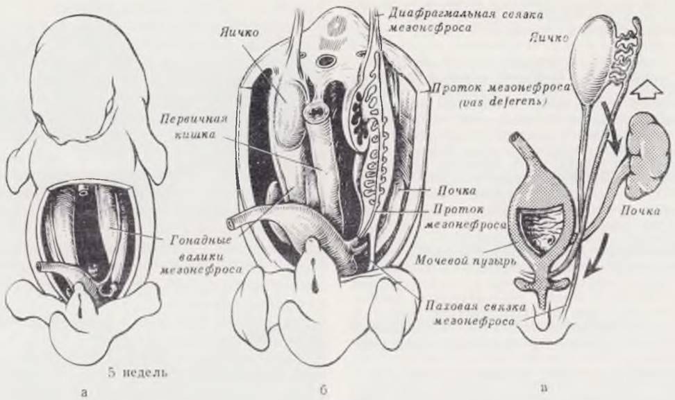
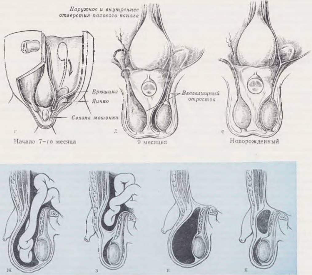
The stages of embryogenesis and possible variants of pathology are shown in Fig. 71.
Class="center">Fig. 71. Stages of testicular development and descent. Formation of the vaginal process of the peritoneum and the consequences of failed obliteration.
Gonads develop as ridge-like thickenings (genital ridges) on the ventral borders of the mesonephros while the latter still Functions as the primary excretory organ (a). In the developing male embryo, indifferent gonads progressively differentiate into Testes. Rapidly increasing in volume, the mesonephros bulges into the coelom, pushing the peritoneum ahead of it. At this stage, the peritoneum forms folds at both ends of the mesonephros. One is termed the diaphragmatic ligament (b), while the other extends toward the caudal end of the coelom, acquires a fibrous Structure, and is designated as the inguinal ligament of the mesonephros (b, c). The developing testis displaces the regressing mesonephros and becomes covered by its peritoneal layer. Following the degeneration of the mesonephros, its inguinal ligament is termed the "testicular ligament," forming the cranial part of the gubernaculum testis, which plays a crucial role in testicular descent. Simultaneously, in the inguinal region on each side of the body, at the sites of testicular ligament attachment, coelomic evaginations develop—the vaginal processes of the peritoneum (processus vaginalis). From the outer layer of the processus vaginalis, fibrous strands (d) extend toward the skin of the growing scrotum; these, together with the testicular ligament, form the gubernaculum. The latter does not elongate in proportion to the growth of the surrounding pelvic structures, thereby "pulling" the testis downward. Concurrently with the development of the scrotum, the testes shift in a caudal direction (c, e). By the 5th month, they lie adjacent to the deep inguinal ring; during the 7th month, they begin to pass through the inguinal canal (d); and by the 8th month, they reach the scrotum. During descent, the testis slides downward beneath the peritoneal lining and enters the scrotum, becoming "wrapped" in a doubled peritoneal layer (e). By birth, the processus vaginalis obliterates in the majority of boys (f). Failures of obliteration lead to the development of congenital inguinal hernias (g, h) or Spermatic Cord cysts (i, j).
In girls, Nuck's diverticulum (the homologue of the vaginal process of the peritoneum) is obliterated by birth in the vast majority of cases, which is why hernias occur 10 to 12 times less frequently in females than in males.
The inguinal canal in newborns is short, relatively wide, and directed almost straight from back to front. Its length ranges from 1 to 1.5 cm, with a relatively large diameter of the superficial inguinal ring (up to 1.5 cm according to R. I. Venglovsky, 1903). Even a minor increase in intra-abdominal pressure during crying, straining, or other exertion is sufficient for abdominal organs to stretch the hernia ring and prolapse into the preformed hernial sac.
Clinical presentation. The signs of an inguinal hernia are typical and usually manifest on the 10th to 15th day of life. Parents or caregivers discover a round or oval Swelling in the inguinal region that enlarges during crying and agitation, and decreases or disappears at rest. Frequently, the swelling goes unnoticed for some time due to its small size and the prominent subcutaneous fat layer in the inguinal regions of newborns. Occasionally, strangulation serves as the initial manifestation of the hernia. In 45% of cases, the protrusion descends into the scrotum, causing Asymmetry. In girls, the protrusion most commonly occupies the area of the superficial inguinal ring, but it may extend downward to involve the entire labium majus.
Diagnosis of an inguinal hernia is based on a detailed medical history, external inspection, and careful Palpation. On palpation, the hernial protrusion is painless, smooth, and elastic in consistency. Its contents can usually be easily reduced back into the abdominal cavity with a characteristic gurgling sound. The superficial inguinal ring is examined with the little finger inserted via the invaginated scrotum. The spermatic cord is palpated between the index finger and thumb on each side simultaneously with both hands. A finding of a thickened spermatic cord on one side indicates the presence of a hernia. The condition of the testes in the scrotum and the prominence of the cremasteric reflex are also assessed.
In girls, the inguinal region is palpated, paying particular attention to the inguinal canal. Difficulties in diagnosis may arise when uterine appendages descend into the region of the labium majus. Rectal examination with simultaneous palpation of the hernial protrusion area by the other hand helps clarify the diagnosis. A cord-like structure extending into the canal is characteristic of a hernia. The contralateral inguinal region must always be examined.
Positive findings upon examination, combined with a refined medical history, make the diagnosis of an inguinal hernia unquestionable.
Differential diagnosis is primarily conducted with communicating hydrocele of the testis. Hydrocele is characterized by a decrease in the swelling in the mornings after Sleep. Confidence in the diagnosis increases when prolonged, gentle pressure on the scrotum causes the fluid accumulation to gradually diminish and disappear. Other distinguishing features include the absence of tension, fluctuation, and positive transillumination results. Isolated hydrocele of the testis is oval in shape, more tense, and does not change shape under pressure. Differentiating a spermatic cord cyst is more challenging. Its painless nature, well-defined smooth borders, oval shape, elastic consistency, volume that remains unchanged under gentle pressure, and incomplete reduction into the abdominal cavity help establish the correct diagnosis.
When differentiating from cryptorchidism or testicular ectopia combined with a hernial protrusion, attention is focused on the primary sign—the absence of the testis in the scrotum.
An inguinal hernia is sometimes confused with an enlarged Pirogov-Rosenmüller Lymph node, which is located inferiorly and below the inguinal ligament, as well as with various tumors of the inguinal region (dermoid cyst, angioma, etc.).
Treatment. Surgical repair is the method of choice for inguinal hernia. Modern advancements in pediatric surgery, anesthesiology, and intensive care make it possible to perform operations in children of any age. However, elective herniorrhaphy in a newborn should be performed at a pediatric surgical facility equipped with a neonatal surgery center. In cases of uncomplicated inguinal hernia accompanied by prematurity, Pyoderma, diaper dermatitis, Pneumonia, or other conditions, Surgical treatment is indicated at an older age (typically after 6 months).
Various herniorrhaphy techniques are used in newborns, ranging from simple amputation of the hernia sac (O. S. Bokastov, 1937) to complex reconstructive Methods involving the transposition of the spermatic cord and reinforcement of the posterior wall of the inguinal canal (A. P. Biezins, 1964). However, the majority of pediatric surgeons close the inguinal canal with 1 or 2 sutures According to the Bassini-Cherny method (or as it is commonly called, the Roux-Krasnobaev method). Abroad, techniques based on the Ferguson method are widely utilized.
The primary goal of herniorrhaphy in newborns is to eliminate the communication between the patent processus vaginalis and the peritoneal cavity. Narrowing the superficial inguinal ring and reinforcing the anterior abdominal wall are of secondary importance.
The technique consists of simple transection of the hernia sac, high dissection, and ligation of its neck without incising the aponeurosis of the external oblique Muscle. The close anatomical alignment of the deep and superficial inguinal rings facilitates this task. The distal portion of the transected hernia sac is left in place without ligation. Concerns that a cyst might form in the remaining membranes are exaggerated (T. V. Krasovskaya, 1970; Duhamel, 1957).
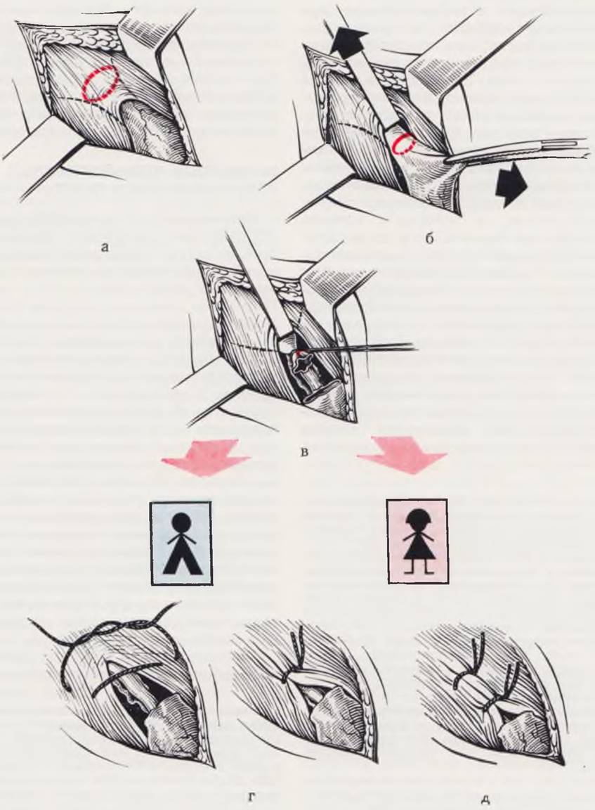
The surgical technique is illustrated in Fig. 72. The infant is placed in the supine position. General anesthesia is administered. A 1.5 — 2 cm incision is made along the skin crease in the inguinal region to expose the aponeurosis of the external oblique muscle and the superficial opening of the inguinal canal. The hernia sac is located, and its neck is isolated at the superficial inguinal ring by elevating the anterior wall of the inguinal canal with a Farabeuf retractor. The testicular vessels and vas deferens are gently separated from the sac using a blunt instrument. The bundle of inferior epigastric vessels serves as a landmark; their appearance indicates that the neck of the hernia sac has been reached. The hernia sac is opened between clamps, its contents are inspected and reduced into the abdominal cavity. The neck of the hernia sac is transfixed, ligated as high as possible, and finally transected. The superficial inguinal ring is usually not narrowed. If necessary, a single Lembert-type suture is placed on the crura of the superficial ring. This technique helps avoid injury to the trophic Branches of the ilioinguinal nerve passing through them. The wound is closed in layers with primary closure. Subcutaneous absorbable stitches are placed in the skin.
Fig. 72. Herniorrhaphy in a newborn.
The superficial inguinal ring is exposed (a). The neck of the hernia sac is mobilized (b). The hernia sac is transected transversely; the proximal stump is transfixed and ligated, while the distal stump is left unligated (c). When the superficial ring is wide in boys, a single suture is placed across its crura (d). In girls, the opening of the inguinal canal is closed tightly with 1 — 2 stitches (e).
In girls, after isolation, transection of the hernia sac, and management of its neck, the inguinal canal is closed tightly.
In the postoperative period, primary attention is paid to the condition of the sutures in the inguinal region. Feeding the infant is initiated 4 hours after surgery. Subcutaneous absorbable sutures do not require removal.
Complications. Incarceration is one of the most frequent complications of inguinal hernia in children, with over 50% of incarcerations occurring during the first 3 months of life. At the Neonatal Center of the Department of Pediatric Surgery at the Central Institute for Advanced Medical Training (TSOLUV), 84 children with inguinal hernias have been operated on over the past 9 years, 48 of whom presented with incarceration.
Incarceration occurs without apparent cause. A previously reducible hernia suddenly becomes irreducible. Consequently, the internal organs that have herniated into the sac (intestinal loops, omentum, uterine adnexa in girls, etc.) are compressed within the aponeurotic ring. A lack of timely intervention leads to impaired Blood Circulation and Necrosis of the hernia contents.
The clinical signs of incarceration are characteristic: crying, screaming, refusal to feed, and occasionally reflexive vomiting. Parents can usually pinpoint the exact onset of these symptoms. The hernia bulge in the groin becomes tense, painful, and irreducible. Later, hyperemia and scrotal edema appear. During the first hours of incarceration, there may be a bowel movement, sometimes mixed with blood; later, constipation, gas retention, and vomiting develop, which are signs of intestinal obstruction. However, diarrhea may also be observed (Rickham, 1969). The combination of vomiting (frequently Bile-stained) and diarrhea can lead to diagnostic errors.
In typical cases, the diagnosis of incarcerated hernia is straightforward. Diagnostic doubts arise when the parents cannot report a pre-existing hernia, or when incarceration is the very first manifestation of the hernia.
Incarcerated hernia must be differentiated from acute inguinal lymphadenitis (where the primary site of infection can typically be found on the lower extremities or buttocks); an acutely developing cyst of the spermatic cord, especially if located near the superficial opening of the inguinal canal; and an acutely presenting communicating hydrocele of the spermatic cord and testicular tunics.
Significant differential diagnostic challenges arise in cases of incarceration within the deep or superficial inguinal ring involving an ectopic or cryptorchid testis, Torsion of the spermatic cord, or inflammation of the vermiform Appendix residing within the hernia sac.
Inguinal hernias are less common in girls but complicate more frequently than in boys. The contents of the hernia sac in girls typically consist of the uterine adnexa, which descend into the labium majus and are prone to rotation and rapid necrosis. Differential diagnosis is primarily conducted with cysts of the canal of Nuck, femoral hernias, and inguinal lymphadenitis.
Thus, a history of a pre-existing hernia combined with the aforementioned signs allows for an accurate diagnosis of incarceration. In doubtful cases, the diagnosis should lean toward an incarcerated inguinal hernia, particularly in girls.
Treatment. Given the current development of pediatric surgery and its expanded capabilities, the choice of management for an incarcerated hernia is perhaps not as critical as it was in previous years. Since incarceration within the first 8 — 12 hours rarely leads to necrosis of the hernia contents, our clinic employs a set of conservative measures (S. Ya. Doletsky, 1950) designed to achieve spontaneous reduction of the incarcerated hernia (hypertonic microenema, promedol and atropine, repeated warm baths, etc.). Exceptions include: newborns in whom more than 12 hours have elapsed since incarceration, making spontaneous reduction unlikely; newborns with inflammatory signs in the scrotal and inguinal regions; girls, because the uterine adnexa comprising the hernia contents undergo rotation and rapidly undergo necrosis upon incarceration; and boys, in whom an incarcerated hernia is associated with cryptorchidism or ectopic testis, as the risk of testicular necrosis is similarly high in such situations. Conservative therapy is successful in 43% of newborn cases. After 2 — 3 days, these children undergo elective surgery in the "cold" period.
Surgical technique. After exposing the superficial inguinal ring, isolating and opening the hernia sac between clamps, the condition of the hernia contents is assessed, and they are reduced into the abdominal cavity. Special attention is paid to the incarcerated bowel loops to rule out retrograde injury. If the viability of the bowel is questionable, measures are taken to restore peristalsis (novocainization of the mesentery, wrapping the loops in Sponges soaked in warm isotonic saline, etc.). Any suspicious segment of the bowel is reperitonealized. Non-viable bowel is resected with an end-to-end anastomosis using a single-layer suture.
The viable bowel is returned to the abdominal cavity. The need to widen the neck and incise the aponeurotic ring rarely arises. The neck of the hernia sac is secured, ligated, and managed as described above.
Subsequently, the entire spermatic cord is inspected. Special attention is paid to the testis, gently returning it into the scrotum. If the hernia incarceration was associated with cryptorchidism, herniorrhaphy is concluded with mandatory orchidopexy (mobilization and fixation of the testis). The Bailey-Keetley-Sokolov method is preferred. The wound is closed in layers. Leaving a rubber drain is mandatory.
In the postoperative period, if the child's condition is stable, feeding is initiated after 4 hours, administered fractionally, similar to the protocol following pyloroplasty. Following surgery accompanied by bowel resection, continuous nasogastric suction and intravenous parenteral Nutrition are established. Corrective therapy is administered until the symptoms of intestinal obstruction are resolved.
The prognosis for inguinal hernia is favorable. There are no fatalities associated with elective surgery. However, mortality rates of 0.8 — 2.5% can occur in cases of incarcerated hernia and are associated with general complications (such as pneumonia).
Congenital gastrointestinal obstruction
Congenital gastrointestinal obstruction is one of the most common conditions requiring emergency surgery in newborns and infants during their first weeks of life. According to the data from the neonatal surgery department of Rusakov Children's Hospital, children admitted for emergency surgery present with a diagnosis or suspected diagnosis of intestinal obstruction in 50–60% of cases across various years. Depending on The Nature and form of the obstruction to the passage of intestinal contents, the clinical manifestations of congenital obstruction may first appear in both neonates and older children. The causes of congenital obstruction encompass A wide variety of developmental anomalies, which can be conventionally divided into four main groups: 1) Malformations of the intestinal tube; 2) malformations of the intestinal wall; 3) disturbances of intestinal rotation; and 4) malformations of other abdominal organs that impair the normal passage through the gastrointestinal tract.
The most frequent malformations of the intestinal tube are stenosis and atresia.
Their occurrence is caused by an arrest in the development of the intestinal tube during the solid and vacuolization stages. A certain role is also played by Impaired blood supply to specific segments of the intestine during early embryonic development (Dickson, 1970; Shafie, Rickham, 1970, et al.). The validity of this theory is supported by clinical statistics showing that 95–96% of intestinal atresias and stenoses are localized in the duodenum and Small Intestine.
Depending on the extent and Nature of the obstruction, a distinction is made between segmental intestinal atresia (which can be single or multiple) and membranous atresia, with or without an opening in the center of the membrane.
An unusual type of Small bowel atresia is the "apple-peel" (or "Christmas tree") atresia, a term first proposed by Santulli in 1961.
This anomaly occurs in extremely premature infants and is characterized by a common mesentery attached in an "apple-peel" configuration, accompanied by severe hypoplasia of the distal bowel. There are 15 documented cases of this pathology in literature (Dickson, 1970).
The second cause of congenital gastrointestinal obstruction comprises Developmental anomalies of the walls of the Esophagus, Stomach, and intestines.
Gastrointestinal dysmotility in newborns is primarily associated with the immaturity of the neural elements within the intestinal wall (Bughaigis, Emery, 1971). These authors established and histologically proved that by birth, only about one-third of the Neurons in the intestinal wall have developed, whereas the complete ganglionated System of the bowel matures by the age of 5, coinciding with the completion of Auerbach's plexus development.
Typical developmental Anomalies of the wall of the cardial esophagus that present with a clinical picture of esophageal-level obstruction are chalasia and achalasia. The Essence of the defect lies in the inadequate development of the neural elements in the wall of the cardial esophagus. In chalasia, there is an underdevelopment of sympathetic nerve fibers, causing the lumen of the cardial esophagus to lose its normal tone and remain patent (patulous). Gastric peristalsis leads to the reflux of gastric contents, resulting in vomiting and the potential development of esophageal mucosal erosions. Achalasia occurs due to the underdevelopment of the parasympathetic system. In these cases, the esophagus is spastic, leading to the clinical picture of cardiospasm. A characteristic feature of both chalasia and achalasia is the disappearance of obstruction symptoms as the neural elements mature.
One of the most frequent developmental defects of the pyloric wall causing a Clinical presentation of obstruction is pyloric stenosis. This condition is characterized by congenital hypertrophy of all layers of the pylorus (particularly the muscular layer). A recognized factor is the abnormal development or absence of neural (parasympathetic) ganglia in the pyloric wall (A. G. Pugachev, 1964, et al.).
A typical anomaly of the duodenal wall that leads to the clinical picture of complete high intestinal obstruction in the first days of life is true megaduodenum. In this condition, the intestinal lumen remains preserved throughout its length. The core of the disorder is the absence or abnormal development of parasympathetic nerve plexuses in a segment of the duodenal wall. The segment with impaired innervation is spastic, lacks peristalsis, and acts as an obstruction to the passage of intestinal contents. The proximal bowel segment becomes overloaded with contents and undergoes secondary dilation.
Impaired normal development of neural elements throughout the small intestine leads to the development of so-called neurogenic ileus (V. Tošovský, O. Vichytil, 1957) in the infant's first days of life. Intestinal obstruction in these infants develops As a result of absent peristalsis in the affected segment.
A developmental defect of the large bowel wall that causes a picture of complete or partial obstruction—often in the first days and weeks of life—is Hirschsprung's disease. Morphological and histochemical studies (Ehrenpreis, 1946; Swenson, 1958; S. Ya. Doletsky, 1956, 1972; Yu. F. Isakov, 1958, 1971, et al.) have reliably established that Hirschsprung's disease is based on a malformation of the colonic wall elements, characterized by the complete absence or deficiency of intramural nerve ganglia in certain segments of the colon, the presence of abnormal nerve fibers and ganglia, and impaired conduction in the reflex arcs of the intestinal wall. In 85% of patients, the distal colon is affected, though involvement of the entire intestinal wall is possible (a circumstance that must be considered when creating a colostomy). The abnormally developed segment is spastic, loses its ability to peristaltic motion, and serves as an obstruction to the normal transit of intestinal contents.
Thus, among intestinal wall malformations causing a clinical picture of gastrointestinal obstruction, impaired innervation (primarily parasympathetic) at various levels of the digestive tract is of paramount importance.
One of the frequent causes of congenital intestinal obstruction is impaired intestinal rotation. As noted previously, the intestine transitions from its initial embryonic position to its normal position through rotation. Arrested rotation at various stages leads to numerous anomalies that hinder or prevent the passage of intestinal contents, resulting in clinical intestinal obstruction.
Thus, in cases of arrested rotation at the end of the first or beginning of the second period, an infant is born and may live their entire life with malrotation of the intestine. In this anomaly, the intestine remains fixed to the posterior abdominal wall at only a single point—the Water/144.html">Origin of the superior mesenteric artery. The loops of the small intestine in malrotation are located predominantly in the right half and lower PARTS OF THE abdominal cavity, the cecum is situated in the epigastric region or left hypochondrium anterior to the duodenum, and the colon lies in the left half of the abdominal cavity. This position and fixation of the intestine create conditions for midgut volvulus around the superior mesenteric artery and the development of intestinal obstruction. When rotation is arrested during the second period, the cecum, located in the upper abdominal quadrants anterior to the duodenum, becomes fixed to the posterior abdominal wall and compresses the duodenal lumen. Depending on the degree of compression, the infant develops symptoms of complete or partial high intestinal obstruction at various times after birth. A combination of midgut volvulus around the superior mesenteric artery and duodenal compression by abnormal peritoneal bands of the cecum is also possible—known as Ladd's syndrome (see Fig. 65).
An anomalous venous ring may also be a cause of duodenal obstruction (Fuchs, Himer, 1972).
A rare variant of rotational abnormality is reverse rotation (clockwise). In such cases, the colon lies posterior to the duodenum, which may lead to its compression and the development of large bowel obstruction.
Rotational arrest during the third period and abnormal mesenteric development are accompanied by the formation of mesenteric defects, into which intestinal loops may herniate, subsequently leading to obstruction and even intestinal necrosis.
Among malformations of other abdominal organs that impair normal passage through the intestinal tube in the first days and weeks of life, the most common are pyloric stenosis caused by aberrant vessels, annular Pancreas, and meconium ileus.
Pyloric compression may occur due to an aberrant position of the portal vein or hepatic artery, which in such cases run anterior to the duodenum and compress it. Duodenal compression is also possible due to anomalies of the gastroduodenal or mesenteric arteries, among others.
Annular pancreas is a congenital anomaly in which the HEAD of the pancreas forms a ring encircling the duodenum, and it can likewise cause intestinal obstruction. The ring formed by the pancreatic tissue band may be completely or partially closed, which determines the degree of duodenal compression and the clinical manifestations of the anomaly.
In some cases, congenital obstruction may be caused by the compression of intestinal loops herniating into the pleural cavity or Mediastinum due to congenital diaphragmatic defects and hernias. The clinical picture depends on the size of the diaphragmatic defect and the degree of compression of the herniated bowel loops.
A specific form of intestinal obstruction in newborns is meconium ileus. This is a manifestation of a systemic, genetically inherited (recessive) disorder—cystic fibrosis (mucoviscidosis)—characterized by the hypofunction of all exocrine glands (Blanock et al., 1965; Kalagogly, 1971). In cystic fibrosis, the most pronounced lesions are found in the pancreas (congenital cystinofibrosis/cystic fibrosis of the pancreas) and the bronchial ciliated epithelium. The pathologically altered pancreas secretes an insufficient amount of weakly active pancreatic juice, causing the meconium to acquire a putty-like consistency and obstruct the intestinal lumen. The site of obstruction is typically located 10–15 cm proximal to the ileocecal valve.
Clinical presentation. As outlined above, congenital intestinal obstruction can be caused by various Developmental Anomalies and Diseases of the intestine and other abdominal organs. Depending on the Location and degree of the obstruction to the passage of intestinal contents, the clinical presentation of congenital gastrointestinal obstruction and the timing of symptom onset can be diverse yet simultaneously similar across different malformations. Consequently, the Classification of Congenital obstruction is based on the anatomical site and severity of the obstruction. Based on these criteria, we divide patients with congenital obstruction into two groups: those with complete obstruction and those with partial obstruction. In turn, both groups are subdivided into three subgroups: a) patients with high intestinal obstruction (gastric outlet, duodenum, proximal small intestine); b) patients with small bowel obstruction; and c) patients with large bowel obstruction (low intestinal obstruction).
Causes of complete high intestinal obstruction may include developmental defects of the intestinal tube (intestinal atresia), the intestinal wall (true megaduodenum), disorders of intestinal rotation, and developmental anomalies of other abdominal organs, such as abnormal vascular positioning (Fig. 73), arteriomesenteric obstruction (Fig. 74), annular pancreas (Fig. 75), etc. The clinical manifestations of these defects are identical in the majority of cases.
The initial sign of complete high intestinal obstruction is typically vomiting. Vomiting in these patients occurs as early as the second half of the first day of life. The initial portions of vomitus mostly consist of swallowed Amniotic Fluid and gastric contents. Since such vomiting in newborns is a fairly common occurrence, maternity ward staff usually do not pay due attention to the first episode. After 1 — 1 1/2 h, vomiting recurs, and bile appears In the second or third portion of the vomitus (the obstruction to the passage of intestinal contents is located below the major duodenal papilla in most cases). Persistent vomiting at the end of the 1st to the beginning of the 2nd day of life, along with the presence of bile in the vomitus, serves as a reliable indicator of high intestinal obstruction.
Fig. 73. Stages of portal vein formation. Variant with a preduodenal location.
Venous blood outflow from the primitive gut initially originates from the vitelline Veins OF THE yolk sac, which merge into the main paired vitelline-mesenteric veins and empty into the venous sinus. On their way to The Heart, these veins lie adjacent to the developing Liver (a).
The umbilical (allantoic) veins are incorporated into the lateral body walls along their entire length from the body stalk to the venous sinus (b).
Growing cords of hepatic tissue divide the proximal part of the vitelline-mesenteric veins into a network of small vessels, which in turn grow into the liver tissue. As the liver increases in volume, it fuses with the lateral body wall. At the fusion site, vessels develop that connect the umbilical veins with the hepatic vascular plexus. The initial vitelline-mesenteric trunks are connected by transverse anastomoses (b, c).
With the regression of the yolk sac and the growth of the intestine, the vitelline portion of the vitelline-mesenteric veins disappears; the mesenteric branches fuse and develop in accordance with the growth of the intestinal tract. Parts of the umbilical veins within the umbilical cord and inside the embryo's body fuse. The segments of the umbilical veins that emptied into the venous sinus degenerate. Blood flowing from the umbilical cord into the liver passes through a network of anastomosing sinusoids which, as they develop, form a large vessel passing through the liver parenchyma—the venous duct (ductus venosus) (d).
The initial vitelline-mesenteric trunks transform into the unpaired portal vein due to the disappearance of the initial left vessel cranial to the middle transverse anastomosis and the right vessel. The right umbilical vein ceases to channel blood into The Liver and regresses, although a portion of it persists and drains blood from the body wall (e).
If the degeneration processes of the respective sections of the vitelline-mesenteric trunks are disrupted, the forming portal vein comes to lie anterior to the duodenum and may cause its obstruction after birth (f, g).
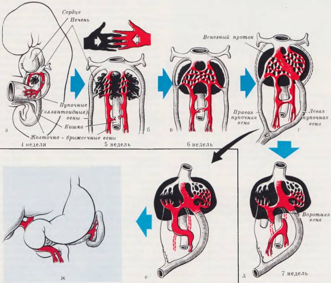
Fig. 74. Diagram of arteriomesenteric obstruction.
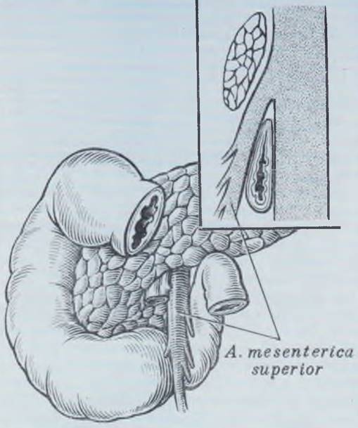
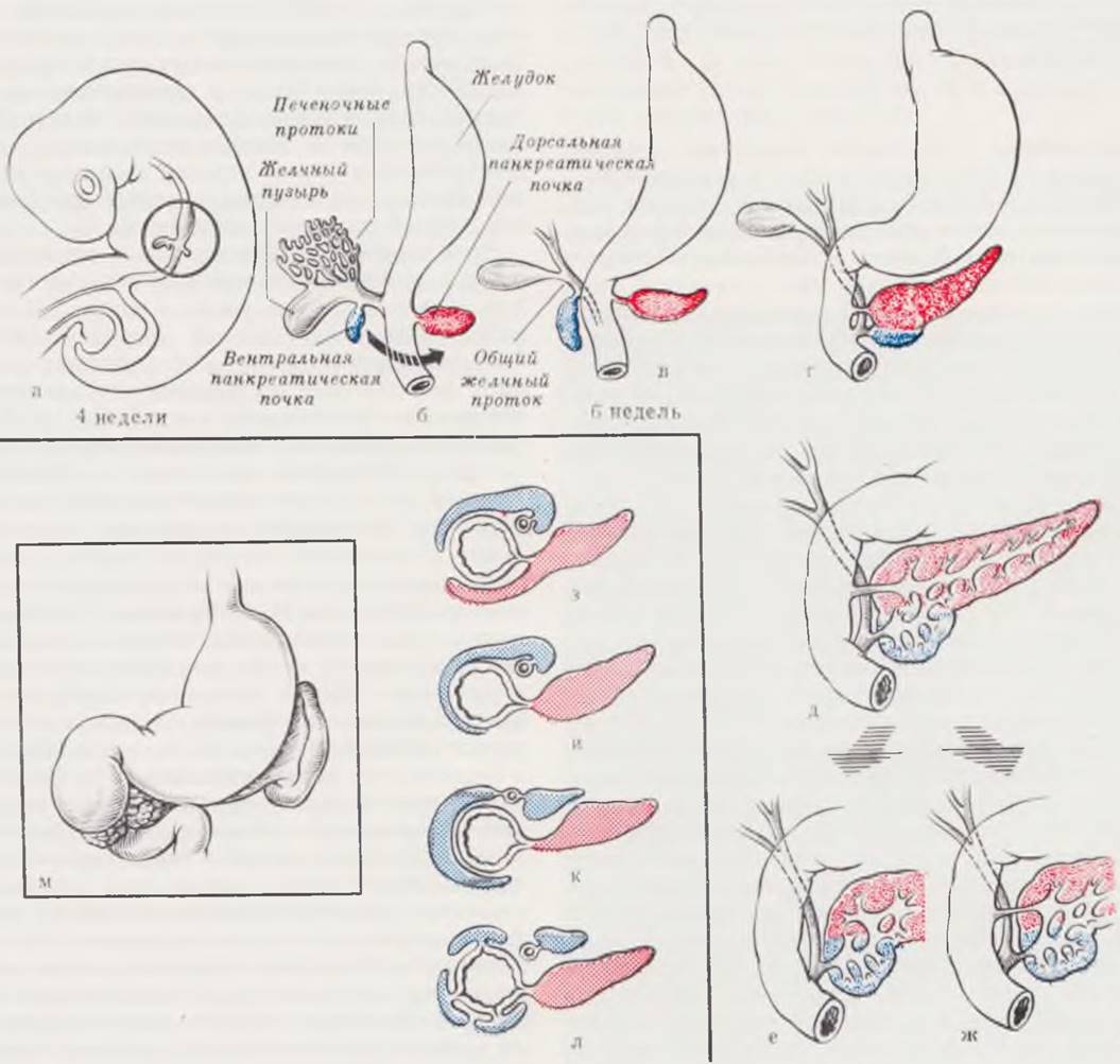
Fig. 75. Stages of pancreas formation. Variants of annular pancreas.
The pancreas develops from two buds: the ventral and dorsal pancreatic buds (a). The ventral bud grows in a caudal direction and shifts downward as the hepatic diverticulum elongates (compare b, c, d), appearing as an outgrowth from the common bile duct. Due to the Rotation of the intestine and the rightward bending of the common bile duct, the ventral bud begins to grow to the right of the duodenum, approaches the dorsal bud, and soon fuses with it (d). The definitive pancreas is primarily formed from the dorsal pancreatic bud, whereas the ventral bud contributes only to a portion of the pancreatic head (e). During development, the ventral duct persists (f) and, through anastomoses, drains secretions from all parts of the gland. The proximal part of the dorsal duct usually undergoes atrophy, but may also persist (g).
Of clinical interest are cases of annular pancreas formation, which cause compression and obstruction of the duodenum (m). Hypothetical Variants of the formation of the pancreatic tissue ring are shown in Figs. h — l (h — after Ticken; i — after Zecco; k — after Baldwin, l — after Erimoglu).
Upon examination of the infant during this period, their general condition is only mildly affected. Various rales may appear in the Lungs as a result of aspiration of vomitus. The external appearance of the abdomen depends on the timing of the examination. If the examination is performed while The Stomach and duodenum are filled with contents, a prominence of the epigastric region is observed. The rest of the abdomen is scaphoid, and Percussion yields a dull sound. If the examination is conducted after vomiting, retraction of the abdomen is noted in all quadrants. Stool in patients with congenital high intestinal obstruction typically occurs a few times. Unlike normal stool, it consists of grayish meconium plugs and is scanty in amount. A simple examination of these patients' meconium using Farber's test allows the diagnosis of obstruction to be confirmed or ruled out with a high degree of certainty. In complete obstruction, desquamated epithelial Cells—which the fetus swallows along with amniotic fluid—are absent in the meconium. These symptoms make it possible to suspect, and with a positive Farber test to establish, the diagnosis of intestinal obstruction. Further clarification of the diagnosis is carried out using radiological imaging.
Clinical manifestations of small bowel obstruction develop somewhat later than those of complete high intestinal obstruction and are more severe. The first symptom of the condition, as in complete high intestinal obstruction, is vomiting, which typically begins at the end of the first or the beginning of the second day of life. The initial portions of vomitus consist of swallowed amniotic fluid and gastric contents. Subsequent portions become mixed with bile, and later with intestinal contents. The child's condition progressively deteriorates; exicosis and toxicosis increase due to the intensive absorption of toxic contents from the afferent loop of the small intestine, accompanied by marked electrolyte imbalance and acid-base disturbances. Upon examination as early as the beginning of the 2nd day of life, the condition is evaluated as moderately severe or severe. Aspiration pneumonia may develop. The abdomen is distended in the upper regions, and dilation of the venous network on the anterior abdominal wall is noted. Peristalsis of dilated intestinal loops can frequently be visualized. Stool may occur 1 — 2 times and is characteristically scanty.
Complete large bowel obstruction is characterized by a slower onset of clinical symptoms against a relatively satisfactory General condition of the infant. In most cases, the initial symptom is the absence of stool and a progressive, gradual enlargement of the abdomen. The lack of stool usually alerts the physician, and a cleansing enema is prescribed. In patients with colonic atresia, when small amounts of fluid are introduced, it quickly flows back out. If the obstruction is due to Hirschsprung's disease, especially with an extended or double aganglionic zone, the fluid readily passes into the dilated segment of the intestine during the enema and is expelled with difficulty or not at all. In these patients, an enema may yield copious stool and flatus. The abdomen is generally distended, with visible dilation of the anterior abdominal wall veins and visible peristalsis of stretched intestinal loops. Vomiting sets in on the 2nd or even 3rd day of life. The vomitus initially contains milk and gastric contents, followed by admixture with bile, and later with intestinal contents.
The rather characteristic clinical picture of complete congenital intestinal obstruction outlined above, combined with the findings from the physical examination, generally allows for a diagnosis with a fairly high degree of confidence. Further refinement is achieved through radiological examination, which will be discussed below.
Diagnosing congenital partial intestinal obstruction presents significantly greater challenges. The clinical manifestations are characterized by diversity and Variability of symptoms, as well as a wide range of onset times. The most frequent causes of partial intestinal obstruction in newborns include pyloric stenosis, prolapse of the gastric mucosa into the duodenum, membranous forms of duodenal atresia with a central aperture in the membrane, and disorders of intestinal rotation.
Congenital hypertrophic pyloric stenosis is one of the most common causes of partial obstruction in infants during the first weeks and months of life. In a series of observations reported by Benson (1970)—1,465 surgeries over 29 years—the following patterns were established: a familial and hereditary predisposition was identified in 6.9%. Out of 22 pairs of twins, surgery was performed on both in 5 pairs. The male-to-female ratio was 4 : 1. Premature infants accounted for 2.7%. The Significance of the ethnic factor was not established.
Based on the clinical course and the timing of the initial symptoms, N. S. Mankina (1968) distinguishes acute and protracted Stages of the disease. The acuteness and time of onset of pyloric stenosis symptoms depend on the degree of pyloric narrowing and the compensatory capabilities of the infant's stomach. The initial symptoms of the disease most commonly appear in the 2nd to 3rd week of life as frequent and persistent regurgitation. Gradually, regurgitation becomes more copious and progresses to characteristic projectile vomiting. The vomitus generally contains no bile and exceeds the volume of milk ingested during the last feeding. Frequently, the vomitus has a sour odor resulting from stagnation of gastric contents. Prolonged, persistent vomiting severely deteriorates the infant's condition and leads to rapid emaciation. Upon examination, the child's condition depends on the stage of the disease.
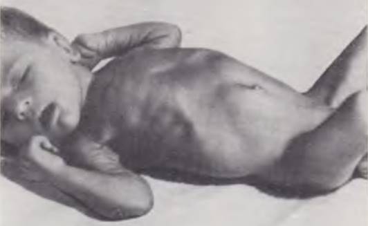
In recent years, thanks to timely medical consultation and intensive conservative therapy, patients with advanced forms of pyloric stenosis are rarely encountered, and a satisfactory general appearance of the patient—differing from classical descriptions of pyloric stenosis—may mislead the physician. Particular attention during the examination of an infant with pyloric stenosis is paid to the abdomen. Pyloric stenosis is characterized by the "hourglass" sign, caused by deep peristalsis of the distended stomach (Fig. 76). Sometimes, even before consulting a doctor, the mother notices that "ripples" or waves are moving across the baby's abdomen.
Fig. 76. An infant with pyloric stenosis. The obstruction in the pyloric region causes deep, strangulating gastric peristalsis—the hourglass sign.
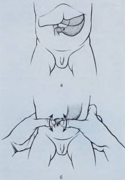
The diagnosis is refined by palpating the pylorus. Palpation of the pylorus should be performed with warm hands, prior to feeding the infant. First, the infant is calmed and given a pacifier dipped in a glucose solution. Pyloric palpation is carried out on the right side at the lateral edge of the rectus abdominis muscle, directly beneath the liver. In cases of significant gastric distension, the pylorus is displaced inferiorly and to the right (Fig. 77).
Fig. 77. Diagram illustrating the anatomical location and palpation of the pylorus in pyloric stenosis. The pylorus may lie beneath the liver (a) or, when the stomach is distended, at the umbilical level. Bimanual palpation is employed (b).
According to the Center for Neonatal Surgery at Rusakov Hospital, the pylorus can be successfully palpated in over 90% of cases. In all patients, the diagnosis was confirmed intraoperatively. If palpation of the pylorus is unsuccessful while clinical suspicion of pyloric stenosis remains, the diagnosis is confirmed using radiological imaging.
Cytology/practical/136.html">DIFFERENTIAL DIAGNOSIS OF pyloric stenosis is primarily performed to rule out pylorospasm. The effective use of antispasmodic therapy and neuroplegic drugs in pylorospasm generally helps clarify the diagnosis. One should also bear in mind the possibility of gastric mucosa prolapse into the pyloric canal. In cases of pyloric area obstruction in neonates within the first hours of life, pyloric and prepyloric atresia may occur. Thomson et al. (1968) reported on 23 patients with this rare pathology, considering pyloroplasty with resection of the obstruction to be the appropriate management.
Diagnosing partial intestinal obstruction caused by malrotation, membranous atresia, and other anomalies presents considerable challenges. The clinical manifestations and time of onset in such cases are so diverse that perhaps the most typical feature of congenital partial intestinal obstruction can be considered the atypical nature of its clinical presentation. The primary symptoms of these malformations include regurgitation, intermittent vomiting, abdominal pain, and periodic stool retention.
Vomitus usually contains an admixture of bile, since the obstruction to the passage of intestinal contents in most patients is located distal to the major duodenal papilla (ampulla of Vater). These clinical manifestations recur periodically and worsen with feeding irregularities, the onset of dyspeptic symptoms, etc. Clinical examination in these instances does not reveal the cause of the obstruction. The diagnosis is refined through radiography, which is decisive in diagnosing complete or partial intestinal obstruction.
Radiological findings. Examination in all cases begins with plain radiography of the chest and abdomen with the infant in an upright position. Radiographs are taken in the anteroposterior (AP) and, if necessary, lateral projections. When interpreting plain radiographs, the timing of gas appearance in various sections of the intestine is taken into account (Table 18).
Table 18. Timing of gastrointestinal tract filling with gas in newborns
Time |
after birth |
|||||||||
Gastrointestinal tract segment |
15 min |
30 min |
45 min |
1h |
2h |
3 — 4h |
3 — 6h |
6 — 8 h |
||
Stomach |
+ |
+ |
+ |
+ |
+ |
+ |
+ |
+ |
||
Duodenum |
— |
+ |
+ |
+ |
+ |
+ |
+ |
+ |
||
Proximal small intestine |
— |
— |
— |
+ |
+ |
+ |
+ |
+ |
||
Distal small intestine |
— |
— |
— |
— |
— |
+ |
+ |
+ |
||
Cecum |
— |
— |
— |
— |
— |
+ |
+ |
+ |
||
Transverse colon |
— |
— |
— |
— |
— |
— |
+ |
+ |
||
Descending colon |
— |
— |
— |
— |
— |
— |
+ |
+ |
||
Note. A plus sign indicates the presence of air, and a minus sign indicates its absence.
The pattern of gas distribution throughout the intestine often suggests or resolves the presence, level, and nature of the obstruction to intestinal contents. A reliable sign of high complete duodenal obstruction (regardless of the specific cause) on an upright plain abdominal radiograph is the "double bubble" sign (Fig. 78, a)—i.e., the presence of a fluid level in the right hypochondrium (projection area of the duodenum) in the absence of gas in other parts of the abdominal cavity. This radiographic picture of two fluid levels with no gas elsewhere allows for a high degree of confidence in diagnosing complete high intestinal obstruction, obviating The Need for further investigations. If a single fluid level is present in the stomach with no gas in other abdominal areas, it is advisable to insert a gastric tube and partially aspirate the contents. This allows the duodenum to decompress, enabling air to enter it so that a subsequent radiograph clearly reveals the typical double bubble sign (Fig. 78). If a second fluid level is still not detected after aspiration, an obstruction in the gastric outlet (prepyloric atresia) should be suspected. If the obstruction is localized at the duodenojejunal junction, one or even two additional small fluid levels may be visualized beneath the gastric shadow.
Fig. 78. Plain abdominal radiographs of newborns with complete duodenal obstruction. Typical double bubble sign (a). Gastric gas bubble with an overfilled duodenum (b). Appearance of a second fluid level in the duodenum following evacuation of gastric contents. Gas in the Large Intestine after an enema (c).

A characteristic sign of complete small bowel obstruction on an upright plain radiograph is the multiple fluid levels sign (Fig. 79), accompanied by the absence of gas in distal abdominal segments and an appropriate clinical presentation. This qualification is necessary because multiple intestinal fluid levels also occur in various types of paralytic (dynamic) ileus, which is frequently observed in neonates. Dynamic ileus is characterized (Fig. 80) by a uniform distribution of fluid levels throughout the abdominal cavity. Furthermore, the infant must have an underlying primary condition, the identification of which is usually not particularly difficult.
Fig. 79. Plain abdominal radiograph of a newborn. Diagnosis: small bowel atresia. Multiple fluid levels in the loops of the proximal (afferent) small intestine.
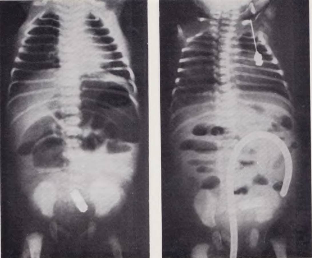
Fig. 80. Plain abdominal radiograph of a newborn. Paralytic ileus. Fluid levels are evenly distributed throughout the abdominal cavity. A tube is visible in the large intestine.
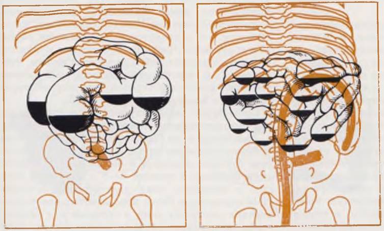
The radiographic presentation of complete small bowel obstruction is fairly characteristic in most cases, and additional studies are generally not indicated. In doubtful cases, the diagnosis is refined using contrast enema examination with air or a contrast medium. In small bowel atresia, the large intestine is markedly narrowed and hypoplastic.
A similar radiographic picture is observed in patients with meconium ileus. The site of intestinal obstruction in meconium ileus is typically localized in the distal ileum (10–12 cm proximal to the ileocecal valve). In most infants with meconium ileus, upright plain radiographs reveal gas-distended loops of the small intestine with fluid levels. The "soap bubble" sign in the terminal ileum, resulting from the mixture of gas with viscous meconium, is typical of meconium ileus. According to our data and those of Herzog (1968) and Kalagogly (1971), the radiographic findings in meconium ileus in most patients differ little from those of small bowel atresia, and patients are taken to surgery with a diagnosis of atresia.
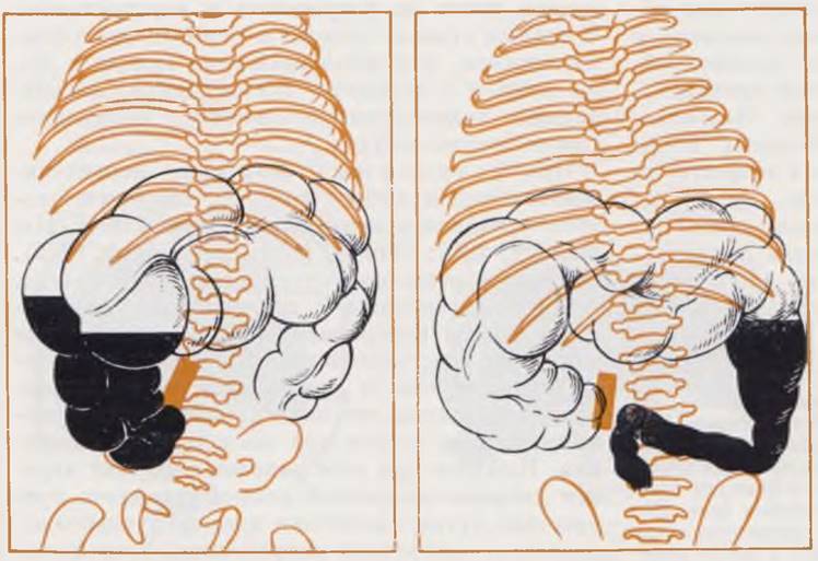
In patients with large bowel obstruction, most commonly caused by Hirschsprung's disease, plain radiographs reveal A large number of distended intestinal loops and fluid levels throughout the entire abdominal cavity. Due to intestinal distension, the Diaphragm is elevated and its excursion is limited. The mediastinal shadow is displaced upward, and the heart appears flattened against the diaphragm (Fig. 81, a). When Hirschsprung's disease is suspected, the diagnosis is confirmed by contrast barium enema (400 g per 1000 ml of a 1% sodium chloride solution). Thirty to forty milliliters of the barium suspension prepared in this manner is administered into the colon. The aganglionic zone is markedly narrowed, its walls are rigid and irregular, and the transition point from the narrowed segment to the dilated funnel-shaped portion is clearly defined (Fig. 81, b).

Fig. 81. Plain abdominal radiograph of a 2-day-old infant. Intestinal loops are markedly distended with gas (a). Barium enema of the same patient (b). A 15 cm long aganglionic zone. Diagnosis: Hirschsprung's disease.
Various types of congenital obstruction are accompanied by overdistension of the wall of the proximal bowel segment filled with intestinal contents, leading to impaired blood supply, necrosis, and subsequent perforation. In such patients, a plain abdominal radiograph reveals free gas in the peritoneal cavity (Fig. 82, a). While a large amount of gas is easily detected, small amounts may lead to diagnostic errors (Fig. 82, b).
Thus, plain radiography of the abdomen and chest in patients with congenital intestinal obstruction is an extremely valuable diagnostic imaging method. Plain radiographs allow clinicians to establish a diagnosis with a high degree of certainty in virtually all patients with complete intestinal obstruction of various etiologies, detect cases of hollow organ perforation, and suspect Hirschsprung's disease, hiatal hernias, etc.
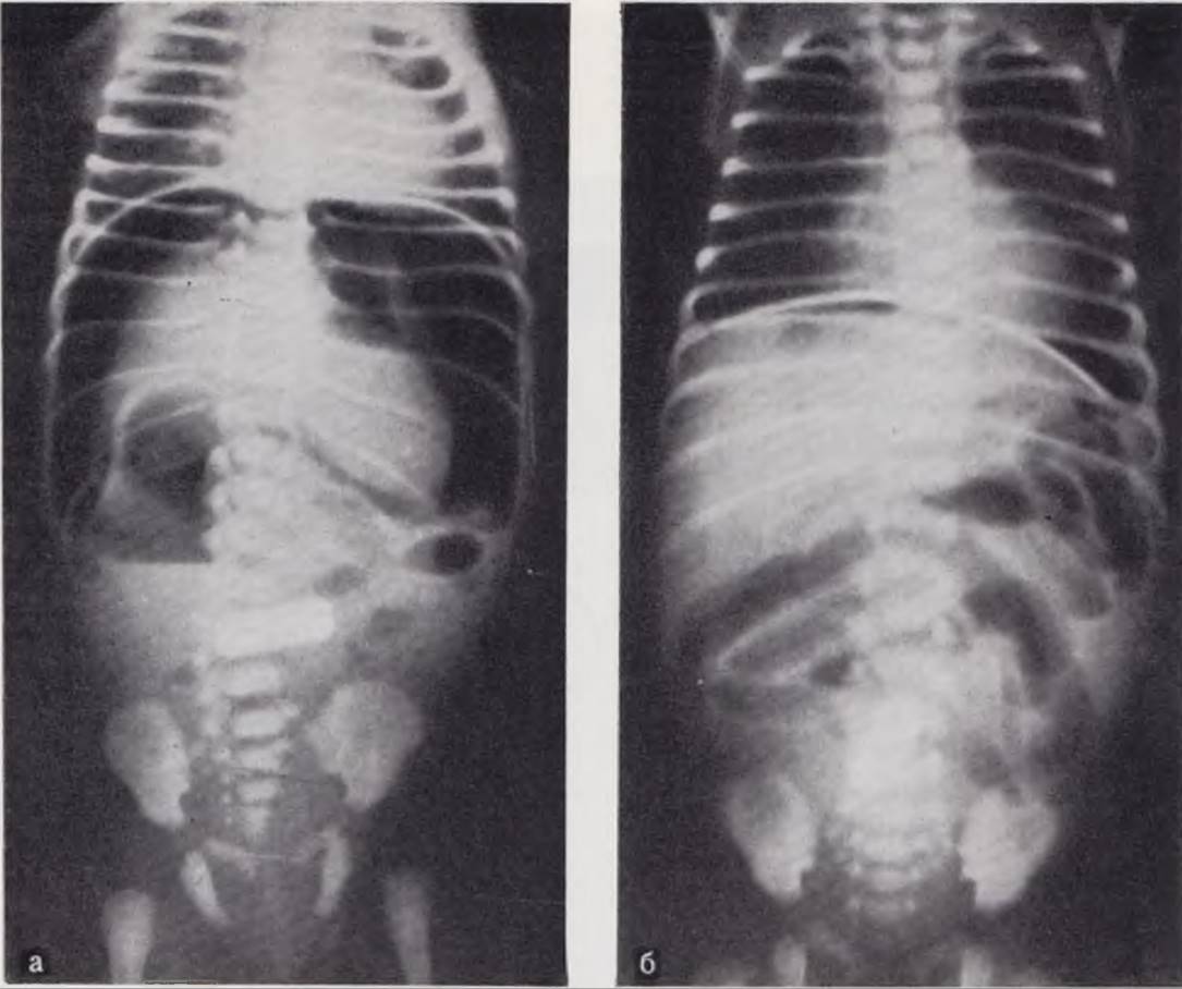
Fig. 82. Plain abdominal radiograph of a newborn with intestinal perforation due to bowel wall necrosis in atresia. A large amount of free gas is present in the abdominal cavity (a). A small amount of gas reveals a narrow crescent sign above the diaphragm (b).
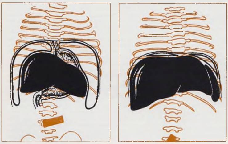
If the aforementioned symptoms are absent on the plain radiograph—which most commonly occurs in cases of partial obstruction—The sequence of further diagnostic Procedures is crucial. Since intestinal malrotation is the most frequent cause of partial intestinal obstruction in newborns, we recommend contrast-air irritography following plain radiography. Under fluoroscopic guidance, with the patient in a vertical position, air is introduced into the rectum using a Richardson bulb, filling the colon and making it clearly visible. Under normal anatomical conditions, the cecal dome is located in the right iliac fossa. The ileocecal valve typically prevents air from entering the small intestine. In patients with intestinal malrotation, the cecum and ascending colon are located in the left half of the abdomen, the epigastrium, or beneath the liver. Identifying these anatomical variants of the cecal dome position allows for the diagnosis of malrotation. There is no need for further diagnostic studies in such cases.
If air contrast enema reveals the cecal dome in its normal anatomical position and the diagnosis remains unclear, a gastrointestinal contrast study is performed. For contrast examination of the gastrointestinal tract in newborns, iodolipol, barium suspension, or water-soluble iodine-containing agents are used (Astley, 1952). We prefer barium suspension as, when used correctly, it poses no danger to the patient and provides a clear outline of the mucosal contours and pattern. This does not preclude The Use of iodolipol (which is preferable when tracheoesophageal fistulas are suspected) and water-soluble contrast media. When intestinal obstruction is suspected, 15 to 20 ml of barium suspension is usually sufficient per examination. The standard suspension is mixed with breast milk. If the infant feeds well, the suspension is given via a nipple. In cases of swallowing disorders, infant weakness, etc., the barium suspension is administered through a nasogastric tube. In the first oblique position, the passage of the contrast suspension through the esophagus is monitored. The position, shape, and lumen width of the esophagus are evaluated in all patients. When hiatal hernias are present, the stomach or a portion of it is located in the mediastinum. In achalasia, a persistent stenosis of the cardiac part of the esophagus and esophageal dilation are observed. The barium suspension is retained above the site of stricture for a prolonged period. A limited suprastenotic dilation symptom—the "arrowhead" sign—is identified. Subsequent portions of the esophagus fill with barium, allowing the degree of dilation to be determined. As the contrast agent passes into the stomach, the narrowed segment of the esophagus and its extent can be visualized.
Conversely, chalasia is characterized by the free passage of the contrast agent into the stomach. When pressure is applied to the epigastric region, increasing intra-abdominal pressure, the contrast suspension easily refluxes into the esophagus.
Next, the stomach and gastric outlet are examined. The most frequent cause of obstruction at this level is pyloric stenosis. As previously noted, in most patients, we confirm the diagnosis of pyloric stenosis by palpation of the hypertrophied pylorus. In doubtful cases, contrast radiography remains decisive for diagnosis. Its results allow for a definitive diagnosis only when the absolute sign—organic narrowing of the pyloric canal—is identified (V. F. Baklanova, M. A. Filippkin, 1969). The retention of barium suspension and gastric dilation can be caused by numerous factors and are not absolute signs of pyloric stenosis (Barret, 1928). The examination is performed with the infant in the vertical position. Palpation of the stomach facilitates the visualization of the narrowed pyloric channel, which is best observed in oblique positions. In mild forms of stenosis, an elongated, narrowed pyloric canal can be visualized. In cases of marked muscular hypertrophy and spasm, the contrast medium fills only the initial part of the pyloric canal, manifesting as the "beak" sign. A characteristic feature of pyloric stenosis is "sterile" peristalsis. The peristaltic wave fails to propel the contrast medium into the duodenum, fading out in the antrum of the stomach. The presence of these symptoms provides the basis for diagnosing pyloric stenosis.
Duodenal stenosis is characterized by gastric dilation, with a large amount of fasting contents found in the stomach. Due to increased pressure within the duodenum, the pyloric channel appears gaping in most cases. In stenosis, the duodenum is dilated, and the duodenal bulb is difficult to demarcate from other segments. The contrast medium passes freely through the dilated pylorus and is arrested at the site of the stenotic lesion.
Partial duodenal obstruction can also be caused by other etiologies (annular pancreas, vascular anomalies, intestinal malrotation). These and other factors lead to mechanical compression of the intestine followed by clinical signs of obstruction and may yield identical radiographic findings. The radiographic diagnosis of intestinal rotation disorders has been described in the greatest detail. In cases of disturbances during the second period of rotation, the typical horseshoe shape of the newborn duodenum is disrupted. Its inferior portion fails to form a leftward duflexion and continues directly into the descending part. The transition of the duodenum into the jejunum occurs in the right upper quadrant of the abdominal cavity. The small intestine lies in the lower right quadrant. Secondary dilation of the duodenum and a fluid level within the dilated segment are frequently noted.
Specific radiographic signs of duodenal compression by an abnormally formed pancreatic head or Blood Vessels do not exist. In such cases, radiographic examination allows for the confirmation of an obstruction at the duodenal level, which serves as the basis for diagnosing stenosis.
The radiographic manifestations of superior mesenteric artery syndrome (arteriomesenteric duodenal obstruction) are quite distinct. On plain radiographs, these patients often exhibit a horizontal fluid level in the duodenum. The contrast agent fills the duodenum up to the point of its crossing with the superior mesenteric artery, where a sharp cutoff of the contrast shadow is observed. Distal to the obstruction, the intestine is of normal size, and its patency is unimpaired. The diagnosis is refined using the Hayes maneuver: the patient is placed prone on a fluoroscopic table. Reducing the tension of the mesentery and, consequently, the mesenteric artery restores intestinal patency, allowing the barium suspension to pass freely into the small intestine.
If no pathology is detected in the duodenal area, monitoring of the contrast suspension's passage through the intestine is continued. Follow-up examinations are best performed at 3 and 6–8 hours. Normally, after 3 hours, the contrast suspension should be located in the small intestine. The persistence of barium residue in the stomach indicates impaired passage of intestinal contents. In partial small bowel obstruction, the contrast suspension fills the dilated proximal segment. Identifying the site of narrowing provides the basis for the diagnosis. By 6–8 hours, the contrast suspension should normally reach the large intestine, the patency and anatomical position of which were previously determined by air-contrast barium enema.
Adhering to the recommended sequence of radiological examination in patients with suspected congenital intestinal obstruction allows for a diagnosis to be established and a treatment plan to be formulated within 6–8 hours in the vast majority of cases, with minimal radiation exposure to the patient. The need for a more prolonged study arises only in diagnostically complex, atypical cases among a small number of patients admitted with or suspected of having intestinal obstruction.
After establishing the diagnosis and providing appropriate preoperative preparation, the infant undergoes surgery.
Treatment. Relief of the obstruction to the passage of intestinal contents in congenital gastrointestinal obstruction is possible only through surgical intervention in the vast majority of cases. Exceptions include chalasia, achalasia, and compensated forms of Hirschsprung's disease.
In chalasia, expectant management and the Prevention of potential esophageal mucosal erosion (feeding the infant in an upright position, followed by maintaining an elevated head position) yield favorable outcomes in most patients. Gradual "maturation" of nerve elements leads to the resolution of the gaping cardia. In cases of persistent achalasia in newborns, the method of choice is dilation of the cardiac portion of the esophagus using bougies or specialized dilators (Swenson, 1958).
Compensated forms of Hirschsprung's disease respond to conservative treatment (systematic siphon enemas with a 1% saline solution). These children undergo surgery at an older age.
For all other patients with obstruction, emergency surgery is the method of choice. Issues related to preoperative preparation in newborns and surgical approaches to the abdominal organs are detailed in the respective chapters. We will focus on the surgical techniques employed for various types of obstruction.
Pyloric stenosis. Surgery is performed under general anesthesia. Local anesthesia is indicated in debilitated infants with severe pneumonia. Pneumonia is not an absolute contraindication to surgery; on the contrary, normalizing these patients' nutrition postoperatively allows for a more rapid and effective resolution of pneumonia. In the operating room, immediately after the administration of anesthesia once the infant is asleep, a confirmatory palpation of the pylorus is performed. This is easily accomplished with relaxed abdominal wall musculature.
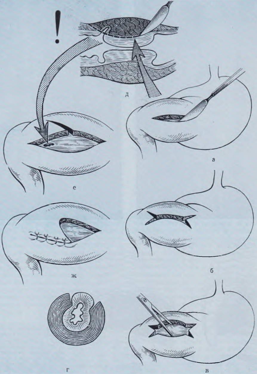
Surgical technique. An incision is made along a skin crease, followed by muscle-splitting along the fiber direction over the projected LOCATION OF THE pylorus. The Pyloric part of the stomach with the thickened pylorus is delivered into the wound. The method of choice is the Fredet-Ramstedt pyloromyotomy as modified by Braunstein in the avascular zone of the pylorus (Fig. 83). When incising the serosa and muscular layer, extreme care must be taken at the transition point of the hypertrophied pyloric muscle into the duodenum, where mucosal injury is possible. Next, using forceps or a mosquito hemostat, the edges of the muscle are gently separated until the mucosa herniates into the wound. Auxiliary incisions ("whiskers") prevent potential mucosal injury and allow for a more thorough Separation of the muscle edges and better protrusion of the mucosa. Bleeding from the edges of the pyloric wound is controlled by suturing the bleeding vessel from the serosal side at a distance of 1–2 mm from the wound edge. After checking the integrity of the mucosa and achieving hemostasis, the incised pylorus is returned to the abdominal cavity. In case of accidental mucosal injury, the defect is closed with a catgut suture on an atraumatic needle. If the mucosal tear is extensive, it is covered with a mobilized seromuscular flap, or the incised pylorus muscle is repaired with U-sutures and a pyloromyotomy is performed on the opposite side of the pylorus. The wound is closed layer-by-layer with nylon sutures.
Fig. 83. Diagram of pyloromyotomy for pyloric stenosis.
a — Fredet-Ramstedt operation; b, c — Fredet-Ramstedt operation as modified by Braunstein; d — bulging of the mucosa after separating the edges of the seromuscular layer incision; e — injury to the duodenal mucosa; f, g — seromuscular flap transposition operation for mucosal injury.
In the postoperative period, the goal is to normalize nutrition as quickly as possible. If the mucosal membrane has not been injured, oral feeding can be initiated 2 to 4 hours after the completion of surgery and anesthesia. Feeding begins with 5 ml of chilled tea or glucose solutions, followed by expressed breast milk. In the absence of vomiting, The amount of milk is gradually increased, and full breastfeeding can be resumed by the 4th day.
If the mucosal membrane has been injured, oral feeding is withheld for the first 24 hours after surgery. Subsequently, depending on the child's condition, oral feeding is gradually introduced. Breastfeeding is typically resumed on the 5th to 6th day.
Duodenal obstruction. The causes of duodenal obstruction are divided into correctable and uncorrectable. The former include stenoses and membranous forms of atresia, external compression of the bowel, etc., while the latter include true megaduodenum, annular pancreas, and vascular anomalies.
For correctable forms of obstruction, the method of choice is surgery aimed at eliminating the obstruction (Hedenberg, 1967; Dinner, 1969; Gourevitsch, 1971). This can be achieved by incising the bowel, excising the membrane, and subsequently performing a transverse suture closure. The incision is initiated just below the site of the membrane and extended into the dilated segment of the intestine, which helps prevent luminal narrowing during subsequent suturing. For membranous forms of duodenal atresia, Rickham eliminates the obstruction by creating a transverse duodenoduodenostomy. External obstructions (such as an abnormally formed mesocolon or tumor-like formations) are corrected using standard surgical techniques.
For uncorrectable forms of obstruction, the method of choice is the creation of a bypass, isoperistaltic duodenojejunostomy using a short loop (Free, Gerald, 1968; Moshe et al., 1968; Guglielmi et al., 1971).
Surgical technique. An opening is created in the transverse mesocolon, to the edges of which the dilated duodenum is sutured. The side of the small intestine is anastomosed to the side of the duodenum using a single-layer suture. The width of the newly formed anastomosis should be at least 2.5 cm. Resection of the anomalous segment of the duodenum followed by primary anastomosis is theoretically possible, but technically challenging. Due to the high risk of injury to the pancreatic ducts, bile ducts, and other structures, it has not gained Practical Application in neonatal surgery.
Gastroenterostomy in neonates is inappropriate, as it may be complicated by hemorrhagic gastrojejunitis. Two such cases were described by Stuart (1970).
Last update: 10/08/2026
Editorial and Educational Adaptation: This material has been compiled based on the primary/original source text. The project team performed an editorial review, corrected technical inaccuracies, structured sections, and adapted the content for an educational format.
What was processed:
- elimination of formatting defects (OCR errors, structural breaks, corrupted characters);
- editorial organization of content;
- standardization of terminology in accordance with academic sources;
- verification of factual statements against the original source text.
All mentions of the author, publication year, and origin of the primary text have been preserved in accordance with the source.