Human Anatomy - H. I. Koliadenko 2009
The Musculoskeletal System
Structure, Shape, and Functions of Bones
The Structure AND Functions of bones are studied by the science of Osteology.
The human Skeleton accounts for 1/5 to 1/7 of the total body mass. It comprises 203–206 bones, of which 164–166 are paired and 36–40 are unpaired (Fig. 14). The skeleton performs protective, supportive, and several other functions. For example, protective functions are performed by the cranium, which houses the Brain; THE Vertebral Column, in whose canal the Spinal Cord lies; and The thoracic cage, formed by the Sternum, Ribs, and thoracic spine, which protects the Lungs, Heart, aorta, INFERIOR VENA CAVA, Esophagus, and other Organs.
The BONES OF THE limbs perform supporting and lever functions, which facilitate diverse movements in space as well as labor activities.
Furthermore, skeletal bones are classified by shape. Depending on their shape, they are categorized as tubular, spongy, irregular, long, flat (broad), short, and pneumatic.
Long bones form the Skeleton of the limbs. Flat or broad bones enclose vital cavities (cranium, pelvis), while short and irregular bones form the spine, wrist, and FOOT.
A tubular bone consists of a body, known as the diaphysis, and two extremities, known as the epiphyses: the proximal epiphysis, located closer to the HEAD, and the distal epiphysis, situated at the opposite end of the bone relative to the first.
Inside the shaft, tubular bones contain cavities filled with adipose tissue, known as yellow Bone Marrow.
Flat and short bones lack cavities.
A bone consists of compact substance (substantia compacta) and spongy substance (substantia spongiosa) (Fig. 15). The former is located externally and forms a dense and robust structure. The spongy substance in tubular bones is found in the epiphyses and consists of bony trabeculae, the orientation of which depends on the pull of the Muscles acting upon them. Cytology/practical/86.html">Red bone marrow lies between the trabeculae of the spongy substance.
Class="center">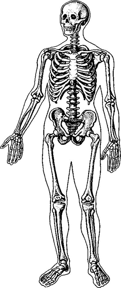
Fig. 14. Human skeleton
Every tubular bone is covered externally by a sheath called the periosteum. The periosteum consists of two layers of Connective Tissue. The outer layer is fibrous, formed by interwoven thick bundles of Collagen fibers, through which Blood Vessels and nerves pass into the depth of the bone. The outer layer performs a protective function. The inner layer, known as the osteogenic (cambial) layer, consists of bundles of collagen and elastic fibers, as well as a significant number of osteoblasts that multiply during bone growth and differentiate into osteocytes.

Fig. 15. Proximal end of the Femur:
a — frontal section:
1 — spongy substance; 2 — compact substance; 3 — medullary cavity; b — diagram of trabecular arrangement in the spongy substance
Bone vascularization (blood supply) is provided by nutrient canals located within the compact bone substance, through which Blood Vessels enter. They penetrate the Bone tissue, branching into smaller vessels that transition into the Haversian canals. Most nutrient canals run between the diaphysis and the epiphysis of the bone.
Flat (broad) skeletal bones (e.g., scapula, hip bone, sternum), like tubular bones, are covered externally and internally by a thin layer of compact substance, between the plates of which lies the spongy substance. A specific structural feature of the flat bones of the cranium is that the inner plate of compact substance—known as the glassy plate (lamina vitrea) due to its properties—easily fractures upon trauma, leading to injury of the cerebral blood vessels.
The wrist, foot, and spine are composed of short and irregular bones. These bones are built of spongy substance covered externally by a layer of compact substance.
Bones containing air-filled cavities are termed pneumatic bones. These include the cranial bones: frontal, temporal, ethmoid, sphenoid, and Maxilla.
□ Bone Marrow. The medullary cavities of tubular bones are filled with bone marrow. Between the trabeculae of the spongy substance in the epiphyses lies red bone marrow, which is a soft red mass with branching blood vessels. Its framework is reticular tissue, within the meshes of which hematopoietic stem Cells and red Blood Cells are located.
Bone marrow is of vital importance to human physiology as it produces erythrocytes, granulocytes, and other blood cells. Yellow bone marrow is located in the diaphyses of tubular bones; it consists of adipose tissue, plays an important role in METABOLISM, and does not take part in hematopoiesis. However, in cases of massive blood loss or infectious diseases, yellow bone marrow resumes hematopoietic functions. The Human Body contains approximately 1,500 cm3 of red bone marrow and an equal amount of yellow bone marrow. In infants and adolescents, red bone marrow fills the entire bone cavity, and only after the age of 20–25 does the red bone marrow in the medullary cavity of the diaphysis become replaced by yellow bone marrow.
□ Chemical composition of Bones. Bones are composed of organic and inorganic substances. Dried and defatted bone consists of 1/3 organic substances and 2/3 inorganic substances. Organic substances are represented by ossein and osseinmucoid, while inorganic substances consist of Water and mineral salts. In a living adult bone, water accounts for nearly 50%, fat for 15.7%, other organic substances for 12.8%, and inorganic substances for 21.5%. Of the Inorganic Compounds, 95% are calcium salts. The adult skeleton contains about 1,200 g of calcium salts (99% of the total amount in the human body), 530 g of phosphorus, and 11 g of magnesium.
Organic substances impart flexibility and elasticity to the bone, whereas inorganic substances provide hardness and rigidity.
The bones of the human skeleton serve as a reservoir for inorganic substances, which are released into the blood as needed and utilized by Tissues and organs during their metabolic processes.
The chemical composition of bones changes with age. It depends on the person's diet and occupational activity. In the case of dietary calcium deficiency (or calcium metabolism disorders), bones become soft and may deform under body weight. This is more commonly observed in childhood, as children's bones contain more organic matter than inorganic matter. In elderly individuals, the proportion of organic components decreases, making bones more brittle and thus more susceptible to fractures compared to younger people.
□ Bone GROWTH AND DEVELOPMENT. Bone develops from condensed mesenchyme, whose cells undergo intensive proliferation. Bone development proceeds via two main pathways. One group of bones develops directly from the mesenchyme (membranous stage); these are termed primary, or dermal, bones. Membranous bones include the bones of the Skull roof, face, and clavicles. All other bones—those of the cranial base, trunk, and limbs—are referred to as cartilaginous, or secondary, bones, meaning they pass through both a membranous and a cartilaginous stage before finally transforming into bone (ossification stage).
Direct bone development occurs via two mechanisms: perichondral ossification, when ossification centers form on the Cartilage surface, and endochondral ossification, when they emerge and develop within the cartilage tissue. In shape, they resemble the future bone.
Bone growth is driven by osteoblasts, which secrete osteoid and calcium salts, forming a thin layer of bone tissue around themselves. Simultaneously, osteoclasts break down bone tissue, creating a central cavity within the bone. This cavity expands from the diaphysis toward the epiphyses, forming a tubular structure through the action of bone-resorbing substances.
Subsequently, the bone cavity fills with embryonic mesenchyme, which gradually differentiates into reticular connective tissue, and ultimately transforms into bone marrow.
Elongational bone growth occurs via the epiphyseal cartilage plate located between the diaphysis and epiphysis of long tubular bones, which gradually thins out. This process continues until ages 20–25, after which the diaphysis firmly fuses with the epiphysis, and longitudinal growth ceases. Appositional growth (thickening) occurs through the periosteum. Periosteal osteoblasts proliferate to form bone lamellae that are deposited On the surface—a process known as apposition, whereas the expansion of bone tissue during longitudinal growth is called intussusception. Even after growth ceases, bone remodeling continues throughout the entire lifespan of the Organism. A person's occupation leaves its mark on Bone Structure. Corresponding to functional demands, the trabeculae of spongy bone tissue arrange themselves along lines that best ensure resistance to compression and tension. The orientation of Haversian canals in compact bone also adapts. In individuals engaged in sports and physical labor, bone thickening develops at sites of Muscle attachments that endure higher mechanical loads than other areas. Excessive mechanical stress on a bone disrupts existing Haversian systems and prompts The formation of new ones.
In old age, bone Osteoporosis develops, leading to the formation of significant cavities between the trabeculae of spongy bone. Similar spaces also form between the osteons of the Haversian system, accompanied by Changes in the chemical COMPOSITION OF THE bone, characterized by a redistribution of organic and Inorganic Components.
Bones possess a high regenerative capacity, which is greater the younger the organism is. In the event of fractures, regeneration is driven by osteoblast proliferation. Initially, a bone callus forms at the fracture site, which subsequently ossifies and is replaced by a solid bony union.
In the embryo, certain bones initially develop as multiple separate elements. For instance, the hip bone develops from three distinct bones: the ilium, ischium, and pubis. In childhood, they are united by synchondrosis, but between the ages of 14 and 16, they fuse via synostosis into a single monolithic pelvic bone. Similarly, tubular bones in children develop in three parts: a diaphysis and two epiphyses (proximal and distal). Initially joined immovably by synchondrosis, by ages 20–25 they fuse into a single bone via synostosis.
Flat bones ossify in a manner similar to the epiphyses of tubular bones.
The rate of bone growth and development depends on mechanical loading and Nutrition. Ossification occurs more rapidly in areas subjected to greater mechanical pressure; for example, bones of the lower limbs ossify faster than those of the upper limbs. Furthermore, in bones experiencing rhythmic mechanical loading at optimal levels, ossification proceeds more swiftly. Conversely, excessive loads inhibit bone growth. Physical exercises, particularly jumping and stretching activities, accelerate bone growth by thickening the compact bone and increasing trabecular spaces within the spongy bone.
Last update: 08/08/2026
Editorial and Educational Adaptation: This material has been compiled based on the primary/original source text. The project team performed an editorial review, corrected technical inaccuracies, structured sections, and adapted the content for an educational format.
What was processed:
- elimination of formatting defects (OCR errors, structural breaks, corrupted characters);
- editorial organization of content;
- standardization of terminology in accordance with academic sources;
- verification of factual statements against the original source text.
All mentions of the author, publication year, and origin of the primary text have been preserved in accordance with the source.