Human Anatomy - G. I. Koliadenko 2009
The Musculoskeletal System
Articulations of the Skeleton
The Doctrine of bone connections is called Arthrology or syndesmology.
Depending on functional loading, the bones in the human Skeleton are connected movably, semimovably, and immovably. Limited-movement and immovable joints are called synarthroses.
Movable joints are synovial (diarthroses). These include joints (articulationes synoviales).
Immovable joints (synarthrosis)—fibrous joints—are types of connections in which bones appear to be fused together by means of a particular type of Connective Tissue, according to which they received their names: syndesmosis, synchondrosis, synostosis, and syncinesia / syncarcosis (Fig. 16).
Syndesmosis is a bone connection via Fibrous connective tissue. Fibrous joints include, for example, the Ligamentous apparatus of the pelvic bones with the sacrum, as well as the BONES OF THE hand and FOOT. Connective-tissue membranes between the diaphyses of the forearm and leg bones are characterized by lesser strength. Syndesmoses also include cranial sutures, etc.
Synchondrosis is a bone connection via cartilaginous tissue. The mobility and strength of cartilaginous joints depend on the type of Cartilage tissue and the thickness of the cartilage. The thicker the cartilaginous layer between bones, the more movable the joint. Hyaline cartilage is characterized by greater elasticity, whereas fibrous cartilage has less. Regarding strength, the inverse relationship applies: hyaline joints (costal cartilages with the Sternum) have less strength, while fibrous cartilage (connections of vertebrae in THE Vertebral Column) has greater strength.
Synostosis is a connection via Bone tissue. For instance, the pelvic bones (ilium, pubis, and ischium) are connected to each other by cartilaginous tissue up to the age of 15–16, after which, in adulthood, the cartilage is replaced by bone tissue, forming an immovable joint known as a synostosis. The same applies to the sacrum, which consists of 5 vertebrae in children, whereas in adults it forms a single monolithic bone. This category also includes the bones of the cranial vault in individuals over thirty years of age, when connective-tissue sutures transform into bony ones.
Syncarcosis is a bone connection via Muscle tissue. For example, the scapula is connected to the spine by the rhomboid muscle and the levator scapulae muscle; this bone is connected to the upper limb by a group of shoulder girdle Muscles (teres Major and minor, subscapularis, supraspinatus, and infraspinatus).
Class="center">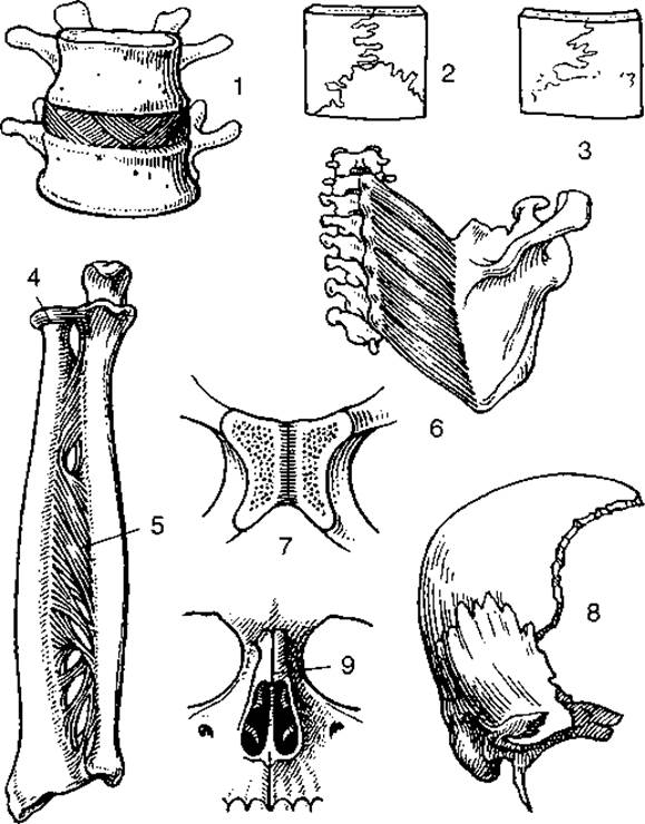
Fig. 16. Immovable bone joints:
1 — intervertebral disc — synchondrosis; 2 — serrate suture; 3 — Ossification of the serrate suture — synostosis; 4 — annular ligament — syndesmosis; 5 — interosseous membrane — syndesmosis; 6 — bone connection via muscles — syncarcosis; 7 — amphiarthrosis — symphysis; 8 — squamous suture; 9 — plane suture
In humans, the most common type of bone connections are synovial joints, or articulations.
A joint comprises primary and auxiliary structures. The primary structures include the articular surfaces of the articulating bones, the Joint Capsule, and the joint cavity. The epiphyses of the bones have smooth surfaces covered with a thin layer of hyaline cartilage. The joint capsule, or articular capsule, consists of two membranes (layers): the outer, or fibrous, layer, which is a continuation of the periosteum, and the inner, or synovial, layer, featuring a branching network of Blood Vessels; the inner surface of the capsule bears fine outgrowths—villi—that secrete synovia, a viscous fluid that lubricates the articular surfaces. In some joints, such as the knee, the synovial membrane forms folds containing adipose deposits. In joints like the shoulder and knee, the synovial membrane forms outpouches filled with synovial fluid that protrude between the fibers of the fibrous membrane beyond the capsule, serving as a cushion for the tendons of nearby muscles or those passing over the joint surface.
Atmospheric pressure within the joint cavity is negative, which helps hold the articulating bones together.
In some joints, apart from the primary structures, auxiliary formations are present: articular cartilages (discs, menisci), sesamoid bones, articular Lips, and ligaments.
Discs and menisci are formed of fibrous cartilage and are located inside the joint cavity. An articular disc lies between the articular surfaces of the bones and fuses at its edges with the joint capsule, dividing the joint into two chambers (e.g., the temporomandibular joints). Menisci are situated along the margins of the articular surfaces of bones (e.g., the knee joint). Both discs and menisci ensure greater congruence (matching) of the articulating bones, thereby enhancing joint strength and stability.
Certain joints contain sesamoid bones (such as the Patella in the knee joint). Within a joint, sesamoid bones play an auxiliary role, serving as attachment sites for muscle tendons and ligaments. Additionally, they act as pulleys during Muscle Action.
Articular lips are located around the articular surfaces of bones, thereby enlarging the articular surface (e.g., of the scapula).
Joint ligaments secure the bones within the joint or restrict their range of motion (e.g., in the knee and hip joints).
Joint shapes and movements. Every movement of a given part of The Human Body is determined by the specific shape of the joints. Throughout Human Evolution, various joint shapes have formed, namely: ball-and-socket, cotyloid (nut-like), ellipsoid, hinge, saddle, pivot, plane, and combined joints (Figs. 17, 18).
If articular surfaces are viewed as segments of geometric figures, one can imagine that Movements of the articulating bones occur around the axes of rotation of these segments. Consequently, the axis of rotation is an imaginary line passing through the center of the joint around which one bone rotates relative to another. To determine The Nature of movements in joints, three mutually perpendicular axes are used: transverse (or frontal), anteroposterior (or sagittal), and vertical. All movements in synovial joints are considered relative to the anatomical position of the body.
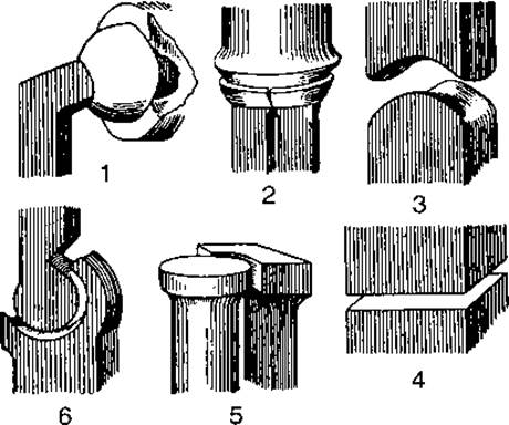
Fig. 17. Various joint shapes:
1 — ball-and-socket; 2 — ellipsoid; 3 — saddle; 4 — plane; 5 — pivot; 6 — hinge
Depending on the number of axes around which movements can occur, joints are classified as uniaxial, biaxial, and triaxial. Triaxial joints include ball-and-socket joints, which possess a wide range of motion. A ball-and-socket joint is defined as one in which the articular surface of one connecting bone approximates a sphere in shape, while the other bone features a concave articular fossa (e.g., the shoulder joint).
The nut-shaped (spheroidal) joint differs from the ball-and-socket joint solely by a deeper articular fossa, which is enlarged by an articular lip, thereby somewhat restricting its range of motion (for example, the hip joint).
Biaxial joints include ellipsoid and saddle joints. In an ellipsoid joint, the articular surfaces are elongated and resemble a segment of an ellipsoid. Ellipsoid joints are most commonly formed by multiple bones (for instance, the radiocarpal joint).
The articular surfaces of a saddle joint are concave in the longitudinal direction and convex in the transverse direction, resembling two saddles fitted together. An example is the carpometacarpal joint of the thumb. Notably, this joint is unique to humans and evolved As a result of labor activity.
Uniaxial joints comprise pivot (trochoid) and hinge (ginglymus) joints. A pivot joint is formed by bones where one has an articular surface resembling a cylinder segment, while the other is concave. Movements in this joint occur either in the transverse plane (flexion and extension) or vertically (rotation, depending on the orientation of the HEAD's axis). Examples include the proximal and distal radioulnar joints. If the cylindrical articular surface features a groove and the corresponding concave surface features a ridge, such a joint is termed a hinge joint (for example, the humeroulnar and interphalangeal joints).
Plane joints possess nearly flat articular surfaces resembling a segment of a sphere with a large radius, lacking distinct articular heads and sockets. Movements in plane joints involve a slight gliding of one articular surface over another, rendering them minimally mobile; hence, they are also referred to as amphiarthroses. Examples of this joint type include the carpometacarpal joints (except for the thumb), tarsometatarsal joints, and others.
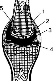
Fig. 18. Diagram of a joint:
1 — periosteum; 2 — articular capsule; 3 — synovial membrane; 4 — articulating bone surfaces covered with articular cartilage; 5 — cartilage
In addition to Classification by the shape of their articular surfaces, joints are categorized as simple, compound, and combined. A simple joint is formed by two bones, such as the interphalangeal JOINTS OF THE hand. If a joint incorporates three or more bones, it is termed a compound joint. An example of a compound joint is the elbow, which is formed by three bones and three articulations sharing a common joint capsule.
Combined joints refer to those that are morphologically independent yet functionally interdependent. For instance, the temporomandibular joints function exclusively as a paired, simultaneous unit.
Between freely movable and slightly movable bone connections, there exists a transitional form known as a hemiarthrosis, or symphysis, featuring a small cavity within the cartilage. Such structures connect certain sternal bones (the manubriosternal symphysis), vertebrae (intervertebral symphyses), and pubic bones (Pubic Symphysis). For enhanced stability, the intervertebral and pubic symphysis are reinforced by numerous ligaments. During childbirth, the pubic bones can slightly separate under pressure, thereby enlarging the pelvic outlet.
Last update: 08/08/2026
Editorial and Educational Adaptation: This material has been compiled based on the primary/original source text. The project team performed an editorial review, corrected technical inaccuracies, structured sections, and adapted the content for an educational format.
What was processed:
- elimination of formatting defects (OCR errors, structural breaks, corrupted characters);
- editorial organization of content;
- standardization of terminology in accordance with academic sources;
- verification of factual statements against the original source text.
All mentions of the author, publication year, and origin of the primary text have been preserved in accordance with the source.