Human Anatomy - G. I. Koliadenko 2009
Musculoskeletal System
Skeleton of the Trunk
Structure of the Spine
THE Vertebral Column, or spine (columna vertebralis), forms the foundation of the entire body and consists of 31–35 vertebrae connected by movable and immovable joints.
The vertebral column has 5 regions: the cervical—7 vertebrae, thoracic—12 vertebrae, lumbar—5 vertebrae, sacral—5 vertebrae fused into a single bone (the sacrum), and coccygeal—4–5 vertebrae.
A vertebra (vertebra) (Fig. 19) has a body (corpus), behind which lies an arch (arcus). The body and arch enclose the vertebral foramen (foramen vertebrale). The foramina of adjacent vertebrae form the vertebral canal, which houses the Spinal Cord. At the junctions of the arch with the body, notches (upper and lower) are formed. The notches of adjacent vertebrae bound the intervertebral foramina through which Spinal Nerves exit. Seven processes extend from the arch: a posterior, unpaired spinous process (processus spinosus), 2 lateral transverse processes (processus transversus),
2 superior articular processes (processus articularis superior), and 2 inferior articular processes (processus articularis inferior). Through their articular processes, the vertebrae connect with one another to form the vertebral column. The vertebrae of each region possess specific features that distinguish them from the vertebrae of other regions.
The cervical region consists of seven cervical vertebrae (vertebrae cervicales), which have small bodies and bifid spinous processes (except for the first and seventh vertebrae). The transverse processes, together with rib rudiments, form the transverse foramen through which the vertebral artery ascends to the Brain.
The first cervical vertebra, or atlas (atlas), articulates with The Skull and is ring-shaped, lacking both a body and a spinous process. It consists of anterior and posterior arches, between which lie thickenings known as the lateral masses.
The anterior arch replaces the vertebral body and features an anterior tubercle on its anterior surface and a facet for the dens (fovea dentis) on its posterior surface for articulation with the dens of the second cervical vertebra. The posterior arch bears a posterior tubercle, which is the remnant of a spinous process.
Short and thick transverse processes extend from the lateral masses, which feature superior and inferior articular facets that replace the articular processes.
Class="center">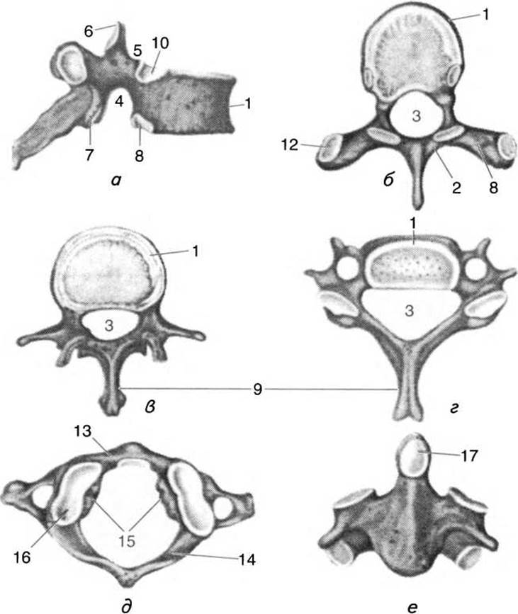
Fig. 19. Vertebrae:
a, b — eighth thoracic vertebra (lateral and superior views); c — third lumbar vertebra (superior view); d — sixth cervical vertebra (superior view); e — first cervical vertebra, or atlas (superior view); f — second cervical vertebra, or axis (anterior view); 1 — vertebral body; 2 — vertebral arch; 3 — vertebral foramen; 4 — inferior vertebral notch; 5 — superior vertebral notch; 6 — superior articular process; 7 — inferior articular process; 8 — transverse process; 9 — spinous process; 10 — superior costal facet; 11 — inferior costal facet; 12 — transverse costal facet; 13 — anterior arch of atlas; 14 — posterior arch of atlas; 15 — lateral mass of atlas; 16 — superior articular facet of atlas; 17 — dens of axis
The II cervical vertebra, or axis (axis), is named after its function, as the first vertebra and the skull rotate around it like an axis. Extending upward from the irregularly shaped vertebral body is a small, thick process—the dens. It should be considered as a part of the body of the first vertebra that fused phylogenetically with the body of the second vertebra. The dens features small anterior and posterior articular surfaces that articulate with the first vertebra. Instead of superior articular processes, two articular surfaces facing upward and laterally are located on either side of the dens; the inferior articular processes are similar to those of other vertebrae, while the spinous process is short and thick.
The anterior tubercle of the transverse process of the VI cervical vertebra is of significant practical importance because it is slightly more developed than in other cervical vertebrae, and the common carotid artery passes anterior to it (hence it is called the carotid tubercle). In case of an injury to the carotid artery, it is necessary to quickly compress the vessel against this tubercle to stop the bleeding.
The thoracic region of the vertebral column consists of twelve thoracic vertebrae (vertebrae thoracicae). Their tall, triangular bodies bear costal facets that articulate with the articular surfaces of the rib heads. Because most Ribs articulate with the bodies of two vertebrae, they have two "hemi-facet" costal depressions: a superior costal facet on the upper edge of the vertebra and an inferior one on the lower edge, which together form a "full" costal facet for the HEAD of the rib.
An exception is the I thoracic vertebra, which features a "full" costal facet for the first rib on its upper edge and a "hemi-facet" for the second rib on its lower edge. The principal diagnostic Features of the X, XI, and XII thoracic vertebrae are the "full" articular facets on their bodies.
The superior and inferior articular processes are flat, oriented in the frontal plane and nearly vertically, with their articular surfaces facing forward and backward, respectively.
The transverse processes are located posterior to the articular processes, are well developed, thickened at their ends, and bear an articular facet on their anterior surface for articulation with the rib tubercle. It should be noted that the transverse processes of the XI and XII thoracic vertebrae lack such facets.
The spinous processes are significantly longer than those of the cervical vertebrae and are triangular in shape. In the upper thoracic vertebrae, they are oriented more horizontally; in the middle ones, they run almost vertically downward; and in the lower ones, they resume a horizontal orientation.
The vertebral foramina of the thoracic vertebrae are circular in shape and smaller in size than those in the cervical and lumbar regions.
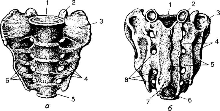
Fig. 20. Sacrum:
a — anterior view:
1 — Base of the sacrum; 2 — superior articular process; 3 — lateral part; 4 — transverse lines; 5 — apex of the sacrum; 6 — pelvic sacral foramina; b — posterior view:
1 — sacral canal; 2 — superior articular process; 3 — auricular surface; 4, 5, 8 — sacral crests (intermediate, median, lateral); 6 — sacral foramen; 7 — apex of the sacrum
The lumbar region of the vertebral column consists of five vertebrae (vertebrae lumbales). They differ from the cervical and thoracic vertebrae in their dimensions (the body is large and oval-shaped), mass, and the absence of costal facets and transverse foramina.
The articular processes of the lumbar vertebrae are robust, oriented in the sagittal plane, and positioned vertically. The superior articular processes feature a small thickening known as the mamillary process. The transverse processes are thin and flat, with accessory processes located at the base of each. Most anatomists view these as rudimentary ribs. The spinous processes of these vertebrae are large, long, broad and plate-like, thickened at their extremities, and lie almost horizontally.
The sacral region of the vertebral column consists of five sacral vertebrae, which fuse into a single bone (os sacrum) between the ages of 17 and 25.
In shape, the sacrum (Fig. 20) resembles a wedge with its base directed upward and its apex downward and curved slightly forward. The base of the sacrum resembles a vertebral body and articulates with the last (V) lumbar vertebra at an angle, forming the sacral promontory (promontorium), which projects into the pelvic cavity. Posterior to it lies the upper opening of the sacral canal, which runs through the entire sacrum and is a continuation of the vertebral canal. Extending upward from the posterior part of the base are two small, flat superior articular processes that, together with the inferior articular processes of the last lumbar vertebra, form the lumbosacral joint. The apex of the sacrum terminates in the sacral cornua, between which lies the sacral hiatus.

Fig. 21. Male (top) and female pelvis:
1 — ilium; 2 — iliac fossa; 3 — sacrum; 4 — acetabulum; 5 — coccyx; 6 — ischium; 7 — Pubic Symphysis; 8 — subpubic angle; 9 — ischial tuberosity; 10 — obturator foramen; 11 — pubis; 12 — pelvic inlet; 13 — sacroiliac joint; 14 — anterior superior iliac spine; 15 — iliac crest
The sacrum features pelvic and dorsal surfaces, as well as lateral parts. The pelvic surface is markedly concave and forms part of the posterior wall of the lesser pelvis. Near its margin are four or five pairs of pelvic sacral foramina, through which pass the NERVES OF THE Sacral Plexus and Blood Vessels.
The dorsal surface of the sacrum is convex. Running along the midline is the median sacral crest, formed by the fusion of the spinous processes of the sacral vertebrae. Flanking it are the low intermediate sacral crests, resulting from the fusion of the superior and inferior articular processes, lateral to which lie the lateral sacral crests, formed by the fusion of the transverse processes.
The massive lateral PARTS OF THE sacrum are formed by the fusion of the transverse processes of the sacral vertebrae and rudimentary sacral ribs. The upper halves of the lateral parts bear auricular surfaces that articulate with the iliac bones to form the sacroiliac joints. Posterior to these surfaces are the sacral tuberosities, which serve as attachment sites for ligaments.
In women, the sacrum is broader, less curved, and shorter (Fig. 21) than in men.
The coccygeal region of the vertebral column (coccygis) consists of 4–5 coccygeal vertebrae, which fuse in middle-aged adults. The shape of the coccyx resembles a bird's beak, with its base directed upward and slightly curved. Its superior surface articulates with the sacrum, toward which two coccygeal cornua project. In women, the coccyx is wider and less curved than in men.
Last update: 08/08/2026
Editorial and Educational Adaptation: This material has been compiled based on the primary/original source text. The project team performed an editorial review, corrected technical inaccuracies, structured sections, and adapted the content for an educational format.
What was processed:
- elimination of formatting defects (OCR errors, structural breaks, corrupted characters);
- editorial organization of content;
- standardization of terminology in accordance with academic sources;
- verification of factual statements against the original source text.
All mentions of the author, publication year, and origin of the primary text have been preserved in accordance with the source.