Human Anatomy - H. I. Koliadenko 2009
Musculoskeletal System
Skeleton of the Trunk
Articulations and Development of the Spinal Bones
THE Vertebral Column contains all types of synarthroses: synchondroses, syndesmoses, synostoses, and diarthroses. The Occipital bone connects via its articular condyles with the superior articular facets of the atlas, forming the paired, ellipsoid atlanto-occipital joint. Movements occur simultaneously in both the right and left joints. These movements include flexion and extension around the transverse axis (nodding movements), as well as abduction and adduction—movements around the sagittal axis that tilt the HEAD to the right and left.
Between the atlas and the axis (second cervical vertebra) lie three independent joints (Fig. 22), which are also functionally integrated into a single combined joint. The lateral atlantoaxial joint (right and left) is formed by the superior articular surfaces of the axis and the inferior articular facets of the atlas.
The median atlantoaxial joint is formed by the articular surfaces of the anterior arch of the atlas and the anterior articular surface of the dens of the axis. The three aforementioned joints are reinforced by the alar ligaments, the apical ligament of the dens, and the tectorial membrane. This joint facilitates head rotation; its axis is vertical and coincides with the axis of the dens. Structurally, this is a cylindrical joint.
Intervertebral discs composed of fibrocartilage lie between the vertebrae. The fibers run in a circular direction, and the center of the disc contains The Nucleus pulposus—a remnant of the notochord. The height of the discs along the vertebral column varies: they are thinner in the upper regions and thicker in the lumbar region. The first intervertebral disc lies between the second and third cervical vertebrae, while the last is located between the fifth lumbar vertebra and the sacrum. There is no disc between the First and Second cervical vertebrae.
Planar, slightly movable joints are found between the articular processes. In the lumbar region, the joints between the articular processes are also cylindrical in shape. The vertebrae of the sacrum and coccyx are united by synostoses.
The syndesmoses of the spine consist of long and short ligaments. The long ligaments include the anterior and posterior longitudinal ligaments. The anterior longitudinal ligament runs along the anterior surface of the vertebral bodies from the anterior tubercle of the first cervical vertebra to the sacrum. The posterior longitudinal ligament is located within the vertebral canal along the posterior surface of the vertebral bodies, extending from the second cervical vertebra to the sacrum.
The short ligaments include the ligamenta flava (yellow ligaments), which connect the laminae of adjacent vertebrae and contain yellow elastic fibers. The interspinous ligaments connect the spinous processes, while the intertransverse ligaments join the transverse processes of adjacent vertebrae. The supraspinous ligament anchors the tips of all spinous processes, and its continuation in the neck region forms the broad ligamentum nuchae (nuchal ligament). It extends from the spinous process of the seventh cervical vertebra to the external occipital protuberance. This ligament contains a significant amount of elastic fibers.
The length of the spine averages 73–75 cm in men and 69–71 cm in women. The adult vertebral column features physiological curves: lordoses and kyphoses (Fig. 23). Lordoses are anteriorly convex curves in the cervical and lumbar regions, whereas kyphoses are posteriorly convex curves in the thoracic and sacral regions. These curves enhance the resilience of the vertebral column, acting as Shock absorbers that cushion the body against jolts during walking, running, jumping, and other activities.
Class="center">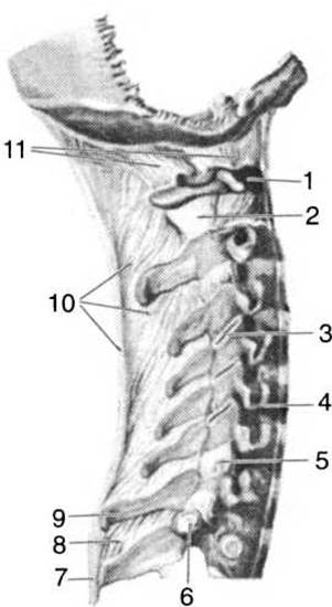
Fig. 22. Ligaments and JOINTS OF THE cervical spine:
1 — atlas; 2 — capsule of the atlantoaxial joint; 3, 5 — zygapophysial (facet) joints; 4 — anterior longitudinal ligament; 6 — costal facet of the transverse process of a thoracic vertebra; 7 — supraspinous ligament; 8 — interspinous ligament; 9 — seventh cervical vertebra (vertebra prominens); 10 — ligamentum nuchae; 11 — posterior atlanto-occipital membrane
The newborn vertebral column has only a single curve—the sacral Kyphosis. During the child's GROWTH AND DEVELOPMENT at 1.5–2 months of age, the cervical lordosis forms first as the infant begins to hold its head up. By 5–7 months, the thoracic kyphosis appears, during which time the child is able to sit, and by 10–12 months, the lumbar lordosis develops as the child begins to walk.
The formation of the spinal curves is finally completed only by the age of 18–20 years. In the horizontal position, the vertebral column is 2–3 cm longer than in the vertical position because body weight flattens and compresses the intervertebral discs. The length of the vertebral column relative to total body height is approximately 70%. In children and women, the vertebral column is relatively longer compared to body height than in men. This depends not only on the height of the vertebral bodies but also on the height of the intervertebral discs. With age, their height decreases. In newborns, they account for 50% of the spinal length, whereas in adults, this figure is 25%.
If the load on a child's Skeleton is uneven at an early age (carrying excessive loads, sitting incorrectly at a desk), a spinal curvature known as Scoliosis may develop. Therefore, It is important to frequently change BODY POSITION AND encourage active physical movement in children through various games and gymnastic exercises, which help keep the ligaments and intervertebral discs elastic for a long time. This promotes healthy Muscle Development and strengthening, increases the overall endurance and flexibility of the vertebral column, and reduces fatigue. All of this highlights the benefits of physical culture as a health-promoting factor not only for children, but for people of all ages.
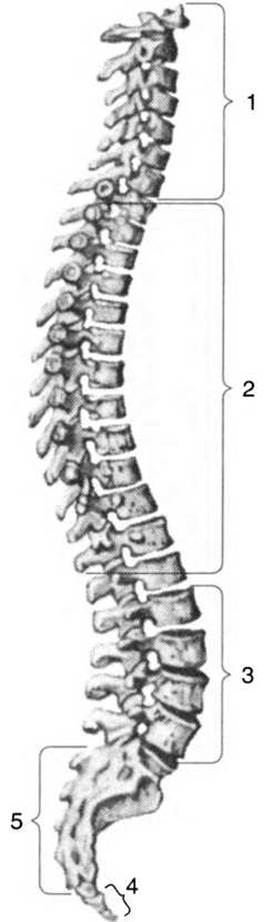
Fig. 23. The vertebral column:
1 — cervical lordosis; 2 — thoracic kyphosis; 3 — lumbar lordosis; 4 — coccyx; 5 — sacral kyphosis
Movements of the vertebral column comprise four types: flexion and extension (forward and backward, around the transverse axis); lateral flexion (bending to the right and left, around the sagittal axis); circular motion or rotation (around the vertical axis); and springy, shock-absorbing movements enabled by the physiological curves.
The cervical region of the vertebral column is the most mobile, followed by the lumbar region; the thoracic region is less mobile, while the sacral and coccygeal regions are completely immobile.
Last update: 08/08/2026
Editorial and Educational Adaptation: This material has been compiled based on the primary/original source text. The project team performed an editorial review, corrected technical inaccuracies, structured sections, and adapted the content for an educational format.
What was processed:
- elimination of formatting defects (OCR errors, structural breaks, corrupted characters);
- editorial organization of content;
- standardization of terminology in accordance with academic sources;
- verification of factual statements against the original source text.
All mentions of the author, publication year, and origin of the primary text have been preserved in accordance with the source.