Human Anatomy - Kotsan I. Y. 2009
Skeleton of the Head
Bones of the Neurocranium
The cerebral cranium (cranium cerebrale, neurocranium) has an ellipsoidal shape and forms the cavity that houses the Brain. The cerebral cranium is formed by 8 bones: 4 unpaired (occipital, frontal, sphenoid, and ethmoid) and 2 paired (temporal and parietal).
The Occipital bone (os occipitale) forms the posterior and inferior walls of the cranium (Fig. 30). In humans, it is formed by the fusion of several bones that exist independently in some animals. The occipital bone consists of four parts surrounding the foramen magnum: anteriorly—the basilar part; laterally—the lateral parts; and posteriorly—the occipital squama. All these parts develop separately during Embryogenesis and fuse into a single bone only between the ages of 3 and 6.
The basilar part (pars basilaris) is wedge-shaped. By the age of 18–20, it fuses completely with the body of the Sphenoid bone. The superior (cerebral) surface of the basilar part is smooth, concave in the form of a groove, and, together with a part of the body of the sphenoid bone, forms the clivus (clivus). On the Inferior surface of the basilar part, there is the pharyngeal tubercle (tuberculum pharyngeum), to which the fibrous membrane of the Pharynx attaches. The sulcus of the inferior petrosal sinus (sulcus sinus petrosi inferioris) runs along the lateral margin of the basilar part.
Class="center">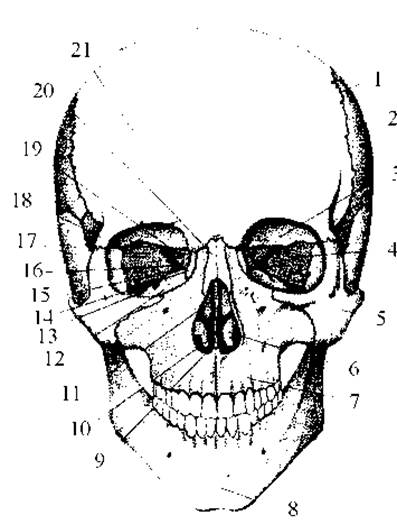
Fig. 28. Human Skull. Anterior view
1 — coronal suture, 2 — Parietal bone, 3 — orbital part of the Frontal bone, 4 — orbital surface of the greater wing of the sphenoid bone, 5 — Zygomatic bone, 6 — Inferior nasal concha, 7 — Maxilla, 8 — mental protuberance of the Mandible, 9 — Nasal cavity, 10 — Vomer, 11 — perpendicular plate of the Ethmoid bone, 12 — orbital surface of the maxilla, 13 — inferior orbital fissure, 14 — Lacrimal bone, 15 — orbital plate of the ethmoid bone, 16 — superior orbital fissure, 17 — squamous part of the Temporal bone, 18 — zygomatic process of the frontal bone, 19 — optic canal, 20 — Nasal bone
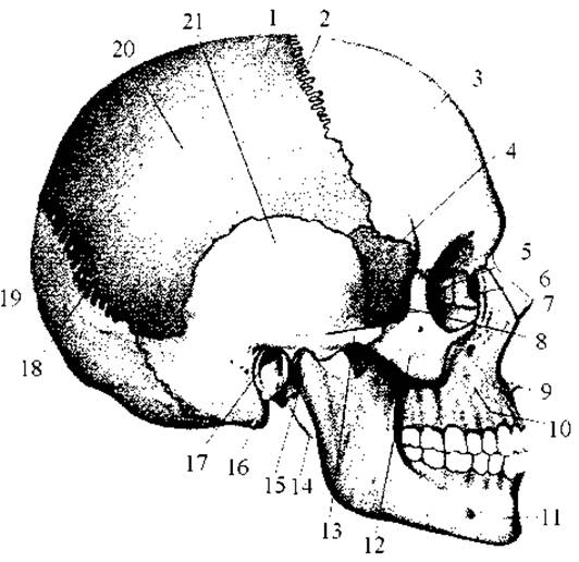
Fig. 29. Human skull. Lateral view
1 — parietal bone, 2 — coronal suture, 3 — frontal tuber, 4 — temporal surface of the greater wing of the sphenoid bone, 5 — orbital plate of the ethmoid bone, 6 — lacrimal bone, 7 — nasal bone, 8 — temporal fossa, 9 — anterior nasal spine, 10 — body of the maxilla, 11 — mandible, 12 — zygomatic bone, 13 — zygomatic arch, 14 — styloid process, 15 — condylar process of the mandible, 16 — mastoid process, 17 — external acoustic meatus, 18 — lambdoid suture, 19 — squama of the occipital bone, 20 — superior temporal line, 21 — squamous part of the temporal bone
The lateral part (pars lateralis) is paired, irregularly shaped, and gradually widens posteriorly into the occipital squama. On the inferior surface of each lateral part is the occipital condyle (condylus occipitalis), which articulates with the first cervical vertebra. The substance of the occipital condyle is pierced by the hypoglossal canal (canalis hypoglossi), through which the Hypoglossal nerve passes. Posterior to the occipital condyle lies the condylar fossa (fossa condylaris), at the bottom of which is the condylar canal (canalis condylaris), traversed by an emissary vein. On the lateral margin of the lateral part of the occipital bone, there is the jugular notch (incisura jugularis), which, together with the corresponding notch of the petrous part of the temporal bone, forms the jugular foramen (foramen jugulare). Nerves and the Internal jugular vein pass through it. Posteriorly, the jugular notch is bounded by the jugular process (processus jugularis), next to which, on the cerebral surface of the lateral part, runs the sulcus of the sigmoid sinus (sulcus sinus sigmoidei).
The occipital squama (squama occipitalis) is the largest part of the occipital bone. It appears as a broad plate with a concave internal surface and a convex external one. The external relief of the occipital squama is shaped by the attachment of Muscles and ligaments, whereas its internal relief corresponds to the shape of the brain.

Fig. 30. Occipital bone
a — anterior and superior view: 1 — sulcus of the superior sagittal sinus, 2 — internal occipital protuberance, 3 — internal occipital crest, 4 — sulcus of the sigmoid sinus, 5 — basilar part, 6 — jugular notch, 7 — foramen magnum, 8 — sulcus of the transverse sinus, b — posterior and inferior view: 1 — external occipital protuberance, 2 — superior nuchal line, 3 — inferior nuchal line, 4 — external occipital crest, 5 — foramen magnum, 6 — occipital condyle, 7 — pharyngeal tubercle, 8 — probe inserted into the hypoglossal canal.
On the external surface of the squama, in the center, lies the external occipital protuberance (protuberantia occipitalis externa), to which the nuchal ligament attaches. Extending downwards from the external occipital protuberance to the posterior margin of the foramen magnum is the external occipital crest (crista occipitalis externa). Arching laterally (to the right and left) from the external occipital protuberance is the superior nuchal line (linea nuchalis superior), along which the trapezius muscles attach on both sides. Parallel to the superior nuchal line, approximately at the level of the middle of the external occipital crest, the inferior nuchal line (linea nuchalis inferior) extends laterally. Additionally, a barely noticeable highest nuchal line (linea nuchalis suprema) may be present above the external occipital protuberance.
On the internal surface of the occipital squama lies the internal occipital protuberance (protuberantia occipitalis interna), which serves as the center of the cruciate eminence (eminentia cruciformis). Extending to the right and left of the internal occipital protuberance is the sulcus of the transverse sinus (sulcus sinus transversi), which continues as the sulcus of the sigmoid sinus (sulcus sinus sigmoidei). Extending upwards from the internal occipital protuberance is the sulcus of the superior sagittal sinus (sulcus sinus sagittalis superioris). Downwards, the internal occipital protuberance narrows and transitions into the internal occipital crest (crista occipitalis interna). These sulci and the internal occipital crest divide the internal surface of the occipital squama into four fossae. The two superior cerebral fossae (fossa cerebralis) are in contact with the occipital lobes of the cerebral hemispheres, while the two inferior cerebellar fossae (fossa cerebellaris) accommodate the cerebellar hemispheres.
The frontal bone (os frontale) in adults is unpaired and situated in the anterior region of the cranium (Fig. 31). It consists of four parts: the frontal squama, two orbital parts, and one nasal part. In early embryogenesis, all these parts form separate cranial bones that fuse into the frontal bone during the postnatal period.

Fig. 31. Frontal bone
a — anterior view: 1 — frontal squama, 2 — frontal tuber, 3 — temporal line, 4 — zygomatic process, 5 — superciliary arch, 6 — nasal part, b — posterior and inferior view: 1 — sulcus of the superior sagittal sinus, 2 — zygomatic process, 3 — orbital part, 4 — supraorbital notch, 5 — opening (aperture) of the frontal sinus, 6 — fossa for the lacrimal gland, 7 — frontal crest.
The frontal squama (squama frontalis) is the largest part of the frontal bone. In humans, the frontal squama is broad and vertically oriented, whereas in fossil hominids and apes it is narrower and inclined backwards. It features a convex external surface (facies externa), the lateral parts of which transition into the temporal surfaces (facies temporales), and a concave internal surface (facies interna). Inferiorly, the squama is separated from the right and left orbital parts by the paired supraorbital margin (margo supraorbitalis), which presents the supraorbital notch (incisura supraorbitalis) or, less commonly, the supraorbital foramen (foramen supraorbitale), transmitting the supraorbital artery and the lateral branch of the supraorbital nerve. In the medial part of the supraorbital margin is a depression—the frontal notch (incisura frontalis), or sometimes the frontal frontale (foramen frontale), which also transmits nerves and Blood Vessels. Laterally, the supraorbital margin terminates in the zygomatic process (processus zygomaticus), which articulates with the zygomatic bone. Extending upwards and backwards from the zygomatic process is the temporal line (linea temporalis), which separates the anterior part of the external surface from its temporal surface. Slightly above each supraorbital margin lies an arched elevation—the superciliary arch (arcus superciliaris)—which medially transitions into a smooth area, the glabella (glabella). Considerably higher than the superciliary arches, approximately in the middle of each half of the frontal squama, is a slanting rounded elevation—the frontal tuber (tuber frontale). Frontal tubers are a characteristic feature of the human skull, absent in both apes and fossil hominids. In the middle of the external surface of the frontal squama, the frontal suture (sutura frontalis) is visible—the remnant of the fusion of the paired frontal bone in early childhood.
The internal surface (facies interna) of the frontal squama is concave and gradually transitions into the horizontally oriented orbital PARTS OF THE frontal bone. Along the midline of the internal surface of the squama runs the sulcus of the superior sagittal sinus (sulcus sinus sagittalis superioris), which inferiorly transitions into the frontal crest (crista frontalis). Near the Base of the crest lies the foramen caecum (foramen caecum), which anchors a process of the dura mater. The bottom of the foramen caecum sometimes contains a smaller opening through which an emissary vein passes.
The orbital part (pars orbitalis) of the frontal bone is paired, representing a horizontal plate whose inferior surface faces the orbital cavity and superior surface faces the cranial cavity. The right and left orbital parts are separated by the deep ethmoidal notch (incisura ethmoidalis), which accommodates the cribriform plate of the ethmoid bone. On the superior (cerebral) surface of the orbital parts, clearly defined digital impressions (impressiones digitatae) and cerebral eminences (jugai) (juga cerebralia) are visible. The inferior surfaces of the orbital parts, forming the superior walls of the orbits, are smooth, concave, and bear traces of the auxiliary structures of the eye: in the lateral region of the inferior surface lies the fossa for the lacrimal gland (fossa glandulae lacrimalis), lodging the lacrimal gland; in the medial region, a small depression is visible—the trochlear fossa (fovea trochlearis)—where the fibrous-cartilaginous trochlea attaches, through which the tendon of the superior oblique Muscle of the eye loops. Adjacent to the trochlear fossa, a small trochlear spine (spina trochlearis) is frequently found, providing additional attachment for the fibrous-cartilaginous trochlea of the superior oblique muscle.
The nasal part (pars nasalis) of the frontal bone is horseshoe-shaped and bounds the ethmoidal notch anteriorly. In the posterior Regions of the nasal part, There is a series of depressions that cover the air Cells of the ethmoid bone opening upwards. In the anterior regions of the nasal part of the frontal bone is the nasal spine (spina nasalis), which contributes to The formation of the nasal septum. Flanking the nasal spine are the apertures of the frontal sinus (aperturae sinus frontalis), which lead into the right and left parts of the frontal sinus. The frontal sinus (sinus frontalis) varies in size in adults. Lined with mucous membrane and filled with air, the cavity of the frontal sinus is divided by a vertically oriented intersinus septum (septum intersinuale frontale) into two unequally sized parts that communicate with the nasal cavity via the frontal sinus apertures and the ethmoidal infundibulum.
The sphenoid bone (os sphenoidale) occupies a central position at the base of the cranium. It articulates with all the BONES OF THE cranial base, which is why it is also anatomically referred to as the main bone of the skull. In shape, the sphenoid bone resembles a butterfly (Fig. 32). Accordingly, it is subdivided into the following parts: the body, greater and lesser wings, and pterygoid processes.
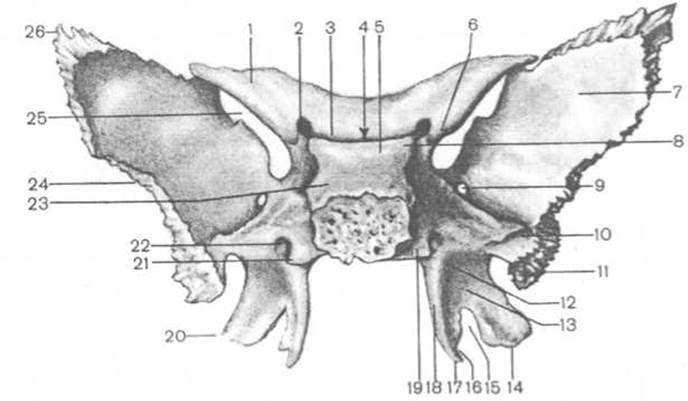
Fig. 32. Sphenoid bone (posterior view)
1 — lesser wing, 2 — optic canal, 3 — chiasmatic sulcus, 4 — hypophysial fossa, 5 — dorsum sellae, 6 — anterior clinoid process, 7 — cerebral surface, 8 — posterior clinoid process, 9 — foramen rotundum, 10 — sulcus for auditory tube, 11 — spine of sphenoid bone, 12 — scaphoide fossa, 13 — pterygoid fossa, 14 — lateral plate of pterygoid process, 15 — pterygoid notch, 16 — sulcus of pterygoid hamulus, 17 — pterygoid hamulus, 18 — medial plate of pterygoid process, 19 — carotid sulcus, 20 — pterygoid process, 21 — sphenoidal lingula, 22 — pterygoid canal, 23 — body, 24 — greater wing, 25 — superior orbital fissure, 26 — parietal angle.
The body (corpus) of the sphenoid bone is shaped like an irregular cube. Inside the body lies the sphenoidal sinus (sinus sphenoidalis), which is divided into two parts by the intersinus septum (septum intersinuale sphenoidale). Each part communicates with the nasal cavity through the aperture of the sphenoidal sinus (apertura sinus sphenoidalis).
The superior or cerebral surface of the body of the sphenoid bone faces the cranial cavity and features a depression resembling a Turkish saddle (sella turcica). In the center of the sella turcica is the hypophysial fossa (fossa hypophysialis), which houses the Pituitary Gland (hypophysis). Anterior to the sella turcica lies a small tuberculum sellae, while posteriorly stands the relatively high dorsum sellae. The lateral parts of the dorsum sellae project anteriorly, forming the posterior clinoid processes (processus clinoidei posteriores). Near the base of the dorsum sellae, on the right and left, runs the carotid sulcus (sulcus caroticus). Externally and slightly posterior to the carotid sulcus is the sphenoidal lingula (lingula sphenoidalis), which transforms the carotid sulcus into a deep groove. Along with the apex of the pyramid of the temporal bone, this groove bounds the internal carotid aperture, through which the Internal Carotid Artery emerges from the carotid canal into the cranial cavity.
The anterior surface of the body of the sphenoid bone extends into a small sphenoidal crest (crista sphenoidalis), which continues onto the inferior surface of the body as a sharp sphenoidal rostrum (rostrum sphenoidale). To the right and left of the crest are irregularly shaped bony plates—the sphenoidal conchae (conchae sphenoidales), which bound the apertures of the sphenoidal sinus (aperturae sinus sphenoidalis).
The lesser wings (alae minores) of the sphenoid bone extend laterally as thin, horizontally positioned plates from the anterosuperior angles of its body. At the base of each lesser wing are the anterior clinoid process (processus clinoideus anterior), to which a part of the dura mater attaches, and the optic canal (canalis opticus), through which the Optic nerve and ophthalmic artery pass. The lesser wings present superior and inferior surfaces, as well as anterior and posterior margins. The superior surface faces the cranial cavity, while the inferior surface contributes to the Formation of the superior wall of the Orbit. The anterior margin of the lesser wing is thickened and serrated, articulating with the orbital part of the frontal bone. The posterior margin is smooth, concave, and projects freely into the cranial cavity. The space between the lesser and greater wings is called the superior orbital fissure (fissura orbitalis superior). Through it, the oculomotor, trochlear, and abducent nerves (III, IV, and VI cranial nerve pairs) and the first branch of the Trigeminal nerve (V pair) pass from the cranial cavity into the orbit, whereas the superior ophthalmic vein enters the cranial cavity from the orbit.
The greater wings (alae majores) originate with a broad base from the lateral surface of the body of the sphenoid bone. Each greater wing has four surfaces: cerebral, orbital, temporal, and maxillary.
The cerebral surface (facies cerebralis) is slightly concave and exhibits cerebral eminences, digital impressions, and arterial sulci. The orbital surface (facies orbitalis) is flat, quadrilateral in shape, and forms the lateral wall of the orbit. The temporal surface (facies temporalis) of the greater wing is convex and forms the medial wall of the temporal fossa. At the lower part of the temporal surface, a horizontally oriented infratemporal crest (crista infratemporalis) is visible, dividing it into two parts. The upper, larger part is oriented almost vertically and forms part of the wall of the temporal fossa. The lower part is oriented almost horizontally and forms the superior wall of the infratemporal fossa. The maxillary surface (facies maxillaris) of the greater wing is a small, triangular area located between the orbital surface and the pterygoid process.
Near the body at the base of the greater wing lie three apertures: anteriorly, the foramen rotundum, through which the maxillary nerve (II branch of the trigeminal nerve) passes into the pterygopalatine fossa; posterior and lateral to it is the foramen ovale, through which the mandibular nerve (III branch of the trigeminal nerve) passes into the infratemporal fossa; and posterior and lateral to the latter is the small foramen spinosum, through which the middle meningeal artery enters the cranial cavity.
The pterygoid processes (processus pterygoidei) originate from the sphenoid bone near the base of the greater wings and project vertically downward. The base of the pterygoid process is pierced anteroposteriorly by the pterygoid canal (canalis pterygoideus), through which the nerve of the same name passes. Each process consists of two plates: lateral (lamina lateralis) and medial (lamina medialis). Anteriorly, the plates of the pterygoid process are fused, whereas posteriorly they diverge to enclose the pterygoid fossa (fossa pterygoidea). Inferiorly, the plates are separated by the pterygoid notch (incisura pterygoidea). The medial plate of the pterygoid process is somewhat narrower and longer than the lateral one and terminates inferiorly in the pterygoid hamulus (hamulus pterygoideus). A sharp bend of the hamulus forms the sulcus for the pterygoid hamulus (sulcus hamuli pterygoidei), over which loops the tendon of the tensor veli palatini muscle. The posterosuperior margin of the medial plate widens near the base to form the oblong scaphoid fossa (fossa scaphoidea), which serves as the origin for bundles of the tensor veli palatini muscle. Lateral to the scaphoid fossa runs the shallow sulcus for the auditory tube (sulcus tubae auditoriae), which is related to its cartilaginous part. On the medial side of the medial plate is a small vaginal process (processus vaginalis), which lies beneath the body of the sphenoid bone and laterally covers the wings of the vomer.
The ethmoid bone (os ethmoidale) is located anterior to the body of the sphenoid bone and participates in the formation of the walls of the orbits, the anterior cranial fossa, and the nasal cavity. The ethmoid bone consists of the following parts: the cribriform plate, the perpendicular plate, and two ethmoidal labyrinths (Fig. 33).
The cribriform plate (lamina cribrosa) is situated in the ethmoidal notch of the frontal bone and contributes to the formation of the floor of the anterior cranial fossa and the superior wall of the nasal cavity. The entire plate is perforated like a sieve by 30–40 small cribriform foramina (foramina cribrosa)—hence its name. Through these foramina, Olfactory nerve filaments (I pair of Cranial Nerves) and blood vessels pass into the cranial cavity.
The perpendicular plate (lamina perpendicularis) lies in the sagittal plane. It comprises two parts: an upper part, located above the cribriform plate, and a larger, lower part, situated below the cribriform plate. The upper part of the perpendicular plate forms a thickening—the crista galli—which projects into the cranial cavity. The falx cerebri (a fold of the dura mater) attaches to the crista galli. The anteroinferior margin of the crista galli is bounded on each side by an inconstant process—the ala of the crista galli. Together with the frontal bone located anteriorly, these two processes bound the foramen caecum of the frontal bone. The lower part of the perpendicular plate extends vertically downward into the nasal cavity and forms the anterosuperior part of the bony nasal septum.
The ethmoidal labyrinth (labyrinthus ethmoidalis) is a paired Structure located on both sides of the perpendicular plate. Each labyrinth consists of thin bony plates running in various directions to form the walls of numerous ethmoidal cells. The ethmoidal cells (cellulae ethmoidales) contain air, are lined with mucous membrane, and communicate with one another and with the nasal cavity. The cells are subdivided into anterior (cellulae ethmoidales anteriores), middle (cellulae ethmoidales mediae), and posterior (cellulae ethmoidales posteriores) ethmoidal cells. The anterior and middle ethmoidal cells open into the middle nasal meatus, while the posterior ones open into the superior nasal meatus.
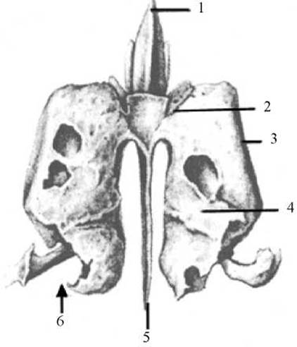
Fig. 33. Ethmoid bone
1 — crista galli, 2 — cribriform plate, 3 — orbital plate, 4 — superior nasal concha, 5 — perpendicular plate, 6 — ethmoidal labyrinth
Laterally, the cells are closed by the orbital plate (lamina orbitalis), which faces the orbital cavity and forms the greater part of its medial wall. It is extremely thin and smooth, which is why it is also referred to as the lamina papyracea.
Medially, the cells of the ethmoidal labyrinths communicate via a series of openings with the nasal cavity and are covered by two thin, curved bony plates—the nasal conchae. The upper part of each concha is attached to the labyrinth cells, while the lower margin hangs freely into the cleft between the labyrinth and the perpendicular plate. The superior nasal concha (concha nasalis superior) is attached superiorly; below it and slightly anteriorly lies the middle nasal concha (concha nasalis media). Occasionally, a poorly developed supreme nasal concha (concha nasalis suprema) is found above the superior concha. A narrow space—the superior nasal meatus (meatus nasi superior)—lies between the superior and middle nasal conchae. Beneath the curved margin of the middle nasal concha is the middle nasal meatus (meatus nasi medius). At its posterior end, the middle nasal concha features a downward-curved uncinate process (processus uncinatus), which articulates with the ethmoidal process of the inferior nasal concha. Posterior to the uncinate process, one of the largest cells of the ethmoidal labyrinth—the ethmoidal bulla (bulla ethmoidalis)—bulges into the middle nasal meatus. Slightly anterior and inferior to the ethmoidal bulla lies the semilunar hiatus (hiatus semilunaris), which, together with adjacent bones, forms the ethmoidal infundibulum (infundibulum ethmoidale). Through this infundibulum, the frontal sinus communicates with the middle nasal meatus.
The temporal bone (os temporale) is situated between the sphenoid, parietal, and occipital bones (Fig. 34). It contributes to the formation of the skull base and the lateral wall of the cranial vault. It houses the Organs of Hearing and Equilibrium, and its canals transmit blood Vessels and nerves. Articulating with the mandible via a joint, the temporal bone serves as the support for the masticatory apparatus. Masticatory and Neck Muscles attach to it. Structurally, the temporal bone is one of the most complex bones. It consists of three parts: squamous, tympanic, and petrous (with the mastoid process), which are unfused in newborns. During the first year of life, these parts merge into a single bone; at their fusion sites, sutures and fissures persist throughout life.
The squamous part (pars squamosa), or squamous temporal bone, forms part of the lateral wall of the skull. It appears as an oval, vertically oriented plate with a beveled free superior margin. It overlaps—like fish scales (squama)—the corresponding margin of the parietal bone and the greater wing of the sphenoid bone, and inferiorly articulates with the pyramid, mastoid process, and tympanic part of the temporal bone. The squamous part presents two surfaces: cerebral and temporal, and two free margins: parietal and sphenoidal.
The internal, cerebral surface (facies cerebralis) of the squama is slightly concave and exhibits digital impressions, cerebral eminences, and arterial sulci—traces left by the course of the middle meningeal artery and its branches.
The external, temporal surface (facies temporalis) of the squama is slightly convex and rough. In its posterior region, the sulcus for the middle temporal artery (sulcus arteriae temporalis mediae) runs vertically—marking the course of the artery of the same name. The external surface of the squama participates in forming the temporal fossa (serving as the origin for bundles of the temporalis muscle).
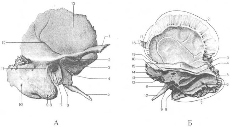
Fig. 34. Temporal bone (right)
A — external view: 1 — zygomatic process, 2 — articular tubercle, 3 — mandibular fossa, 4 — petrotympanic fissure, 5 — styloid process, 6 — tympanic part, 7 — external acoustic meatus, 8 — tympanomastoid fissure, 9 — mastoid process, 10 — mastoid foramen, 11 — suprameatal spine, 12 — sulcus for middle temporal artery, 13 — squamous part; B — internal view: 1 — arcuate eminence, 2 — parietal border, 3 — tegmen tympani, 4 — sulcus for superior petrosal sinus, 5 — sulcus for sigmoid sinus, 6 — mastoid foramen, 7 — occipital border, 8 — external aperture of vestibular aqueduct, 9 — subarcuate fossa, 10 — styloid process, 11 — vaginal process of styloid process, 12 — external aperture of cochlear canaliculus, 13 — sulcus for inferior petrosal sinus, 14 — apex of petrous part, 15 — petrous part, 16 — zygomatic process, 17 — sphenoidal border, 18 — posterior surface of petrous part, 19 — internal acoustic meatus
The sphenoidal border (margo sphenoidalis) of the squamous part of the temporal bone articulates with the greater wing of the sphenoid bone, whereas the parietal border (margo parietalis) articulates with the squamous border of the parietal bone. Posteriorly, the parietal border terminates in the parietal notch (incisura parietalis). The zygomatic process (processus zygomaticus) projects from the squamous part of the temporal bone slightly above and anterior to the external acoustic meatus. The zygomatic process of the temporal bone, together with the temporal process of the zygomatic bone, forms the zygomatic arch (arcus zygomaticus). At the base of the zygomatic process lies the mandibular fossa (fossa mandibularis), which receives the HEAD of the mandibular condylar process, forming the temporomandibular joint. Anteriorly, the mandibular fossa is bounded by the articular tubercle (tuberculum articulare), separating it from the infratemporal fossa. In humans and fossil hominids, the mandibular fossa is deep, and the articular tubercle is more developed than in apes. The upper margin of the temporal squama in humans forms a convex arch, and the squama itself is tall. This shape of the temporal squama is associated with the significant height of the cerebral cranium in modern humans; in apes and fossil hominids, this border is nearly straight.
The tympanic part (pars tympanica) is the smallest region of the temporal bone. It is shaped like a slightly curved plate and forms the anterior, inferior, and posterior walls of the external acoustic meatus (meatus acusticus externus). The external margin of the tympanic part, closed superiorly by the temporal squama, bounds the external acoustic meatus (porus acusticus externus). Anterior to the acoustic meatus, below the mandibular fossa, lies the tympanosquamosal fissure (fissura tympanosquamosa), into which a narrow bony plate of the petrous part of the temporal bone wedges. Consequently, the tympanosquamosal fissure is divided into two: the petrosquamosal fissure (fissura petrosquamosa), located closer to the mandibular fossa, and the petrotympanic fissure (fissura petrotympanica), situated closer to the pyramid of the temporal bone (through the latter fissure pass a branch of the Facial Nerve — the chorda tympani — and small vessels). Posteriorly, the tympanic part fuses with the mastoid process of the petrous part. At the site of this fusion, posterior to the external acoustic meatus, the tympanomastoid fissure (fissura tympanomastoidea) is formed. A flat process of the tympanic part directed inferiorly and embracing the base of the styloid process anteriorly forms the Vagina of the styloid process (vagina processus styloidei).
The petrous part (pars petrosa) is named for the hardness of its Bone tissue. In shape, it resembles a pyramid, and is therefore also referred to as the pyramid of the temporal bone. The pyramid in the cranium is situated in a nearly horizontal plane; its apex is directed anteriorly and medially toward the sella turcica of the sphenoid bone, while its base is directed posteriorly and laterally, continuing into the mastoid process. The petrous part presents three surfaces: anterior, posterior, and inferior, and three borders: anterior, posterior, and superior, which separate these surfaces.
The anterior border of the petrous part (margo anterior partis petrosae) separates the anterior surface of the petrous part from the inferior surface. It features the opening of the musculotubal canal (canalis musculotubarius), which is more clearly visible on the external cranial base. This canal is divided by an incomplete septum into two semicanals: the semicanal for the tensor tympani muscle (semicanalis musculi tensoris tympani) and the semicanal for the auditory tube (semicanalis tubae auditoriae).
The posterior border of the petrous part (margo posterior partis petrosae) separates the posterior surface of the petrous part from the inferior surface. Along this border, on the cerebral surface, runs the sulcus for the inferior petrosal sinus (sulcus sinus petrosi inferioris).
The superior border of the petrous part (margo superior partis petrosae) separates the anterior surface of the petrous part from the posterior surface. Along this border runs the sulcus for the superior petrosal sinus (sulcus sinus petrosi superioris).
The anterior and posterior surfaces of the petrous part face the cranial cavity, whereas the inferior surface faces outward (it is well visible from the external cranial base).
The anterior surface of the petrous part (facies anterior partis petrosae) faces anteriorly and superiorly. Laterally, it transitions into the cerebral surface of the squamous part, from which the petrous part is separated in youth by the petrosquamosal fissure (fissura petrosquamosa). In the middle portion of the anterior surface of the petrous part lies a small arcuate eminence (eminentia arcuata), formed by the bulging of the anterior (superior) semicircular canal of the bony labyrinth of the Inner ear, located within the substance of the petrous part.
Between the arcuate eminence and the petrosquamosal fissure lies a flattened area, the tegmen tympani (tegmen tympani), beneath which the tympanic cavity is located. On the anterior surface of the petrous part, near its apex, there is a small trigeminal impression (impressio trigeminalis), which lodges the trigeminal ganglion. Lateral to the trigeminal impression are two small openings: the hiatus for the greater petrosal nerve (hiatus canalis nervi petrosi majoris), from which the sulcus for the greater petrosal nerve (sulcus nervi petrosi majoris) originates, and further laterally and anteriorly lies the hiatus for the lesser petrosal nerve (hiatus canalis nervi petrosi minoris), from which the sulcus for the lesser petrosal nerve (sulcus nervi petrosi minoris) extends.
The posterior surface of the petrous part (facies posterior partis petrosae) faces posteriorly and medially. Approximately in the middle of the posterior surface is the internal acoustic meatus (porus acusticus internus), which leads into the internal acoustic meatus (meatus acusticus internus). At the Cytology/practical/108.html">Fundus of the internal acoustic meatus are several openings for the facial (CN VII) and vestibulocochlear (CN VIII) cranial nerves, as well as for the artery and Veins OF THE Vestibulocochlear Organ. Superior and lateral to the internal acoustic meatus is a small depression, the subarcuate fossa (fossa subarcuata), which receives a process of the meningeal dura mater. Inferior and lateral to the internal acoustic meatus is a small cleft, the external aperture of the vestibular aqueduct (apertura externa aqueductus vestibuli).
The inferior surface of the petrous part (facies inferior partis petrosae) is rough and uneven, resting on the inferior surface of the cranial base. It features the deep jugular fossa (fossa jugularis), which lodges the superior bulb of the internal jugular vein. On the anterior wall of the jugular fossa is a small groove that terminates in the opening of the mastoid canaliculus (canaliculus mastoideus). The jugular fossa lacks a posterior wall; it is bounded by the jugular notch (incisura jugularis), which, together with the corresponding notch of the occipital bone, forms the jugular foramen (foramen jugulare) in the intact skull. Through it pass the glossopharyngeal (CN IX), vagus (CN X), and accessory (CN XI) cranial nerves, as well as the internal jugular vein. Anterior to the jugular fossa begins the carotid canal (canalis caroticus). Here lies the external opening of the carotid canal (foramen caroticum externum). The internal opening of the carotid canal (foramen caroticum internum) opens at the apex of the petrous part. In the wall of the carotid canal, near its external opening, are two small fossae that continue into fine caroticotympanic canaliculi (canaliculi caroticotympanici), which connect the carotid canal with the tympanic cavity. Between the external opening of the carotid canal and the anterior circumference of the jugular fossa lies a small petrosal fossa (fossula petrosa), to which the petrosal ganglion of the Glossopharyngeal nerve is attached. In the depth of this fossa opens the inferior aperture of the tympanic canaliculus (apertura inferior canaliculi tympanici).
Lateral to the jugular fossa projects the slender and long styloid process (processus styloideus). Posterior to it — between the styloid and mastoid processes — lies the stylomastoid foramen (foramen stylomastoideum), through which the facial nerve (CN VII) emerges from the facial canal of the temporal bone.
The mastoid process (processus mastoideus) is located posterior to the external acoustic meatus and forms the posterior part of the temporal bone. Its external surface is convex and rough, serving as a site for muscle attachment. Inferiorly, the mastoid process transitions into a cone-shaped projection that is readily palpable through the Skin. Medially, the process is bounded by the deep mastoid notch (incisura mastoidea). Medial to the notch and parallel to it runs the sulcus for the occipital artery (sulcus arteriae occipitalis). Near the base of the mastoid process, closer to the posterior border of the temporal bone, lies the inconstant mastoid foramen (foramen mastoideum), through which passes the mastoid emissary vein. On the internal surface of the mastoid process, facing the cranial cavity, runs the broad, S-shaped sulcus for the sigmoid sinus (sulcus sinus sigmoidei). It lodges the dural venous sinus of the same name. Within the process are mastoid air cells (cellulae mastoideae) separated from one another by bony trabeculae, lined with mucous membrane and filled with air. The constant and largest of these is the mastoid antrum (antrum mastoideum), situated in the central part of the process; the mastoid cells open into it, and it communicates with the tympanic cavity.
Internally, the petrous part of the temporal bone contains the bony labyrinth, the tympanic cavity (their structure is detailed in the chapter "Organs of Hearing and Equilibrium"), and numerous canals (Tab. 6).
Table 6 CANALS OF THE TEMPORAL BONE
|
Name of canal |
Origin of canal |
Communications along the course of the canal and its termination |
Structures passing through the canal |
|
1. Carotid canal (canalis caroticus) |
External carotid aperture on the inferior surface of the pyramid |
Caroticotympanic canaliculi (see below). Internal carotid aperture at the apex of the pyramid within the cranial cavity |
Internal carotid artery accompanied by the venous plexus of the same name and the internal carotid (autonomic) plexus |
|
2. Caroticotympanic canaliculi (canaliculi caroticotympanici) |
Begin as openings on the wall of the carotid canal (near its origin) |
Openings on the anterior (carotid) wall of the tympanic cavity |
Caroticotympanic nerves (Branches of the internal carotid plexus): superior caroticotympanic nerve (from the tympanic nerve to the internal carotid plexus), arterial caroticotympanic branches (from the internal carotid artery) |
|
3. Facial canal (canalis facialis) |
Entrance to the facial canal — at the fundus of the internal acoustic meatus |
Along the course of the canal on the anterior surface of the pyramid — hiatus for the greater petrosal nerve; in the lower part — opening into the canaliculus for the chorda tympani (see below). Termination — stylomastoid foramen |
Facial nerve (CN VII); superficial petrosal branch (from the middle meningeal artery) — superiorly, and stylomastoid artery and vein — inferiorly |
|
4. Canaliculus for the chorda tympani (canaliculus chordae tympani) |
Opening in the lower part of the facial canal |
Opening on the posterior (mastoid) wall of the tympanic cavity |
Chorda tympani — branch of the facial nerve. Exits the tympanic cavity via the petrotympanic fissure |
|
5. Tympanic canaliculus (canaliculus tympanicus) |
Inferior aperture of the tympanic canaliculus — in the petrosal fossa on the inferior surface of the pyramid |
Opening on the inferior (jugular) wall of the tympanic cavity, where the canal is interrupted. The nerve runs on its medial (labyrinthine) wall and terminates on the anterior surface of the pyramid at the hiatus for the lesser petrosal nerve |
Tympanic nerve, which upon exiting the tympanic cavity is termed the lesser petrosal nerve (branch of CN IX); entering — superior tympanic artery (branch of the middle meningeal artery) |
|
6. Musculotubal canal (canalis musculotubarius) — divided into two semicanals. Upper — semicanal for the tensor tympani muscle, lower — semicanal for the auditory tube |
Begins at the junction of the anterior border of the pyramid with the temporal squama near the apex of the pyramid |
Terminates with openings on the anterior (carotid) wall of the tympanic cavity |
Tensor tympani muscle and auditory tube |
The parietal bone (os parietale) is a paired bone forming the anterolateral part of the cranial vault (Fig. 35). It is shaped like a quadrilateral plate, convex externally, presenting two surfaces: external and internal; four borders: frontal, occipital, sagittal, and squamous; and four angles: frontal, occipital, sphenoidal, and mastoid.
The external surface (facies externa) of the parietal bone is smooth and convex. The site of greatest convexity is termed the parietal tuber (tuber parietale). Slightly inferior to it, two arch-shaped temporal lines are visible: the superior and inferior temporal lines (lineae temporales superior et inferior). The former serves as the attachment site for the temporal fascia, and the latter for the temporalis muscle.
The internal surface (facies interna) of the parietal bone is concave; its relief is shaped by the apposition of the brain, particularly its dura mater and its vessels. Thus, along the superior (sagittal) border of the parietal bone runs a well-defined sulcus for the superior sagittal sinus (sulcus sinus sagittalis superioris). This sulcus, continuous with the corresponding sulcus of the opposite side, lodges the superior sagittal venous sinus. In the region of the mastoid angle lies a small but deep sulcus for the sigmoid sinus (sulcus sinus sigmoidei) — an impression of the sigmoid and transverse Dural Venous Sinuses. Furthermore, the internal surface of the parietal bone exhibits arborizing arterial sulci (traces of meningeal artery courses), granular foveolae (impressions of arachnoid granulations), digital impressions (impressions of cerebral gyri), cerebral eminences located between the digital impressions, and inconstant venous sulci.
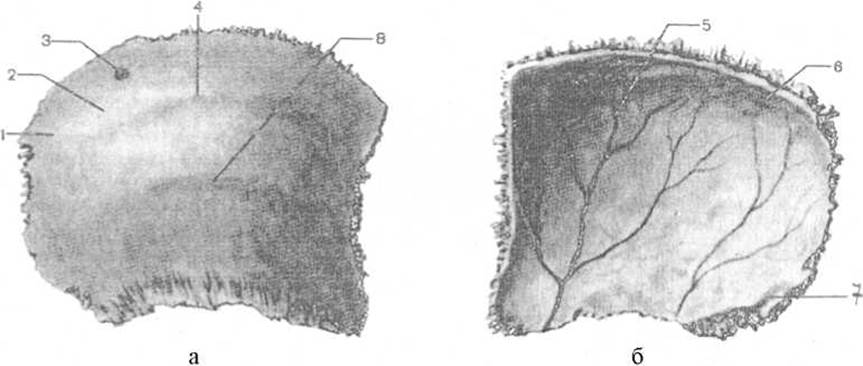
Fig. 35. Parietal bone
a — external surface of the right bone, b — internal surface of the right bone, 1 — external surface, 2 — parietal tuber, 3 — parietal foramen, 4 — superior temporal line, 5 — internal surface, 6 — sulcus for superior sagittal sinus, 7 — sulcus for sigmoid sinus, 8 — inferior temporal line
The squamous (inferior) border (margo squamosus) of the parietal bone, which is arcuately concave and sharp, is overlapped by the temporal squama, forming the squamous suture (sutura squamosa). The other three borders of the parietal bone are serrated in shape.
The sagittal (superior) border (margo sagittalis) of the parietal bone articulates with the corresponding border of the opposite bone, forming the sagittal suture (sutura sagittalis). Near the sagittal border, closer to the occipital angle, the parietal bone features a parietal foramen (foramen parietale) through which a branch of the occipital artery passes to the dura mater of the brain.
The frontal (anterior) border (margo frontalis) of the parietal bone articulates with the parietal margin of the squamous part of the frontal bone, forming the coronal suture (sutura coronalis).
The occipital (posterior) border (margo occipitalis) of the parietal bone articulates with the lambdoid margin of the occipital bone, forming the lambdoid suture (sutura lambdoidea).
Corresponding to its four borders, the parietal bone has four angles.
The frontal (anterosuperior) angle (angulus frontalis) is nearly right-angled in shape (bounded by the coronal and sagittal sutures).
The sphenoidal (anteroinferior) angle (angulus sphenoidalis) is acute (bounded by the coronal and sphenoparietal sutures).
The occipital (posterosuperior) angle (angulus occipitalis) is obtuse (bounded by the lambdoid and sagittal sutures).
The mastoid (posteroinferior) angle (angulus mastoideus) of the parietal bone is also obtuse in shape (bounded by the lambdoid and parietomastoid sutures).
Last update: 08/08/2026
Editorial and Educational Adaptation: This material has been compiled based on the primary/original source text. The project team performed an editorial review, corrected technical inaccuracies, structured sections, and adapted the content for an educational format.
What was processed:
- elimination of formatting defects (OCR errors, structural breaks, corrupted characters);
- editorial organization of content;
- standardization of terminology in accordance with academic sources;
- verification of factual statements against the original source text.
All mentions of the author, publication year, and origin of the primary text have been preserved in accordance with the source.