Human Anatomy - Kotsan I. Y. 2009
Skeleton of the Head
Bones of the Facial Skull
The BONES OF THE facial Skull form the bony framework of the face and the initial sections of the digestive and respiratory systems, as well as providing bony cavities for the Sense Organs (Vision, Olfaction, and gustation). The masticatory Muscles of the HEAD attach to the bones of the facial Skeleton.
The facial skull (viscerocranium) comprises 15 bones: 6 paired (Maxilla, nasal, zygomatic, lacrimal, palatine, and Inferior nasal concha) and 3 unpaired (Mandible, Vomer, and Hyoid bone). Additionally, embryologically, three paired auditory ossicles (malleus, incus, and stapes), located within the tympanic cavity of the Middle ear, also belong to the bones of the facial skull. However, given their specific role in the auditory system, they are described in the chapter "Sense Organs".
The maxilla is located in the upper part of the facial skull. It participates in The formation of the Orbit, the nasal and oral cavities, the infratemporal and pterygopalatine fossae, and also contributes to the masticatory apparatus. The evolutionary shift of grasping Functions from the jaws (as seen in animals) to the hands in humans, driven by labor activity, resulted in a reduction in the size of the maxilla, whereas The Emergence of human speech rendered its Structure more delicate. The maxilla articulates with all the bones of the facial skeleton, as well as with the frontal, ethmoid, and sphenoid bones of the neurocranium.
The maxillary bone consists of a body and four processes: the frontal, zygomatic, alveolar, and palatine processes (Fig. 36).
The body of the maxilla (corpus maxillae) contains the air-filled maxillary sinus (sinus maxillaris), which is lined with mucous membrane. The maxillary sinus, or Highmore's antrum (antrum Highmori), communicates with the Nasal cavity via a wide maxillary hiatus (hiatus maxillaris).
In shape, the body of the maxilla resembles a tetrahedral prism. It features four surfaces: anterior, orbital, infratemporal, and nasal.
The anterior surface (facies anterior) of the maxillary body contributes to facial relief. Unlike the flattened anterior surface of our evolutionary ancestors, in modern humans it is somewhat concave. It is separated from the orbital surface by the infraorbital margin (margo infraorbitalis). Below this margin lies the infraorbital foramen (foramen infraorbitalis), through which Blood Vessels and nerves pass. Inferior to this foramen is the canine fossa (fossa canina), which serves as the origin for the levator anguli oris Muscle. On the anteromedial margin of the anterior surface lies the nasal notch (incisura nasalis), which forms part of the anterior section of the nasal cavity. The inferior margin of the nasal notch projects anteriorly to form the anterior nasal spine (spina nasalis anterior).
The orbital surface (facies orbitalis) of the maxillary body forms the floor of the orbit. In shape, it resembles a triangular, slightly concave plate. Its medial margin articulates with the Lacrimal bone, the orbital lamina of the Ethmoid bone, and the orbital process of the Palatine bone. At the posterior free edge of the orbital surface, which bounds the inferior orbital fissure (fissura orbitalis inferior), the infraorbital groove (sulcus infraorbitalis) begins. Extending anteriorly, this groove deepens and gradually transitions into the infraorbital canal (canalis infraorbitalis), which opens onto the anterior surface of the maxilla via the aforementioned infraorbital foramen. The infraorbital nerve, artery, and Veins run within the infraorbital groove and canal. The floor of the canal contains small openings for the anterior dental canaliculi, known as the anterior alveolar foramina (foramina alveolaria anteriora), through which nerves pass to the anterior Teeth of the upper jaw.
Class="center">
Fig. 36. Maxilla
A — external view: 1 — frontal process, 2 — anterior lacrimal crest, 3 — infraorbital margin, 4 — anterior surface, 5 — infraorbital foramen, 6 — canine fossa, 7 — nasal notch, 8 — palatine process, 9 — anterior nasal spine, 10 — alveolar juga, 11 — alveolar process, 12 — zygomatic process, 13 — orbital surface, 14 — infraorbital groove transitioning into the infraorbital canal; B — internal view: 1 — lacrimal groove, 2 — ethmoidal crest, 3 — conchal crest, 4 — nasal crest, 5 — incisive canal, 6 — alveolar process, 7 — nasal surface, 8 — maxillary hiatus.
The infratemporal surface (facies infratemporalis) of the maxillary body participates in the Formation of the infratemporal and pterygopalatine fossae. This surface features the maxillary tuberosity (tuber maxillare), which bears 2–3 small dental canalicular openings known as the posterior alveolar foramina (foramina alveolaria posteriora), transmitting vessels and nerves to the posterior maxillary teeth. Medial to these foramina runs the vertically oriented greater palatine sulcus (sulcus palatinus major), which contributes to the formation of the canal of the same name.
The nasal surface (facies nasalis) of the maxillary body has a complex relief, forms part of the lateral wall of the nasal cavity, articulates with the palatine bone and the inferior nasal concha, and inferiorly transitions into the superior surface of the palatine process of the maxilla. The nasal surface bears the maxillary hiatus (hiatus maxillaris), through which the maxillary sinus opens into the nasal cavity. Anterior to the maxillary hiatus lies the vertically directed lacrimal sulcus (sulcus lacrimalis). By articulating with the lacrimal bone and the inferior nasal concha, the latter forms the nasolacrimal canal (canalis nasolacrimalis), connecting the orbit to the nasal cavity. Anterior to the lacrimal groove, across the Base of the frontal process, is a ridge called the conchal crest (crista conchalis), to which the inferior nasal concha attaches.
The frontal process (processus frontalis) of the maxilla projects vertically upwards. It forms part of the medial wall of the orbit and the piriform aperture of the Nose. The anterior border of the frontal process articulates with the Nasal bone. Its posterior part articulates with the lacrimal bone, forming the fossa for the lacrimal sac (fossa sacci lacrimalis). The upper serrated tip of the frontal process reaches the nasal part of the Frontal bone. On the lateral surface of the process, the anterior lacrimal crest (crista lacrimalis anterior) runs almost vertically, continuing inferiorly into the infraorbital margin. On the Medial surface of the frontal process is the ethmoidal crest (crista ethmoidalis), which fuses with the anterior part of the middle nasal concha of the ethmoid bone.
The zygomatic process (processus zygomaticus) projects from the upper lateral angle of the maxillary body and articulates with its serrated end to the Zygomatic bone. The inferior margin of the process transitions onto the lateral surface of the maxillary body towards the first molar as a thick buttress (pillar), through which masticatory pressure is transmitted to the zygomatic bone.
The alveolar process (processus alveolaris) is a thick, arch-shaped plate extending inferiorly from the body of the maxilla and terminating in the alveolar arch (arcus alveolaris). The alveolar arch contains eight dental alveoli (alveoli dentales) that house the eight upper teeth. The dental alveoli are separated from one another by interalveolar septa (septa interalveolaria). The three posterior dental alveoli contain interradicular septa (septa interradicularia). On the outer surface of the alveolar process, alveolar juga (juga alveolaria) are visible, being particularly prominent near the anterior teeth.
The palatine process (processus palatinus) is a horizontally oriented plate projecting medially from the inferior margin of the nasal surface of the maxillary body. The upper surface of the palatine process is smooth and forms the floor of the nasal cavity. The lower surface of the palatine process is rough and features posteriorly directed palatine grooves (sulci palatini) in its posterior region, with palatine spines (spinae palatinae) located between them. The palatine grooves lodge vessels and nerves emerging from the greater palatine foramen.
The palatine processes of both maxillae articulate along the midline via the median palatine suture (sutura palatina mediana), forming the greater part of the hard palate. Near the anterior end of the median suture lies the incisive foramen (foramen incisivum), which leads into the incisive canal (canalis incisivus) containing the greater palatine artery and the nasopalantine nerve.
On the free medial edge of the palatine process lies the slightly elevated nasal crest (crista nasalis), which, by articulating with the corresponding crest of the opposite maxilla, forms a low ridge to which the vomer attaches.
The nasal bone (os nasale) is a small paired bone that forms the bony bridge of the nose. In humans, compared to animals, the nasal bone is underdeveloped. In shape, the nasal bone resembles a quadrilateral plate whose longitudinal dimension exceeds its transverse dimension (Fig. 37). The superior margin of the nasal bone is thicker and slightly narrower than the inferior margin, articulating with the nasal part of the frontal bone. The inferior free margin of the nasal bone, together with the anterior margin of the base of the frontal process of the maxilla, bounds the piriform aperture of the nasal cavity. The lateral margin of the nasal bone articulates with the anterior margin of the frontal process of the maxilla. By its medial, somewhat serrated margin, the nasal bone articulates with the corresponding margin of the contralateral nasal bone. The anterior (external) surface of the nasal bone is smooth and bears one or more foramina (for the passage of vessels and nerves). The posterior (internal) surface is slightly concave and features a longitudinal ethmoidal sulcus (sulcus ethmoidalis), transmitting a branch of the anterior ethmoidal nerve.

Fig. 37. Nasal bone (right)
A — external view, B — internal view, 1 — ethmoidal groove

Fig. 38. Zygomatic bone (right, external view)
1 — orbital surface, 2 — zygomaticofacial foramen, 3 — lateral surface, 4 — temporal process, 5 — frontal process.
The zygomatic bone (os zygomaticum) is the strongest of the facial bones. By connecting the bones of the neurocranium and viscerocranium (frontal bone, Temporal bone, maxilla), it helps reinforce the facial skeleton. The zygomatic bone plays a vital role in shaping facial relief. The contour of the middle third of the face largely depends on the shape of the zygomatic bone.
The zygomatic bone features three surfaces—lateral, temporal, and orbital—and two processes: the frontal and temporal processes (Fig. 38).
The lateral surface (facies lateralis) faces laterally and anteriorly and is slightly convex. The temporal surface (facies temporalis) is concave, smooth, and faces toward the temporal fossa. The orbital surface (facies orbitalis) is also smooth and concave, forming the anterolateral floor of the orbit and the lateral border of the infraorbital margin. On the orbital surface is the zygomaticoorbitale foramen (foramen zygomaticoorbitale), which leads into a canal containing the zygomatic nerve. Within the zygomatic bone, this canal branches into two channels traversed by the Branches of the zygomatic nerve. They emerge onto The surface of the zygomatic bone through two openings: the zygomaticofacial and zygomaticotemporal foramina. The zygomaticofacial foramen (foramen zygomaticofaciale) is located on the lateral surface, while the zygomaticotemporal foramen (foramen zygomaticotemporale) is situated on the temporal surface of the zygomatic bone.
The frontal process (processus frontalis) of the zygomatic bone is broad and rather massive, directed superiorly to articulate with the zygomatic process of the frontal bone and the greater wing of the Sphenoid bone (deep within the orbit).
The temporal process (processus temporalis) of the zygomatic bone extends posteriorly and, articulating via a suture with the zygomatic process of the temporal bone, forms the zygomatic arch.
The inferomedial aspect of the zygomatic bone connects to the maxilla via a broad, serrated surface.
The lacrimal bone (os lacrimale) is the thinnest of all cranial bones, situated in the anterior part of the medial wall of the orbit; in shape, it resembles an elongated quadrilateral plate (Fig. 39). Anteriorly and inferiorly, the lacrimal bone borders the frontal process of the maxilla; posteriorly, it meets the orbital plate of the ethmoid bone; superiorly, it articulates with the medial margin of the orbital part of the frontal bone.

Fig. 39. Lacrimal bone (right, external view)
1 — lacrimal groove, 2 — posterior lacrimal crest
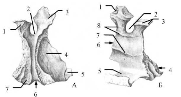
Fig. 40 Palatine bone (right)
A — external view: 1 — sphenoidal process, 2 — sphenopalatine notch, 3 — orbital process, 4 — perpendicular plate, 5 — horizontal plate, 6 — greater palatine groove, 7 — pyramidal process; B — internal view: 1 — orbital process, 2 — sphenopalatine notch, 3 — sphenoidal process, 4 — pyramidal process, 5 — horizontal plate, 6 — perpendicular plate, 7 — ethmoidal crest, 8 — conchal crest
On the lateral surface of the lacrimal bone is the posterior lacrimal crest (crista lacrimalis posterior), which terminates inferiorly in a projection called the lacrimal hook (hamulus lacrimalis). Anterior to the lacrimal crest lies the lacrimal groove (sulcus lacrimalis), which, together with the corresponding groove of the maxilla, forms the fossa for the lacrimal sac (fossa sacci lacrimalis). This fossa extends inferiorly into the nasolacrimal canal (canalis nasolacrimalis), which opens into the inferior nasal meatus (meatus nasi inferior).
The medial surface of the lacrimal bone laterally covers the anterior ethmoidal air Cells.
The palatine bone (os palatinum) is located posterior to the maxilla and contributes to the formation of the walls of the nasal and oral cavities, as well as the orbital and pterygopalatine fossae.
The bone consists of two plates—horizontal and perpendicular—and three processes: orbital, sphenoidal, and pyramidal, which extend from these plates (Fig. 40).
The horizontal plate (lamina horizontalis) is quadrilateral in shape. Its anterior margin is serrated and articulates with the posterior margin of the palatine process of the maxilla, forming the transverse palatine suture (sutura palatina transversa). The posterior margin of the horizontal plate is smooth, free, and transversely concave. The medial margin of the horizontal plate articulates with the corresponding margin of the opposite palatine bone, forming the posterior segment of the median palatine suture. The lateral margin of the horizontal plate fuses with the perpendicular plate of the palatine bone.
The horizontal plate presents two surfaces: an inferior palatine surface and a superior nasal surface. The palatine surface (facies palatina) is rough and features the palatine crest (crista palatina), which runs posterior to the transverse suture. The nasal surface (facies nasalis) of the horizontal plate is smooth. Along the medial margin of the nasal surface lies the nasal crest (crista nasalis), which terminates posteriorly in the posterior nasal spine (spina nasalis posterior). The horizontal plates of the palatine bones and the two palatine processes of the right and left maxillae together form the hard palate (palatum osseum).
The perpendicular plate (lamina perpendicularis) of the palatine bone arises from the horizontal plate at a right angle, projecting superiorly to complete the lateral wall of the nasal cavity posteriorly. Running down the lateral surface of the perpendicular plate is the greater palatine groove (sulcus palatinus major), which, together with the matching grooves of the maxilla and the pterygoid process of the sphenoid bone, forms the greater palatine canal (canalis palatinus major). The greater palatine canal opens onto the hard palate via the greater palatine foramen (foramen palatinum majus). This canal transmits the greater palatine nerve and the descending palatine artery. On the medial (nasal) surface of the perpendicular plate are two prominent horizontal crests: superiorly, the ethmoidal crest (crista ethmoidalis), to which the middle nasal concha of the ethmoid bone attaches, and inferiorly, the conchal crest (crista conchalis), which anchors the posterior portion of the inferior nasal concha.
The orbital and sphenoidal processes of the palatine bone originate from the superior margin of the perpendicular plate. The orbital process (processus orbitalis) is directed anteriorly and laterally, participating in the formation of the orbital floor and partially sealing the ethmoidal cells. The sphenoidal process (processus sphenoidalis) is directed posteriorly and medially, articulating with the Inferior surface of the body of the sphenoid bone. These two processes are separated by the sphenopalatine notch (incisura sphenopalatina), which in the intact skull is roofed over by the body of the sphenoid bone, converting it into the sphenopalatine foramen (foramen sphenopalatinum).
The pyramidal process (processus pyramidalis) of the palatine bone projects downward and backward from the junction of the horizontal and perpendicular plates, fitting into the pterygoid notch of the sphenoid bone. Transmitted through the pyramidal process are the minute lesser palatine canals (canales palatini minores), which open via the lesser palatine foramina (foramina palatina minora) onto the palatine surface of the pyramidal process.
Unlike the superior and middle nasal conchae, which are PARTS OF THE ethmoid bone, the inferior nasal concha (concha nasalis inferior) is an independent bone. In shape, it resembles a thin, long, axially curved bony plate consisting of a body and three processes: lacrimal, maxillary, and ethmoidal (Fig. 41). The superior margin of the inferior nasal concha fuses with the conchal crest of the maxilla and the corresponding crest on the perpendicular plate of the palatine bone; the inferior margin is free and curls laterally. All three processes arise from the superior margin of the body of the inferior nasal concha. Foremost among them is the lacrimal process (processus lacrimalis), which projects superiorly to articulate with the lacrimal bone. The relatively large, laterally directed, and curved maxillary process (processus maxillaris) extends from the superior margin of the inferior concha on its lateral side, forming an acute angle with the bone's body. This angle accommodates the inferior margin of the maxillary hiatus. Projecting from the posterior part of the superior margin of the inferior concha is the ethmoidal process (processus ethmoidalis), which ascends to articulate with the uncinate process of the ethmoid bone.

Fig. 41. Inferior nasal concha (right, lateral aspect)
1 — ethmoidal process, 2 — lacrimal process, 3 — maxillary process
The mandible (mandibula) is the only movable bone of the head skeleton. It develops from two halves that fuse During the first year of a child's life to form a single unpaired bone. It consists of a horizontally positioned body and two rami located almost vertically (Fig. 42).
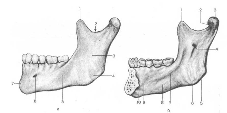
Fig. 42. Mandible
a — external view (left half): 1 — coronoid process, 2 — mandibular notch, 3 — ramus of the mandible, 4 — masseteric tuberosity, 5 — body of the mandible, 6 — mental foramen, 7 — mental protuberance; b — internal view (right half): 1 — coronoid process, 2 — pterygoid fossa, 3 — condylar process, 4 — mandibular foramen, 5 — angle of the mandible, 6 — pterygoid tuberosity, 7 — mylohyoid line, 8 — submandibular fossa, 9 — sublingual fossa, 10 — digastric fossa
The body of the mandible (corpus mandibulae) is shaped like a horseshoe-curved plate. It features inferior and superior margins, as well as external and internal surfaces. The inferior margin of the body is rounded and thickened, forming the base of the mandible (basis mandibulae), while the superior margin forms the alveolar arch (arcus alveolaris). The latter contains tooth sockets (alveoli dentales) for 16 teeth, along with interalveolar septa (septa interalveolaria) and interradicular septa (septa interradicularia).
On the external surface of the alveolar arch are alveolar juga (juga alveolaria), which correspond to the dental alveoli. In the middle of the external surface lies the mental protuberance (protuberantia mentalis), which gradually widens inferiorly and ends in paired mental tubercles (tuberculum mentale). Posterior to the mental tubercle, at the level of the second premolar, is the mental foramen (foramen mentale), which serves as the exit point for the vessels and nerve of the same name. Posterior to the mental foramen begins the oblique line (linea obliqua), which runs backward and upward, terminating near the base of the coronoid process.
In the middle of the internal surface of the mandibular body projects the mental spine (spina mentalis), which serves as the origin for the genioglossus and geniohyoid muscles. Slightly inferior and lateral to the mental spine is the paired digastric fossa (fossa digastrica), where the anterior belly of the muscle of the same name originates. Superior and lateral to the digastric fossa lies the sublingual fossa (fovea sublingualis), which lodges the sublingual salivary gland. Below the sublingual fossa, the mylohyoid line (linea mylohyoidea) begins and runs obliquely upward and backward, giving origin to the mylohyoid muscle. Below the mylohyoid line, at the level of the molars, is the submandibular fossa (fovea submandibularis), which accommodates the Cytology/practical/98.html">Submandibular salivary gland.
The ramus of the mandible (ramus mandibulae) is a broad bone plate extending upward from the posterior end of the body, directed backward and upward, forming the angle of the mandible (angulus mandibulae) with the inferior margin of the body. On the external surface of the mandibular angle is the masseteric tuberosity (tuberositas masseterica), for the attachment of the masseter muscle. On the internal surface of the angle lies the pterygoid tuberosity (tuberositas pterygoidea), for the attachment of the medial pterygoid muscle. Slightly above the pterygoid tuberosity on the internal surface of the ramus is the mandibular foramen (foramen mandibulae), bounded medially by the lingula of the mandible (lingula mandibulae). This foramen leads into the mandibular canal (canalis mandibulae), which runs inside the mandible along its body and opens externally at the mental foramen. The mandibular canal transmits the inferior alveolar artery and nerve. On the internal surface, slightly posterior to the lingula of the mandible, the mylohyoid groove (sulcus mylohyoideus) runs obliquely downward and forward, transmitting the nerve and arterial branch of the same name.
The ramus of the mandible terminates superiorly in two processes: the anterior is the coronoid process (processus coronoideus) (formed under the pull of the powerful temporalis muscle), and the posterior is the condylar process (processus condylaris). Both processes are separated by the mandibular notch (incisura mandibulae). On the internal surface of the coronoid process, near its base, the buccinator crest (crista buccinatoria) runs toward the last molar. The condylar process bears the head of the mandible (caput mandibulae) at its end, which is covered with Cartilage and participates in the formation of the temporomandibular joint. Below the head lies a constricted region—the neck of the mandible (collum mandibulae)—on the anterior surface of which is the pterygoid fossa (fovea pterygoidea), where the lateral pterygoid muscle attaches.
The vomer (vomer) is a trapezoid-shaped bone plate resembling an agricultural tool (hence its name) (Fig. 43). It is located in the nasal cavity and, together with the perpendicular plate of the ethmoid bone, forms the bony nasal septum. The anterior, wedge-shaped pointed part of the vomer is called the cuneiform part of the vomer (pars cuneiformis vomeris). The anterior margin of the vomer connects in its upper part with the perpendicular plate of the ethmoid bone, and in its lower part with the nasal septal cartilage. The posterior margin of the vomer is smooth, separates the choanae, and is designated as the choanal crest of the vomer (crista choanalis vomeris). The posterosuperior margin of the vomer is thickened, bifurcated, and forms two wings of the vomer (alae vomeris), which receive the crest and rostrum of the body of the sphenoid bone. The inferior margin of the vomer fuses with the nasal crest of the maxilla and palatine bone.

Fig. 43. Vomer (lateral view)
1 — wings of the vomer
The hyoid bone (os hyoideum) is located in the anterior region of the neck, between the mandible and the Larynx. Embryologically, it belongs to the facial bones, although it is situated in the neck. The hyoid bone develops from the second and third pharyngeal arches. Reflecting this development, it acquires an arched shape. The bone consists of a body and two pairs of processes: the lesser and greater horns (Fig. 44).
The body of the hyoid bone (corpus ossis hyoidei) resembles a curved bone plate, with an anterior convex surface and a posterior concave surface. The superior margin of the body is sharp, while the inferior margin is thickened.
The greater horns (cornua majora) project backward and slightly outward from both sides of the body. They are thinner and longer than the body and feature slight expansions at their ends. The greater horns articulate with the body of the bone via fibrous or hyaline cartilage, which eventually ossifies.
The lesser horns (cornua minora) project upward, backward, and laterally from the body at the same junction as the greater horns; they are significantly shorter than the greater horns. The lesser horns are connected to the body of the bone by a joint with a lax capsule or by Connective Tissue. It should be noted that the lesser horns ossify only in advanced age and fuse with the
body of the bone after 50 years of age. Ligaments extend from the horns to the styloid processes of the respective temporal bones, effectively suspending the hyoid bone from the cranium. Additionally, the horns serve as attachment sites for the suprahyoid and Infrahyoid muscles.

Fig. 44. Hyoid bone (superior and external views)
1 — greater horn, 2 — lesser horn, 3 — body of the hyoid bone
Last update: 08/08/2026
Editorial and Educational Adaptation: This material has been compiled based on the primary/original source text. The project team performed an editorial review, corrected technical inaccuracies, structured sections, and adapted the content for an educational format.
What was processed:
- elimination of formatting defects (OCR errors, structural breaks, corrupted characters);
- editorial organization of content;
- standardization of terminology in accordance with academic sources;
- verification of factual statements against the original source text.
All mentions of the author, publication year, and origin of the primary text have been preserved in accordance with the source.