Human Anatomy - Kotsan I. Ya. 2009
The doctrine of the nervous system - Neurology
The Nervous system is a collection of Neurons and glial Cells that integrate Organs and systems into a single Organism and ensure its interaction with the environment.
The renowned physiologist I. P. Pavlov noted: "The activity of the nervous system is aimed, on the one hand, at uniting and integrating the work of all PARTS OF THE organism, and on the other hand, at connecting the organism with the environment, at balancing the body's systems with external conditions."
The Structural and functional unit of the nervous system is the neuron, or neurocyte. A neuron is a highly specialized nerve Cell that receives stimuli, transforms them, and transmits them to various Tissues and Organs of the body. Such a complex functional role of the neuron determines the peculiarities of its Structure. It consists of a cell body and processes — dendrites (dendron — tree) and an axon (axis — axis).
The neuron body has a membrane, plasma, Nucleus, Organelles, and special structural elements (tigroid substance and neurofibrils). The sizes of nerve cell bodies range from 4–5 to 130–140 µm.
Dendrites are short processes that branch out into numerous twig-like structures at a short distance from the nerve cell body, thus resembling a tree. Their number varies. Dendrites conduct nerve impulses toward The Cell body.
An axon, or neurite, is always singular in a cell. It is distinguished by its great length, which is measured in centimeters and can reach 1–1.5 m. The axon conducts nerve impulses away from the nerve cell body to another nerve cell or to an effector tissue. The nerve cell is dynamically polarized, meaning it is capable of transmitting a Nerve Impulse in only one direction — from the dendrite through the cell body to the axon (neurite).
The Morphological Characteristics of a neuron are primarily determined by the number of processes extending from it. Based on this feature, neurons are classified as multipolar — with A large number of processes, bipolar — with two processes, and pseudounipolar — with a single process. Unipolar and bipolar cells are round or oval in shape, while multipolar cells are polygonal (Fig. 230).
In multipolar neurons, processes extend from the cell body in various directions; among them, one is always recognized as the axon, and all others as dendrites. The latter can be so numerous that their volume sometimes exceeds the volume of the neuron body several times over. An example of a typical multipolar cell is a motor neuron of the Spinal Cord. The very long axon of this neuron emerges from the Brain and travels within a nerve to a Muscle, where it forms a terminal apparatus. The dendrites of the neuron branch in the Gray matter of the brain and anastomose with the processes of other Nerve Cells.
Class="center">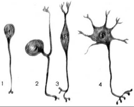
Fig. 230. Types of neurons (according to V. G. Eliseev et al.)
1 — unipolar; 2 — pseudounipolar; 3 — bipolar; 4 — multipolar
A bipolar neuron has processes extending from its opposite poles. Along one of these processes — the dendrite — excitation is conducted from the periphery into the cell, while along the second — the axon — it travels toward the brain. Examples of bipolar neurons include sensory cells in the olfactory organs and the retina of vertebrates.
A pseudounipolar neuron has only a single process that, at a certain distance from the cell, divides into two branches: a peripheral and a central one. The peripheral branch heads toward a specific organ, while the central branch goes to the Central Nervous System. During development, the neuron initially forms two processes that later fuse at their bases, transforming the initially bipolar neuron into a pseudounipolar one. Examples of such neurons are the sensory neurons of spinal ganglia.
Specific structures of the nerve cell include the tigroid substance and neurofibrils.
The tigroid substance (tigroid or Nissl bodies) is located in the body of the nerve cell and at the bases of dendrites, but is absent in axons. Nissl bodies represent elements of the granular Endoplasmic reticulum and polyribosomes, rich in ribosomal ribonucleic acid. The tigroid substance is an essential component of the nerve cell, which changes depending on its functional state. During nervous overfatigue, The amount of this substance sharply decreases, and during intense excitation of the neuron, the tigroid may disappear entirely. A systematic decrease in tigroid and changes in its distribution can also be observed in nerve cells As a result of various pathological processes. This provides grounds to consider the amount of tigroid in the cell, the shape of its clumps, and the pattern of their arrangement as indicators of the physiological state of the neuron.
Neurofibrils appear as very thin threads located in the cell body and its processes. In the cell body and dendrites, neurofibrils form a dense network. In the axon, they intertwine and extend along its length. Neurofibrils are responsible for Intracellular Transport and maintaining cell shape.
In addition to neurons, the nervous system contains neuroglial cells (gliocytes), which perform various Functions: supportive, trophic, protective, and secretory. They form macroglia and microglia.
Macroglia includes astroglia, oligodendroglia, and ependyma.
Astroglia consists of a large number of multipolar cells — astrocytes, whose processes intertwine and provide a supportive framework for neurons. The Cytoplasm of astrocytes is rich in Mitochondria, which indicates their active participation in metabolic processes.
Oligodendroglia is formed by a population of cells called oligodendrocytes, which, unlike astrocytes, have a small number of short processes. Oligodendroglial cells are located near the neuron body and its processes, forming dense clusters around them. The functions of oligodendrocytes are diverse: they participate in the Nutrition of neurons, synthesize protein and lipid substances, play a significant role in nerve regeneration processes, and are involved in the Generation and Conduction of nerve impulses.
The ependyma lines the spinal canal and the brain ventricles. Ependymal cells perform supportive and secretory functions and participate in The formation of CEREBROSPINAL FLUID.
Microglia consists of small, round, or slightly elongated cells with short processes. These cells are capable of active migration and engulfment of various dead elements and foreign particles, thus acting as typical phagocytes and performing a protective function in the nervous system.
The bodies of nerve cells form the gray matter of the BRAIN AND SPINAL cord, as well as the ganglia of vertebrate and invertebrate animals. The connection between the central nervous system and ganglia with organs is carried out through conducting elements — nerves, The basis of which is formed by nerve fibers.
Nerve fibers are processes of nerve cells surrounded by sheaths formed by oligodendrocytes (Schwann cells). A distinction is made between unmyelinated (non-medullated) and myelinated (medullated) nerve fibers.
Myelin, a repeatedly wound double layer of the glial cell Plasma Membrane, forms the inner Sheath of the axon cylinder, while the outer sheath is formed by the cytoplasm and Nucleus of the glial cell. Both types of fibers are externally covered by a basal lamina. At the boundary between two oligodendrocytes, a constriction of the nerve fiber is formed (node of Ranvier), where the myelin sheath is absent.
Depending on their function, three MAIN TYPES OF neurons are distinguished: afferent (sensory or receptor), associative (interneurons), and efferent (motor or secretory).
1. The cell bodies of afferent, sensory, or receptor neurons are always located outside the brain or spinal cord, within the ganglia of the Peripheral Nervous System. One of the processes extending from the nerve cell body runs toward the periphery to a specific organ, terminating in a sensory ending—a receptor—which transforms the energy of an external stimulus into a nerve impulse. The second process enters the central nervous system, specifically the spinal cord or Brainstem, via the dorsal roots of Spinal Nerves or respective Cranial Nerves.
Depending on their Location, receptors are classified into the following types:
exteroreceptors (from Lat. externus — external), located at the interface between the body and the external environment—in the body's external and internal integuments (Skin, mucous membranes)—which perceive pain, Temperature, Touch, pressure, and are also found in Sense Organs;
interoreceptors (from Lat. internus — internal), located in Internal Organs, signaling Changes in the Physical and Chemical state of these organs and their contents;
proprioceptors (from Lat. proprius — one's own), which perceive stimuli originating deep within the body proper (bones, Muscles, tendons, ligaments, fasciae, joint capsules).
2. Associative (intercalated, interneuron, or conductor) neurons mediate the closure or transmission of excitation from a sensory (afferent) neuron to an efferent one. Associative neurons are located entirely within the central nervous system. They frequently form complex neural networks where information and signals arriving from receptors are processed. Associative neurons account for over 90% of the total number of neurons. The cerebral and cerebellar cortex, basal and cerebellar nuclei, as well as the sensory and autonomic nuclei of the spinal cord and brainstem, are all built from the cell bodies of associative neurons.
3. Efferent, effector (motor or neurosecretory) neurons execute a response reaction (motor or secretory) by conducting nerve excitation from the center toward the periphery, to the effector. An effector is the nerve ending of an efferent neuron that transmits a nerve impulse to a working organ (muscle, gland). The cell bodies of efferent neurons are located within the central nervous system or in the periphery (in sympathetic and parasympathetic ganglia).
Neurons in the nervous system form chains, or reflex arcs, which transmit excitation from the site of stimulus reception to the central nervous system and subsequently to the working organ. The transmission of a nerve impulse from one neuron to another occurs at their sites of contact and is facilitated by specialized structures known as interneuronal synapses (from Greek synapsis — connection). Synapses are classified into axosomatic, where the axon terminal of one neuron contacts the body of the next; axodendritic, where an axon contacts the dendrite of another neuron; as well as axoaxonic and dendrodendritic, where processes of the same type make contact, and so forth.
The contact-type relationship at a synapse can either be 'established' or 'disrupted' under various physiological states, thereby ensuring a selective response to a specific stimulus.
The presence of physiological contacts in certain synapses and physiological disjunction in others makes it possible for excitation to be conducted along one of many neuronal chains.
Synapses in which transmission occurs via BIOLOGICALLY ACTIVE SUBSTANCES are called chemical synapses, and the substances mediating this transmission are termed Neurotransmitters (from Lat. mediator — intermediary). Norepinephrine, acetylcholine, and serotonin, among others, act as Transmitters. Neurotransmitter molecules interact with specific Proteins of The cell membrane, altering its permeability to certain ions, which triggers an Action Potential. The impulse reaches the synapse via the presynaptic terminal, bounded by the presynaptic membrane and presynaptic area, and is received by the postsynaptic membrane and postsynaptic area. The synaptic cleft lies between both membranes. The presynaptic terminal is rich in mitochondria and synaptic vesicles containing the neurotransmitter. A nerve impulse arriving at the presynaptic terminal causes the release of the neurotransmitter into the synaptic cleft; it then acts on the postsynaptic membrane, inducing the generation of a nerve impulse in the postsynaptic region. Alongside chemical synapses, electrotonic synapses exist, in which impulse transmission between contacting cells occurs directly via a bioelectric pathway.
The activity of the nervous system, as defined by I. M. Sechenov, is reflex in nature. A reflex (from Lat. reflexus — reflection) is the body's response reaction to a particular stimulus, whether internal or external, occurring with the participation of the central nervous system.
The chain of nerve cells through which a nerve impulse travels from its point of origin (the receptor) to the working organ (the effector) is called a reflex arc. Depending on the number of neurons (nerve cells) forming it, reflex arcs are divided into simple and complex.
A simple reflex arc consists of only two neurons—an afferent (sensory) and an efferent (motor or secretory). Typically, the first neuron is a pseudounipolar sensory neuron whose cell body is located in a spinal ganglion or a sensory ganglion of the cranial nerves. The dendrite of this cell runs as part of the respective spinal or cranial nerve to the periphery, terminating in a receptor that perceives an external (from the environment) or internal (within organs and tissues) stimulus. This stimulus is transformed by the receptor into a nerve impulse, which is transmitted to the nerve cell body, and then travels via the axon (bundles of which form the dorsal or sensory roots of spinal nerves) into the spinal cord, or via respective cranial nerves into the brain. In the gray matter of the spinal cord or a brain nucleus, this process of the sensory cell forms a synapse with the body of the second neuron (efferent, effector). Within the interneuronal synapse, neurotransmitters facilitate the transmission of nerve excitation from the sensory (afferent) neuron to the efferent (motor or secretory) neuron, whose process exits the spinal cord as part of the ventral roots of spinal nerves or the motor (secretory) nerve fibers of cranial nerves, traveling to the working organ to induce Muscle contraction or inhibit/enhance glandular secretion.
The vast majority of reflex arcs do not consist of just two neurons, but have a much more complex structure. Between the two neurons—afferent and effector—there is one or more intercalated (connecting) interneurons. In this case, excitation from the receptor neuron is transmitted via its axon not directly to the effector neuron, but to one or more interneurons. In the spinal cord, interneurons are represented by cells located in the gray matter of the dorsal horns. Some of these cells have axons that run to the motor Cells of the ventral horns of the spinal cord at the same level, closing the reflex arc of that spinal segment. The axons of others may first bifurcate in a T-like fashion within the spinal cord into descending and ascending branches that run to the motor nerve cells of the ventral horns in adjacent, superior, or inferior segments. Each of these ascending and descending branches can then give off collaterals to the motor cells of these and other neighboring segments. Consequently, The stimulation of even a minimal number of receptors is transmitted not only to the nerve cells of a specific spinal segment, but also spreads to the cells of several adjacent segments. As a result, the response reaction manifests as the contraction not of a single muscle, but of several muscles (muscle groups) simultaneously. Thus, in response to a stimulus, a complex, coordinated, yet reflex movement occurs.
Based on the topographical principle, the human nervous system is conventionally divided into the central and peripheral divisions.
The central nervous system (CNS) includes the spinal cord and the brain, which consist of gray and White matter. The gray matter of the spinal and Cerebral Cortex/brain is an accumulation of nerve cell bodies together with the proximal branches of their processes, forming nuclei or nerve centers. White matter consists of nerve fibers (nerve cell processes) covered with a myelin sheath (which gives the fibers their white color). Nerve fibers form the conduction PATHWAYS OF THE spinal cord and brain, connecting individual parts of the central nervous system and various nuclei (nerve centers) with one another.
The peripheral nervous system includes the nerves emerging from the brain and spinal cord (31 pairs of spinal nerves and 12 pairs of cranial nerves) along with their roots and branches, nerve endings, nerve plexuses, and ganglia or nerve nodes formed by neuronal cell bodies.
According to another Classification, based on anatomical and functional principles, the unified nervous system is also conventionally divided into two parts: somatic (animal) and vegetative, or autonomic.
The somatic nervous system innervates mainly the body (soma), specifically the skin, Musculoskeletal System, and sense organs. Through cutaneous sensitivity and sense organs, this division of the nervous system ensures the organism's connection with the external environment.
The vegetative (autonomic) nervous system innervates all internal organs, glands, Cytology/cytology/32.html">Smooth Muscle tissue of organs, skin, Blood Vessels, and The Heart, and also regulates metabolic processes in all organs and tissues. The vegetative nervous system, in turn, is subdivided into two parts: the parasympathetic and the sympathetic. In each of these parts, as in the somatic nervous system, central and peripheral divisions are distinguished.
Last update: 08/08/2026
Editorial and Educational Adaptation: This material has been compiled based on the primary/original source text. The project team performed an editorial review, corrected technical inaccuracies, structured sections, and adapted the content for an educational format.
What was processed:
- elimination of formatting defects (OCR errors, structural breaks, corrupted characters);
- editorial organization of content;
- standardization of terminology in accordance with academic sources;
- verification of factual statements against the original source text.
All mentions of the author, publication year, and origin of the primary text have been preserved in accordance with the source.