BASICS OF MEDICAL BIOLOGY - 2012
Linked Inheritance. Genetics of Sex
Independent assortment of genes can occur only if the genes reside in different chromosome pairs. In any given Organism, the number of genes that assort independently during Meiosis is limited by the number of chromosome pairs. However, the number of organismal traits controlled by genes is exceptionally large, whereas the number of chromosome pairs is small. Assuming that each chromosome carries not just one, but multiple genes, how are genes localized on the same chromosome inherited? T. Morgan answered these questions through his Experiments on the fruit fly Drosophila. Genes are Structural components of Chromosomes. Since there are more genes than chromosomes, we can predict that a single chromosome will contain not one, but several genes that are inherited together. T. Morgan termed this phenomenon Gene linkage. The inheritance of genes located in the same chromosome, which restricts their free assortment, is referred to as gene linkage.
Genes situated on the same chromosome are called linked genes and are predominantly inherited together (linked inheritance). All genes within a single chromosome form a linkage group. Homologous chromosomes share identical linkage groups; therefore, every organism possesses as many linkage groups as it has pairs of homologous chromosomes.
Traits whose genes belong to the same linkage group do not obey THE PRINCIPLE OF independent assortment in a dihybrid cross (AaBb x AaBb) yielding a 9:3:3:1 ratio, nor in a dihybrid test cross (AaBb x aabb) yielding a 1:1:1:1 ratio. Linked genes are not always transmitted together.
The phenomenon of trait linkage was discovered in 1906 by W. Bateson and R. Punnett in experiments with sweet peas by crossing two strains that differed in two pairs of traits: pollen shape and flower color. Instead of the expected segregation In the second generation F2 (9:3:3:1), a segregation ratio close to 3:1 was observed. Thus, the traits did not exhibit independent inheritance.
The phenomenon of gene linkage was analyzed by T. Morgan. In Drosophila, the allele for gray body color (B) is dominant over the allele for black body color (b), and the allele for normal wing length (V) is dominant over the allele for vestigial wings (v). Crossing a homozygous gray fly with normal wings (BBVV) and a black fly with vestigial wings (bbvv) produced first-generation F1 hybrids with gray bodies and normal wings (BbVv), confirming G. Mendel's law of uniformity of first-generation hybrids. The experimental results did not depend on the sex of the recessive homozygote. Next, two dihybrid test crosses were performed. In the first cross, the male was an F1 dihybrid (gray body and normal wings), and the female was homozygous for the recessive alleles (black body and vestigial wings). This cross yielded two phenotypic classes analogous to the parental forms, in equal proportions: 50% gray with normal wings (BbVv) and 50% black with vestigial wings (bbvv). In the second dihybrid test cross, the female was an F1 dihybrid (gray with normal wings), and the male was a recessive homozygote (black with vestigial wings). This cross produced four phenotypic classes in the following ratio:
1) gray with long wings (BbVv) 41.5%;
2) gray with vestigial wings (Bbvv) 8.5%;
3) black with long wings (bbVv) 8.5%;
4) black with vestigial wings (bbvv) 41.5% (fig.)
The results of both dihybrid test crosses did not match the expected phenotypic ratio of 25% gray with normal wings, 25% gray with vestigial wings, 25% black with normal wings, and 25% black with vestigial wings, as occurs in independent assortment (G. Mendel). Morgan explained this deviation from the expected segregation (1:1:1:1) by the fact that the genes for the studied traits (B and V) are located on the same chromosome and are inherited together (linked). The strength of linkage between genes is inversely proportional to the distance between them on the chromosome (Morgan's rule or law).
Class="center">
Fig. 21. Linked inheritance in Drosophila.
Gene linkage can be complete or incomplete. In the first test cross  , complete gene linkage was observed. In the second test cross
  , the number of individuals with phenotypes matching the parental phenotypes predominated (83%), also indicating a tight linkage between alleles B and V. Building upon F. Janssens' discovery of chiasmata in meiosis (1909), T. Morgan explained the appearance of a smaller proportion (17%) of offspring with phenotypes combining traits from both parents as a disruption of linkage resulting from chromosomal crossing-over at a point between genes B and V. Morgan named this crossing-over process crossing-over. The differences in results between the 1st test cross (where the male was the dihybrid) and the 2nd test cross (where the female was the dihybrid) are explained by a biological feature of Drosophila: the absence of crossing-over in male gametogenesis. Therefore, the dihybrid male (BbVv) produces only non-crossover Gametes (BV and bv) of two types at 50% each, whereas the dihybrid female (BbVv) produces 83% non-crossover gametes of two types (41.5% BV and 41.5% bv) and 17% crossover gametes of two types (8.5% Bv and 8.5% bV). Gametes formed As a result of crossing-over are called crossover gametes, whereas those formed without crossing-over are called non-crossover gametes. Accordingly, individuals arising from crossover gametes are termed crossovers, and those formed without them are non-crossovers. Thus, in T. Morgan's experiment, crossovers accounted for 17% of the progeny, and non-crossovers for 83%.
Crossing-over (chromosomal crossover) is the reciprocal exchange of segments between homologous chromosomes. It regularly occurs in prophase I of meiosis at the pachytene stage during gametogenesis (Spermatogenesis and oogenesis). The homologous chromosomes (maternal and paternal) of prophase I align very closely together (synapsis). Each consists of two chromatids. Before separating, the chromatids form X-shaped structures (chiasmata); at the points of intersection, chromatids break and rejoin, but with the maternal segment joining the paternal one rather than another maternal segment. The EXCHANGE OF GENETIC material typically occurs between non-sister chromatids of homologous chromosomes, though it can also occur between sister chromatids. Crossing-over disrupts gene linkage. As a result, recombinant chromosomes emerge with novel gene combinations and new linkage groups, leading to the appearance of offspring with novel combinations of parental genes. Crossing-over is evolutionarily significant as one of the mechanisms generating combinatorial Variability.
In addition to meiotic crossing-over, which regularly takes place during meiosis in gamete formation, genetic exchange between chromatids of homologous chromosomes is occasionally observed in somatic Cells (Mitotic crossing-over). For instance, an autosomal-recessive human mutation known as Bloom syndrome is accompanied by a cytological picture resembling the synapsis of homologous chromosomes and even The formation of chiasmata.
Crossing-over frequency is measured as The ratio of the number of crossovers to the total number of offspring in a test cross and is expressed as a percentage. The recombination frequency between genes is directly proportional to the distance between them. The closer genes are located to one another, the fewer potential crossing-over points exist between them, and conversely, the farther apart they are, the more potential crossing-over points occur between them. Crossing-over between two genes can take place not only at a single point, but also at two (double crossing-over) or even multiple points.
Crossing-over frequency reflects the degree of gene linkage and, for any given pair of genes under identical conditions, is always constant. For other genes on the same chromosome, the crossing-over frequency will necessarily differ, yet it remains constant as well: ranging from fractions of a percent to nearly 50%. The constancy of the percentage of crossing-over between genes is utilized as an indicator of their relative distance. One map unit (centimorgan / morganide) is adopted as the unit of distance between genes. It corresponds to a distance at which the crossing-over frequency equals 1%. In Morgan's experiment, crossovers constituted 17% of the progeny. Hence, the crossing-over frequency between genes B and V is 17%, and the distance between the genes is 17 centimorgans. Based on these data, T. Morgan and his coworkers established METABOLISM/2.html">THE CONCEPT OF the linear arrangement of genes on chromosomes and proposed the principle of constructing chromosome maps.
Key Principles of the Chromosome Theory of Heredity
The chromosome theory of heredity was formulated and experimentally substantiated through studies on the fruit fly (Drosophila melanogaster) conducted between 1910 and 1925 by the American geneticist T. Morgan and his school—A. Sturtevant, H. Muller, and C. Bridges. The core principles of the chromosome theory of heredity are:
1. Genes are arranged linearly along the length of chromosomes; different chromosomes contain varying numbers of genes; the gene set of each non-homologous chromosome is unique.
2. Allelic genes occupy specific and identical loci (positions) on homologous chromosomes.
3. Genes located on the same chromosome form a linkage group, which results in the linkage of certain traits transmitted together (linked) to descendants. The number of linkage groups equals the haploid chromosome number. Linkage is not absolute.
4. During meiosis, which occurs exclusively during gamete formation, the diploid chromosome number is halved (yielding the haploid number). This corresponds to the law of segregation, according to which the genetic material of both parents must segregate and enter different gametes.
5. According to the law of independent assortment, maternal and paternal sets of unlinked genes segregate independently of one another. If unlinked genes reside on different chromosomes, maternal and paternal chromosomes must distribute randomly among gametes during meiosis.
6. Reciprocal recombination can occur between genes of homologous maternal and paternal linkage groups as a result of Crossing Over. This corresponds to the formation of chiasmata during the conjugation of homologous chromosomes in meiosis (genetic crossing over).
7. The strength of linkage between genes is inversely proportional to the distance between them. The closer genes are located on the same chromosome, the stronger their linkage, the lower the frequency of recombination between them, and vice versa. Gene distance is measured in percentages of crossing over. One percent of crossing over corresponds to one centimorgan.
8. Each biological species is characterized by a specific set of chromosomes, known as the karyotype. The Study of the chromosomal mechanism underlying Sex Determination and the Inheritance of Traits linked to
the sex chromosomes, gene linkage, and the correlation of genetic maps with cytological chromosome maps all form the foundation of the chromosomal theory of heredity, which underpins the entire Selection/4.html">Development of Genetics.
Genetic Chromosome Maps
Building upon The Theory of the linear arrangement of genes on chromosomes and utilizing crossing over to determine the distance between them, researchers successfully established their linear sequence on the chromosome. A. Sturtevant compiled the first gene distribution map for one of Drosophila's chromosomes. Subsequently, similar maps were generated for other chromosomes. The unit of distance between genes is defined as 1% crossing over, or 1 centimorgan.
A genetic map is a linear representation indicating the order of genes and the distances between them in percentages of crossing over. It is constructed based on the results of test cross analysis.
Mapping is performed to determine which chromosome pair and what genetic distance (recombination frequency), or specific region of a chromosome, the genes occupy.
Genetic maps have been constructed for numerous organisms. Among angiosperms, the Genetic Maps of maize, barley, rice, and tomatoes are the most extensively studied. In animals, such maps have been developed for Drosophila, and among mammals, for the mouse. It has been proven that this distribution of genes across chromosomes is a universal biological pattern.
In humans, analyzing gene linkage using classical Methods developed in Drosophila is unfeasible due to the impossibility of experimental matings. In recent years, a comprehensive suite of human gene mapping techniques has been developed. Currently, over 15 methods are employed to study Linkage groups and construct chromosomal maps.
Human chromosome mapping is conducted through pedigree analysis and the genetic analysis of Somatic Cell Hybrids.
For a long time, only three autosomal linkage groups and one X-linked group were known. A new era began in 1968 with the successful localization of the Duffy Blood group gene to chromosome 1. All 24 human linkage groups are now known.
The Human Genome contains between 35,000 and 40,000 distinct genes, 16,000 of which have now been mapped onto a skeletal genome map compiled using 1,000 high-polymer DNA markers (J. Schuler et al., 1996).
The identification of linkage groups on chromosomes enables the construction of chromosomal maps, where gene locations are designated as points along the chromosome. The map of the X chromosome has been constructed most comprehensively. X-linked loci—numbering over 200—have been assigned to this chromosome based on pedigree analysis, with many confirmed by somatic cell Hybridization techniques.
Methods for constructing chromosome maps rely on predicting crossing-over probabilities along the entire length of the chromosome and random exchanges between sister chromatids.
Chromosome mapping utilizes the somatic cell hybridization of human cells with cells from other species, predominantly mice. When such hybrid cells are cultured on nutrient media, a gradual loss of Human chromosomes occurs. Once only a single human chromosome remains within the hybrid cell karyotype, it can be identified via differential staining. By subsequently determining the presence or absence of a specific enzyme in that cell, one can deduce that the locus encoding this enzyme resides on that chromosome.
Despite certain challenges in constructing genetic maps and their inherent relativity, detailed map development remains one of the primary methods for analyzing chromosome behavior and fine Structure.
There are already 24 known linkage groups in humans (22 pairs of autosomes, plus X and Y). A significant number of genes have been mapped on human chromosomes.
The compilation of accurate chromosome maps is an essential prerequisite for the further advancement of theoretical Human Genetics. This is of exceptional importance for the detection and Cytology/practical/136.html">Differential Diagnosis OF inherited fetal disorders, early disease diagnosis, the identification of asymptomatic carriers of genetic aberrations, and Genetic Counseling and prognosis.
Current state of Human Genome Research
The international Human Genome Project was initiated in the early 1990s. As a result of painstaking efforts, the human genome was fully sequenced by early 2003, meaning the complete sequence of the three billion Base Pairs making up the DNA of all 23 human chromosome pairs was successfully read. The genetic length of the human genome is 3,000 cM (centimorgans, a genetic distance corresponding to 1% crossing over). The completion of the Human Genome Project facilitated the creation of genetic maps and the reconstruction of The Genome's cytogenetic map.
A genetic map involves establishing the sequential arrangement of genetic markers spaced no more than 1 cM apart along all chromosomes. Such a genetic map makes it possible to map any gene and determine the relative distance between loci.
An exceptional success of the Human Genome Project was the generation of integrated (physical) genome maps. Approximately 40,000 coding sequences have been mapped. The total number of genes is estimated to be between 30,500 and 40,000.
To date, the entire human genome has been cloned as large overlapping fragments. The precise chromosomal location of each fragment has been determined with high accuracy. The term cloning implies that a gene has been mapped, its structure investigated, and the specific mutation causing a given disease identified.
The decoding of the human genome will facilitate The Development of new areas of medicine, the study of The Nature of hereditary and malignant diseases, and the advancement of gene and cell therapy.
Non-chromosomal (Cytoplasmic) Inheritance
T. Morgan's theory of heredity is known as the chromosomal theory, which emphasizes the leading role of nuclear chromosomes in hereditary phenomena. However, heredity is also associated with the Cytoplasm. Unlike chromosomal (nuclear) inheritance, this form is called non-chromosomal or CYTOPLASMIC INHERITANCE. It is governed by genes localized outside the Cell Nucleus—in Mitochondria and Plastids—which possess their own DNA and are capable of autonomous (nucleus-independent) reduplication. The collective set of cytoplasmic genes is called the plasmone, and the genes themselves are termed plasmagenes. Plasmagenes share properties with nuclear genes, being capable of both reduplication and mutation. Cytoplasmic inheritance is transmitted unilaterally, exclusively through the maternal line via the cytoplasm of the egg cells. Examples of maternal inheritance include streptomycin resistance in Chlamydomonas, the direction of shell coiling in freshwater Mollusks (Lymnaea), and variegation in ornamental plants such as four-o'clocks (Mirabilis jalapa) and snapdragons (Antirrhinum majus), as well as cytoplasmic male sterility in maize. In Bacteria, cytoplasmic inheritance is associated with Plasmids—small, extrachromosomal circular DNA molecules. Plasmids vary widely in function: F-plasmids confer The ability to undergo bacterial conjugation, R-plasmids provide multiple drug resistance, and Col-plasmids direct the synthesis of colicins (Proteins with lethal activity).
The main forms of cytoplasmic inheritance are plastid and Mitochondrial Inheritance. An example of plastid inheritance is the transmission of leaf variegation in four-o'clocks and other plants.
Mitochondrial inheritance. Each mitochondrion contains its own DNA. Compared to nuclear DNA, Mitochondrial DNA (mtDNA) is circular, relatively small in size, contains a correspondingly small set of genes, and lacks introns. In humans, mtya contains 16569 nucleotide pairs. The Genetic Code of mitochondrial DNA differs from the universal code: certain triplets encode different Amino Acids, non-identical nucleotide sequences form stop codons, and adenine or cytosine is encountered more frequently in the third position. Most mitochondrial proteins are encoded by nuclear DNA, synthesized in the cytoplasm, and subsequently imported into mitochondria. Only about 2% of mitochondrial proteins are encoded by plasmagenes and synthesized on mitochondrial Ribosomes. Mutations in Mitochondrial Genes are associated with Mitochondrial Diseases.
Each species of organisms has a specific sex ratio at birth; in the majority of organisms, the population sex ratio is 1:1 (or 50% to 50%):
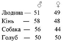
Therefore, G. Mendel and Doncaster proposed the following hypothesis regarding the genetic mechanism of sex determination: one sex is heterozygous (Aa), while the other is homozygous for recessive alleles, thus (see diagram) maintaining a 1:1 sex ratio from generation to generation.

Sex is inherited as a Mendelian trait. Segregation by sex occurs in a 1:1 ratio, just like in a test monohybrid cross.
The sex of animals and plants is genetically determined by a single pair of chromosomes known as sex chromosomes. There are four MAIN TYPES OF sex regulation by sex chromosomes.
1. The XY type, in which the female sex has two X Sex Chromosomes and is homozygous (producing one type of gametes—X), while the male sex possesses a male sex chromosome—Y, which differs from the X chromosome in size and shape. The male sex is heterogametic (producing Two Types of gametes—X and Y). This type is characteristic of mammals, insects, and most dicotyledonous angiosperms.
2. The X0 type, in which the female sex has two X chromosomes and the male has only a single X chromosome.
The X0 type is found among insects and mammals.
3. The ZW type. The female sex has one female sex chromosome W and a second sex chromosome Z, which differs from it in shape and size. Here, the female sex is heterogametic, whereas the male sex has two identical Z sex chromosomes and is homogametic. The ZW type is inherent to certain fish, butterflies, birds, and very rarely occurs among plants.
4. The ZO type, in which the female sex has only one Z chromosome and is heterogametic, while the male sex has two Z chromosomes and is homogametic. This type is known in only one species of lizard.
Modes of Sex Determination:
- progamic type — sex is determined even before Fertilization (in daphnia, females develop under optimal reservoir conditions, whereas males develop at low temperatures);
- syngamic type — sex is determined at the moment of fertilization (in humans, the combination of two X chromosomes forms a female organism, while X and Y chromosomes form a male one);
- epigamic type — sex is determined after fertilization and is shaped under the Influence of Environmental conditions (a typical example is the marine worm Bonellia viridis, the female of which lays fertilized eggs into the Water, where larvae develop from them. If a larva lands on the proboscis of a female, it develops under the action of specific Enzymes into a large-sized female. Conversely, if the larva settles on the bottom of the water body or attaches to another substrate, it develops into a small-sized male);
Sex is also determined depending on whether fertilization has occurred (in bees, females develop from fertilized eggs, whereas males develop from unfertilized ones).
Sex, like any other trait, is genetically determined. In humans, sex is determined at the moment of fertilization (syngamically) and depends on the combination of sex chromosomes from both parents in the zygote. A distinction is made between the homogametic and heterogametic sexes. Homogametic is the sex in which both sex chromosomes are identical and which produces a single type of gametes. Heterogametic is the sex with different sex chromosomes that produces two types of gametes. In humans, mammals, and Drosophila, the female sex is homogametic, and the male sex is heterogametic. During meiosis, a woman (XX) produces one type of egg cell (X), while a man (XY) produces two types of spermatozoa in equal proportions: half of the sperm contain the X chromosome, and the other half contain the Y chromosome.
The sex of the future organism depends on the heterogametic parent individual, which produces two types of gametes: whichever of these gametes takes part in fertilization will determine the sex of the offspring. Whether a girl or a boy is born depends on the sperm type, since egg cells are identical regarding their sex chromosome. Two combinations are possible: 1) if an X egg cell is fertilized by an X-bearing sperm, two X chromosomes combine in the zygote: a female organism (XX) develops from such a zygote; 2) if an X egg cell is fertilized by a Y-bearing sperm, X and Y chromosomes combine in the zygote: a male organism (XY) develops from such a zygote.
Traits whose genes are located not in autosomes, but in sex chromosomes (X and Y), are called sex-linked, and the inheritance of such traits is termed Sex-Linked Inheritance. It was first established by T. Morgan while studying the inheritance of eye color (red and white) in Drosophila.
Top left: genes located in the non-homologous segment of the X chromosome (with respect to the Y chromosome); top right: genes located in the non-homologous segment of the Y chromosome; bottom right and left: genes located in the homologous segments of the X and Y chromosomes.
The first group comprises completely sex-linked genes. They are localized in the non-homologous region of the X chromosome, which lacks a homolog on the Y chromosome, and are inherited exclusively via the X chromosome. These include the recessive genes for hemophilia, color blindness, and the dominant gene for hypophosphatemia (vitamin D-resistant Rickets).
The second group consists of a small number of genes that are also completely sex-linked but located in the non-homologous region of the Y chromosome. They are transmitted from a father to all his sons, because a son receives his Y chromosome solely from his father (e.g., webbed toes, "hairy ears"). Traits whose genes reside in the non-homologous region of the Y chromosome are called holandric.
The third group comprises incompletely (partially) sex-linked genes. They are located in the homologous Regions of the X and Y chromosomes, can be inherited with either the X or the Y chromosome, and can transfer from one to the other during crossing-over.

Fig. 21. Probable localization of incompletely sex-linked genes in the homologous regions of the X and Y chromosomes.
In humans, traits inherited via the Y chromosome can occur exclusively in males, whereas those inherited via the X chromosome can be found in individuals of both sexes. A female can be either homozygous or heterozygous for genes localized on the X chromosome, and recessive alleles of these genes manifest themselves only in the homozygous state. Since males possess only a single X chromosome, all genes localized on it, even recessive ones, are expressed in the phenotype. Such an organism is termed hemizygous.
Genes determining sex traits are not restricted to the sex chromosomes alone. Sex-limited traits can be controlled by genes located in either autosomes or the sex chromosomes of both sexes, yet they are expressed in only one of them. For instance, baritone and bass voices manifest only in males; milk yield and milk fat content, only in cows; egg-laying capacity and egg size, only in chickens.
Sex-influenced traits are characteristics determined by autosomal genes in both males and females, but the dominance of these traits depends on sex Hormones. An example of a sex-influenced trait is the inheritance of pattern baldness in men and women. The baldness gene is dominant, while its recessive allele determines normal Hair. In the homozygous state, the dominant gene (BB) expresses itself almost identically in both men and women, resulting in bald men and women with thinning hair. In recessive homozygotes (bb), the trait's manifestation is likewise identical in both sexes. However, in the heterozygous state (Bb), men are entirely bald, whereas heterozygous women do not exhibit the trait and retain a normal HEAD of hair. In humans, sex also influences such a condition as Gout, characterized by the deposition of uric acid crystals in the joints, causing severe pain. The gene responsible for this disorder is expressed significantly more strongly in the presence of Male Sex Hormones than in the presence of female ones.
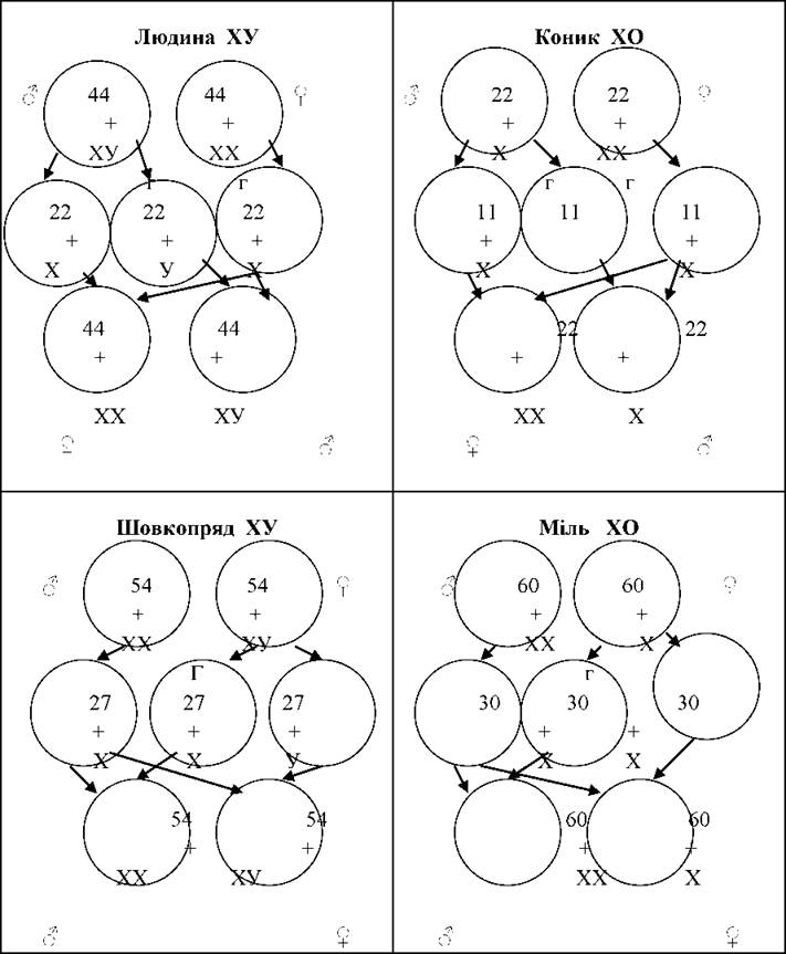
Fig. 22. Diagram of various types of Chromosomal Sex determination.
Sex is the set of organismal properties that ensures the function of reproduction and progeny generation based on the transmission of Genetic information. A distinction is made between Primary and secondary sex characters. Primary sex characters encompass all organismal features that ensure gamete formation, their fusion during fertilization, and structural differences in internal and external reproductive Organs. Secondary sex characters are those that do not directly participate in gametogenesis, yet play a specific role in sexual reproduction (such as Mammary Glands in mammals or plumage in birds). The development of secondary sex characters is systemically regulated by hormonal activity and is consequently directly linked to the function of the primary Gonads.
The physiological theory of sex determination posits that an organism is genetically bisexual and that its genotype contains genes for both sexes, although the ratio and mode of action of these genes likely differ between male and female individuals. Males possess male-determining genes not only on the Y chromosome, but also in small amounts on the X chromosome and in autosomes. Females, much like males, harbor genes for both female and, to a lesser extent, male sex. The process of sex differentiation is driven by sex hormones secreted not only by Endocrine glands, but also by the embryonic gonad: the male embryonic sex hormone is medullarin, and the female hormone is the cortical hormone. The rate of hormonal secretion is controlled by the genic balance. A predominance of male-determining genes leads to elevated secretion of male sex hormones and the Development of the male sex, whereas a predominance of Female Sex Hormones drives female development. Alterations in the secretory activity of either sex hormone can lead to the development of intersexual forms known as hermaphrodites.
Last update: 08/08/2026
Editorial and Educational Adaptation: This material has been compiled based on the primary/original source text. The project team performed an editorial review, corrected technical inaccuracies, structured sections, and adapted the content for an educational format.
What was processed:
- elimination of formatting defects (OCR errors, structural breaks, corrupted characters);
- editorial organization of content;
- standardization of terminology in accordance with academic sources;
- verification of factual statements against the original source text.
All mentions of the author, publication year, and origin of the primary text have been preserved in accordance with the source.