Phycology - Kostikov I.Yu. - 2009-2013
Chapter 21. Cryptomonads – Cryptophyta
The division Cryptophyta comprises about 200 species of unicellular, microscopic, flagellate Algae distributed in both continental Water bodies and marine environments. Until the early 1970s, cryptomonads, along with dinoflagellates and raphidophytes, were grouped within the division Pyrrophyta based on similarities in pigmentation, flagellate Organization, presence of a gullet, and ejective structures. In the 1970s, cryptomonads were separated into an independent division, which, based on a series of cytological features, was considered a distinct phylum representing primitive members of the plant kingdom. Following investigations of the nuclear, chloroplast, and Mitochondrial Genomes, as well as a specialized DNA-containing Structure known as the nucleomorph, it became evident that Cryptophyta are closely related to organisms possessing Cell/35.html">Mitochondria with flat cristae, specifically glaucophytes, red algae, and green algae.
Today, the Cryptophyta are recognized as a division that provides direct Evidence for the plastid origin via chloroplast Endoplasmic reticulum through a "heterotrophic eukaryote + photoautotrophic eukaryote" symbiosm.
Class="center">Taxonomic Features of the division
Pigments and reserve storage products
Photoautotrophic cryptomonads are characterized by the presence of chlorophylls a and c, α- and ε-carotenes (β-carotene, typical of the vast majority of other algal divisions, has not been found in Cryptophyta), phycobilin pigments—phycocyanin and phycoerythrin—as well as two groups of xanthophylls: lutein-cycle xanthophylls (lutein, violaxanthin, zeaxanthin) and several specific xanthophylls (alloxanthin, crocoxanthin, monadoxanthin). Unlike cyanobacteria, glaucophytes, and red algae, phycobilin pigments in cryptomonads are not localized in phycobilisomes, but rather occur directly on the inner surface of thylakoid membranes.
The quantitative ratios of chlorophylls, various phycobilins, and specific xanthophylls vary widely among different genera. This accounts for the great diversity of coloration in cryptomonad algae. Thus, METABOLISM/14.html">Chloroplasts may appear red, blue, yellow, brown, or green. There are also species lacking chloroplasts; these are colorless and feed exclusively heterotrophically.
The primary assimilation product in Cryptophyta is starch. It does not accumulate within the chloroplast, as in green algae, nor in the Cytoplasm, as in glaucophytes, but rather is stored in the periplastidal space, frequently in the vicinity of the pyrenoid. An additional reserve product is oil.
Cytological features
Cryptomonad Cells are typically dorsiventral, obliquely truncated at the anterior end, and feature a deep or shallow gullet and two heterokont flagella emerging from the DORSAL SIDE OF the gullet opening. At the Base of the gullet, freshwater species possess one to two, rarely five to eight, contractile vacuoles. A narrow longitudinal furrow is located on the ventral side.
Cell coverings are highly specialized and are known as the periplast. It consists of a Plasma Membrane bearing rectangular or polygonal protein plates on its inner side. The latter are attached to the Plasmalemma by protein spikes. The outer side of the plasmalemma may be naked, but more frequently it bears a layer of outer protein plates mirroring the shape of the inner ones, or a layer of submicroscopic scales attached to the plasmalemma by fibrillar material (Fig. 21.1). Rectangular plates arranged in regular rows typically impart a characteristic longitudinal striation to The Cell coverings of cryptomonads.

Fig. 21.1. STRUCTURE OF THE periplast in cryptomonad algae: 1 - plasmalemma, 2 - inner protein plate, 3 - protein spikes attaching the inner plate to the plasmalemma, 4 - outer plate, 5 - trichocyst, 6 - membranous capsule, 7 - coiled ribbon of the trichocyst, 8 - membranous capsule of the trichocyst following the discharge of the protein ribbon (schematized after Lucas, 1970; Hausmann, 1979; Kugrens, Lee, 1987).
Trichocysts are located beneath the periplast. Large trichocysts typically line the gullet, whereas smaller ones are scattered beneath the entire cell surface. A trichocyst appears as a membranous capsule enclosing a tightly coiled ribbon. Upon stimulation of the plasmalemma, the ribbon uncoils and discharges outwardly, imparting a reactive thrust to the cell. As a result, the cell can "jump" forward, abruptly slow its movement, or sharply alter its direction. Trichocysts are formed within vesicles of the Golgi apparatus.
The nuclear apparatus is eukaryotic. The Nucleus is typically located in the posterior region of the cell and features a relatively large nucleolus. The outer nuclear membrane is directly continuous with the outer chloroplast membrane (Fig. 21.2). Centrioles are absent; their function is performed by flagellar basal bodies and rhizoplasts, toward which the nucleus migrates at the onset of mitosis. Chromosomes remain condensed during interphase, and their number ranges from 40 to 200.
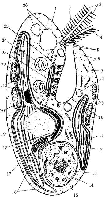
Fig. 21.2. Structure of a cryptomonad algal cell: 1 - periplast, 2 - longer mastigonemate flagellum, 3 - mastigoneme, 4 - shorter hairy flagellum, 5 - flagellar basal bodies, 6 - small trichocysts, 7 - endoplasmic reticulum, 8 - oil droplet, 9 - ventral mitochondrion, 10 - gullet, 11 - double-membraned chloroplast endoplasmic reticulum, 12 — two inner membranes of the chloroplast envelope, 13 - nuclear envelope, 14 - nucleus, 15 - nucleolus, 16 - periplastidal space, 17 - nucleomorph, 18 - starch, 19 - pyrenoid, 20 - eyespot (stigma), 21 - paired-thylakoid lamellae, 22 - dorsal mitochondrion, 23 - Golgi apparatus, 24 - Mop body, 25 - large gullet trichocysts, 26 - contractile vacuoles (schematized after Lucas, 1970, 1982; Santore, Leedale, 1985).
Mitosis is open and bears some resemblance to nuclear division in Prymnesiophytes. Thus, in metaphase, chromosomes form a dense plate where individual chromosomes are indistinguishable. The plate possesses several channels through which bundles of spindle microtubules pass. Microtubules do not converge at the poles, but remain parallel and terminate near the cisternae of The endoplasmic reticulum (Fig. 21.3).
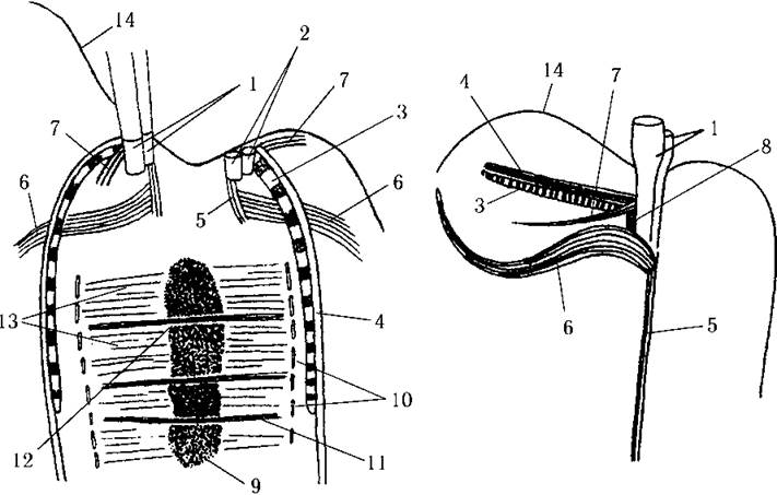
Fig. 21.3. Mitosis (left) and the flagellar ROOT system (right) in cryptomonad algae: 1 - flagellar basal bodies, 2 - replicated pair of basal bodies, 3 - rhizoplast, 4 - microtubular rhizoplast root, 5 - rhizostyle, 6, 7 - simple microtubular roots, 8 - smooth microfibrillar root, 9 - Chromatin plate, 10 - endoplasmic reticulum cisternae, 11 - bundle of microtubules passing through the chromatin plate, 12 - channel in the chromatin plate, 13 - individual spindle microtubules, 14 - periplast (schematized after Oakley, 1978 and Sedova, 1996).
Photosynthetic apparatus. Typically, the cell contains one or two large, parietal chloroplasts enclosed by a four-membraned envelope. The two outer membranes form the chloroplast endoplasmic reticulum. The outer membrane of the chloroplast endoplasmic reticulum is directly continuous with the outer nuclear membrane. The periplastidal space between the two outer and two inner chloroplast membranes contains starch, 80S Ribosomes, and a specialized organelle called the nucleomorph. Although the periplastidal space of cryptomonads possesses its own protein-synthesizing machinery, a periplastidal reticulum is lacking.
Starch is deposited as discrete granules or forms continuous starch plates. The latter is observed only in species possessing a pyrenoid.
The nucleomorph is an organelle delimited from the surrounding periplastidal space by a double membrane which, similar to the nuclear envelope, features pores and contains DNA and RNA. The nucleomorph is located either near the pyrenoid or lies freely within the periplastidal space. Nucleomorph DNA consists of approximately 600 kb of nucleotide pairs, is organized into three chromosomes, and is not associated with Histones. Each nucleomorph chromosome contains Genes encoding ribosomal RNA and at least several Proteins. Nucleomorph division precedes nuclear division. Analysis of nucleomorph genes encoding the 5S and 18S RNA subunits of periplastidal ribosomes has demonstrated that the nucleomorph genome is closely related to the nuclear genome of red algae.
Beneath the inner membranes of the chloroplast envelope lie thylakoids, a pyrenoid, an eyespot, endogenous chloroplast DNA, and chloroplast 70S ribosomes. A girdle lamella is absent. Thylakoids are arranged in pairs. In total, up to several dozen such paired-thylakoid lamellae are observed within the chloroplast. Phycobilin pigments are localized on the inner sides of each pair directly on the membranes, although they are not organized into phycobilisomes. A pyrenoid is present in many cryptomonads. It is typically large and semi-immersed, with its free surface oriented toward the cell interior. Starch is deposited in the region of the periplastidal space immediately adjacent to the pyrenoid. In some cryptomonads, the chloroplast contains an eyespot consisting of several layers of pigment globules appressed to the chloroplast membrane; THE POSITION OF the eyespot is not coordinated with the flagellar apparatus.
The chloroplast DNA is divided into several fragments located in different PARTS OF THE chloroplast stroma, meaning the genophore is dispersed.
Among the Cryptophyta, there are heterotrophic species that have secondarily lost their Photosynthetic Pigments or even the chloroplast, as well as primarily heterotrophic representatives. Secondary heterotrophs—such as those of the genus Chilomonas—retain a reduced plastid, known as a leucoplast.
The mitochondrial apparatus is typically represented by two large, branched mitochondria: a dorsal and a ventral one. Both possess lamellar cristae and are connected to the basal bodies of the flagella via microtubular and microfibrillar roots. The Mitochondrial DNA consists of approximately 50,000 nucleotide Base Pairs.
The flagellar apparatus. Both flagella in cryptophytes are typically directed forward. On the longer flagellum, mastigonemes are arranged in two rows, whereas on the shorter one they are uniseriate. Mastigonemes possess an expanded and a tapered region, distinguishing them from the mastigonemes of all other eukaryotic groups. They are synthesized in the endoplasmic reticulum. Furthermore, the mastigonemes of cryptophytes are covered with organic submicroscopic scales of a characteristic shape. In the transition zone, the flagella feature two transverse plates.
The flagellar root system consists of four microtubular and two microfibrillar roots. The former include the rhizostyle, two simple roots, and a root associated with the rhizoplast. The microfibrillar roots are represented by a cross-striated rhizoplast and a smooth root.
The rhizostyle consists of 6–8 parallel microtubules; it is directed toward the posterior end of the cell, passes close to the nucleus, and presumably provides structural support. The first simple microtubular root is composed of four microtubules and connects the flagellar basal bodies to the dorsal mitochondrion; the second root, formed by two microtubules, envelops the contractile vacuoles. The final microtubular root consists of four to six microtubules and lies closely appressed to the rhizoplast (see Fig. 21.3).
The cross-striated microfibrillar root—the rhizoplast—is composed of the protein centrin; it runs directly beneath the periplast and terminates near the nucleus. At the onset of mitosis, the rhizoplast, together with the flagellar basal bodies, serves as the microtubule-organizing center for the spindle. The smooth microfibrillar root connects to the ventral mitochondrion.
Other Organelles. Specific organelles of cryptophytes are a pair of so-called Mop bodies. These are relatively large, well-defined single-membrane structures located near the gullet and containing predominantly trichocyst remnants. It is hypothesized that Mop bodies are homologous to Lysosomes and function to digest excess trichocysts. Typically, a single Golgi complex is situated between the gullet and the Mop bodies.
Body Structure Types
Almost all cryptophyte algae exhibit a monadoid body structure type. The vast majority of species are capable of shedding their flagella, enveloping themselves in a layer of mucus, and entering a palmelloid state. A single genus, Bjornbergiella, features a filamentous structure type.
Reproduction and Life Cycles
The primary mode of reproduction in Cryptophyta is longitudinal Cell Division in the motile state. One daughter cell inherits both maternal flagella, while the other synthesizes new ones de novo. Asexual reproduction via specialized cells has not been observed in cryptophyte algae.
For a long time, it was believed that sexual reproduction was absent in cryptophyte algae. In 1986, a diplohaplontic life cycle with heteromorphic Morphology/12.html">ALTERNATION OF GENERATIONS was described in Proteomonas sulcata. Both generations—Haploid and Diploid—were represented by monadoid cells and reproduced via binary fission. Diploid cells differed from haploid ones in their larger size. Unfortunately, observing the sexual process and Meiosis was unsuccessful.
In 1988, a hologamous sexual process was discovered in Chroomonas acuta. Following Cell Fusion, karyogamy occurred, resulting in a diploid planozygote. However, the site of meiosis and the type of life cycle remained undetermined. Thus, sexual reproduction does exist in cryptophytes, although the details remain uninvestigated.
The predominant life cycle type for the vast majority of cryptophytes is cyclomorphosis. Under favorable conditions, cells reproduce by division while remaining motile. Deteriorating vegetative conditions lead to the loss of flagella, secretion of mucus, and transition to a palmelloid state, during which cells retain The ability to divide. Upon condition improvement, the cells regenerate their flagella and return to the monadoid state. At the end of the vegetative season, cryptophyte algae may form cysts: the cells lose their flagella, become spherical, envelop themselves in a thick layer of dense mucus, and enter a resting state. Upon the Conclusion of the resting period, the cyst germinates into two daughter monadoid cells (Fig. 21.4).
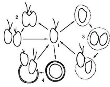
Fig. 21.4. Life Cycle of a cryptophyte alga: 1 - monadoid vegetative cell, 2 - reproduction by division in the motile state, 3 - palmelloid state, 4 - cyst and its germination.
Taxonomic System of the Phylum
All Cryptophyta belong to a single class, Cryptophyceae, and a single order, Cryptomonadales. The Classification of the order into families and genera is based on such features as the presence of a developed gullet, chloroplasts and leucoplasts, and cell coloration.
For instance, the gullet is highly reduced within the family Cryptochrysidaceae, characteristic representatives of which are photoautotrophic algae with brown or reddish chloroplasts belonging to the genus Cryptochrysis. A well-developed gullet is present in all members of the family Cryptomonadaceae, within which genera are distinguished primarily by color. Thus, the genus Cryptomonas features yellow or brown chloroplasts, Rhodomonas has red ones, and Chroomonas has blue ones. In species of the genus Chilomonas, chloroplasts are reduced, with the Plastids persisting as colorless leucoplasts. The sole genus of the family Cyathomonadaceae, Cyathomonas, lacks any plastids and, according to various authors' systems, is viewed either as the most primitive representative of cryptophytes or as the pinnacle of evolution for this phylum (Fig. 21.5).

Fig. 21.5. Selected representatives of cryptophyte algae: 1 - Cryptochrysis commutata, 2 - Cryptomonas skujae, 3 - Chilomonas paramaecium, 4 - Cyathomonas truncata (after Matvienko, Litvinenko, 1977).
Interestingly, Electron Microscopy and molecular biology studies have shown that some colorless algae traditionally classified among cryptophytes (specifically the family Katablepharidaceae) possess tubular rather than lamellar mitochondrial cristae, and therefore should be removed from Cryptophyta and placed into a new, independent taxon of phylum or class rank.
Distribution, Ecology, Significance
Cryptophyte algae inhabit primarily freshwater bodies or marine environments. Exceptions are few—for example, Cryptomonas salina and C. stigmatica are halophiles and frequently occur in the plankton of saline lakes at salt concentrations of 40–90 g/L; Cryptomonas cryophila and C. frigoris are cryophiles that develop On the surface of mountain snow and cause its red and green "blooming".
In freshwater habitats, most cryptophyte algae inhabit the water Column of clean water bodies (such as species of the genus Chroomonas), and less commonly polluted ones (Cryptomonas ovata, C. erosa, and notably colorless Representatives of the genus Chilomonas).
Marine cryptophytes are typically found in supralittoral rock pools and coastal puddles. Several species are also known to develop in open-ocean planktonic communities, particularly in the North Sea.
Cryptophyte algae are predominantly psychrophilic or eurythermal organisms, neutrophils, and less frequently acidophiles. Some species are known to inhabit hydrogen sulfide-rich environments—among decomposing plant debris and in the deep layers of lakes.
Certain Cryptophyta species serve as indicators of organic water pollution; they participate in the self-purification of natural water bodies and the advanced Treatment of wastewater in biological ponds.
Taxonomic Position in the Living World
Based on a combination of cytological features (specifically, the presence of specific bipartite mastigonemes, submicroscopic flagellar scales, mitosis accompanied by The formation of a dense metaphase plate, and a nucleus connected to the chloroplast via the outer membrane of the chloroplast endoplasmic reticulum), cryptophyte algae show similarities to haptophytes. Their chemical reserve composition and lamellar mitochondrial cristae reveal affinities with green and glaucocystophyte algae. The presence of phycobilins links cryptophytes with red algae and glaucocystophytes. However, several essential characteristics clearly distinguish Cryptophyta from the vast majority of other algal divisions, most notably the nucleomorph.
Regarding the features of their nuclear genome, cryptophytes occupy an intermediate position between haptophytes on the one hand, and algae with primary-symbiotic plastids—glaucocystophytes, green, and red algae—on the other; their Mitochondrial Genome also resembles those of green, red, and glaucocystophyte algae. The Chloroplast Genome of cryptophytes is most closely related to the chloroplast genomes of red algae, whereas the nucleomorph genome shows affinity with the nuclear genome of the latter.
Therefore, it is believed that heterotrophic ancestors of Cryptophyta shared a common predecessor with heterotrophic Prymnesiophyta, giving rise to two lineages. The first became the "parental" branch for algae with double-membrane plastids—Glaucocystophyta, Chlorophyta, and Rhodophyta. The second Lineage led to heterotrophic cryptophytes. The latter acquired the capacity for Photosynthesis relatively recently as a result of secondary Symbiosis with red algal cells. The Nucleus of the red alga that gave rise to the cryptophyte chloroplast has survived in the form of a nucleomorph.
Last update: 07/08/2026
Editorial and Educational Adaptation: This material has been compiled based on the primary/original source text. The project team performed an editorial review, corrected technical inaccuracies, structured sections, and adapted the content for an educational format.
What was processed:
- elimination of formatting defects (OCR errors, structural breaks, corrupted characters);
- editorial organization of content;
- standardization of terminology in accordance with academic sources;
- verification of factual statements against the original source text.
All mentions of the author, publication year, and origin of the primary text have been preserved in accordance with the source.