Phycology - Kostikov, I.Y. - 2009-2013
Chapter 16. Brown Algae – Phaeophyta
The phylum Phaeophyta (brown Algae) comprises about 260 genera represented by roughly 2,000 species. All members of this division are multicellular, predominantly macroscopic algae, and are distributed almost exclusively in marine environments. The oldest reliable fossil records of brown algae date back approximately 400 million years, while their probable remains date to around 570 million years ago. Based on molecular markers, the phylum is closely related to Xanthophyta.
Class="center">Taxonomic Features of the phylum
Pigments and reserve nutrients
Brown algae contain chlorophylls a and c, β- and ε-carotenes, and two main groups of xanthophylls: firstly, xanthophylls of the yellow pigment group—fucoxanthin, diatoxanthin, and diadinoxanthin—and secondly, xanthophylls of the lutein series—violaxanthin, zeaxanthin, antheraxanthin, and neoxanthin. Fucoxanthin and violaxanthin accumulate in large quantities, masking the chlorophylls and imparting a bright yellow or brown color to the METABOLISM/14.html">Chloroplasts.
The primary assimilation products are the polysaccharide laminarin and oil, and more rarely, polyhydric alcohols (e.g., mannitol). All assimilation products are stored outside the chloroplasts.
Cytological features
Cell walls consist of distinct layers. The wall is divided into two layers: an outer layer composed of pectic substances and soluble salts of alginic acid (predominantly sodium alginate), and an inner layer with a fibrillar Structure. The framework of the inner layer is formed by Cellulose microfibrils, which create a reticulate skeletal network, along with insoluble calcium alginate.
Alginic acid is a heteropolysaccharide composed of D-mannuronic and L-guluronic acid residues. Alginic acid salts, known as alginates, form gels. The viscosity of the gel is determined by the composition and Proportions of the cations to which the alginic acid binds.
Consequently, due to the high content of pectic substances and alginates, The Cell walls are prone to extensive mucilaginous degeneration and exist in a gel-like colloidal state.
In some brown algae, In addition to alginates, The Cell wall incorporates a sulfated heteropolysaccharide called fucoidan, which consists of oligosaccharide residues of fucose, galactose, mannose, xylose, and glucuronic acid.
Nuclear apparatus. The Nucleus has a typical eukaryotic structure. As in the vast majority of stramenopile algae, the outer membrane of the nuclear envelope continues into the chloroplast Endoplasmic reticulum (Fig. 16.1). A large, prominent nucleolus is clearly visible within the nucleus. Several Golgi complexes and two centrioles are located near the nuclear envelope. During karyokinesis, the latter serve as microtubule-organizing centers for the mitotic spindle. At the onset of nuclear division, they replicate and migrate to opposite poles of the nucleus. An interesting feature of the spindle is The formation of astral microtubule rays around the centrioles, similar to what is observed in Multicellular animals.
Fig. 16.1. Schematic diagram of a brown alga Cell Structure (using Ectocarpales as an example): 1 - endoplasmic reticulum; 2 - laminarin plate around the pyrenoid; 3 - Golgi complex; 4 - vacuole; 5 - nucleolus; 6 - nucleus; 7 - nuclear envelope; 8 - chloroplast DNA; 9 - double membrane of the chloroplast; 10 - girdle thylakoid; 11 - three-thylakoid lamella; 12 - chloroplast endoplasmic reticulum; 13 - protruding pyrenoid; 14 - mitochondrial profile (after Bouck, 1965).
In prophase, the nucleolus disappears, and the nuclear envelope near the centrioles breaks down, forming two large pores. Thus, mitosis in Phaeophyta is semi-closed and centric. Metaphase and anaphase proceed According to the typical eukaryotic pattern. In telophase, the daughter nuclei move apart, the spindle rapidly disintegrates, and then the nuclei approach each other slightly once again. Concurrently, the Plasmalemma forms a Cleavage furrow, separating the nuclei and the protoplast, and initiates the secretion of substances for new cell walls, with pores remaining in the transverse septum.
During zoospore formation at the final stages of cytokinesis in some species (e.g., Chorda tomentosa), centrioles migrate to The surface of the daughter Cells and transform into the basal bodies of the flagella.
Photosynthetic apparatus. The chloroplasts of brown algae are small and numerous. Each chloroplast is enclosed by four membranes, of which the two outer ones form the chloroplast endoplasmic reticulum that merges with the nuclear envelope, and the two inner ones constitute the chloroplast envelope proper. A periplastidial space is situated between the outer and inner membranes.
Thylakoids are typically arranged in groups of three, with a girdle lamella located along the periphery. Many brown algae possess a pyrenoid within the chloroplast. Similar to golden, eustigmatophyte, and yellow-green algae, it is not penetrated by thylakoids. In brown algae of the order Ectocarpales, the pyrenoid is of the protruding type. Laminarin is usually deposited around it, though outside the chloroplast. The laminarin plates are enclosed by their own cytoplasmic membrane. In most other brown algae, the pyrenoid is rudimentary and detectable only by Electron Microscopy, sometimes occurring solely in reproductive cells (zoospores, Gametes) or zygotes.
Chloroplast DNA is organized into a covalently closed circular nucleoid consisting of 110–170 kb of nucleotide pairs and is located beneath the girdle lamella.
In monadal cells, an eyespot (stigma) is also located in the chloroplast stroma. It consists of a layer of pigment globules, and its position is coordinated with the parabasal body situated at the Base of the smooth flagellum. Similar to golden and yellow-green algae, the eyespot acts as a shade shielding the photoreceptor located in the parabasal body.
The mitochondrial apparatus is represented by a branched mitochondrion whose inner membrane forms tubular cristae. Mitochondrial DNA has a circular Organization and consists of approximately 50 kb of nucleotide pairs.
Flagellar stages are represented by zoospores and gametes bearing two flagella of unequal length. Unlike other stramenopile algae, the flagella are located not apically or subapically, but on the ventral side of the cell (Fig. 16.2). The longer flagellum is tinsel-type and covered with tripartite mastigonemes. Mastigonemes are formed within the cisternae of the Endoplasmic reticulum and in vesicles budding off from the outer membrane of the nuclear envelope. They consist of an expanded base, a microtubular middle section, and fibrillar terminal hairs. The shorter flagellum is smooth. In Dictyotales algae, the smooth flagellum is reduced, with only its basal body persisting. In brown algal spermatozoa, a folded structure—the apical papilla (hobok)—is present at the anterior end of the cell, which Functions in Respiration and egg-seeking behavior.
The transition zone of the flagella in brown algae lacks the helical structure characteristic of the vast majority of stramenopile algae. The basal bodies of the flagella are interconnected by a striated connecting fiber.
The flagellar ROOT system is represented by four roots. The first root consists of seven microtubules and is directed toward the anterior end of the cell. The second root extends from the posterior end of the cell to the anterior, connecting in the central region with the basal bodies via two bridging fibers. This root is referred to as bidirectional. It comprises five microtubules and a microfibrillar fiber associated with the posteriorly directed microtubular portion. The third and fourth roots are formed by one or two microtubules and run close to the nuclear envelope; one root turns forward near the nucleus, while the other turns backward.
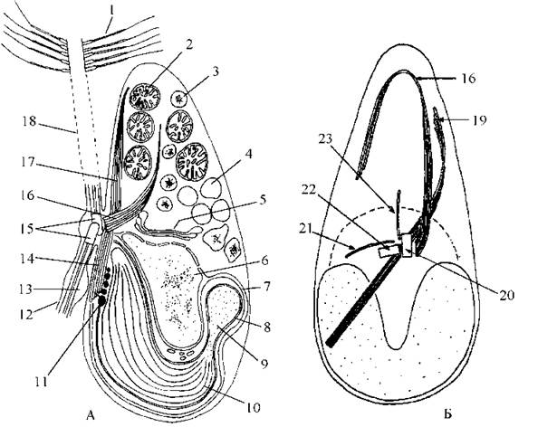
Fig. 16.2. Structure of a brown algal zoospore: A - General structural plan (lateral view); B - flagellar root system (ventral view). 1 - tripartite mastigoneme; 2 - mitochondrial profile; 3 - chrysolaminarin vacuole; 4 - cell sap vacuole; 5 - Golgi apparatus; 6 - nucleus; 7 - chloroplast endoplasmic reticulum; 8 - double-membrane chloroplast envelope; 9 - pyrenoid; 10 - three-thylakoid lamella; 11 - eyespot globule; 12 - short flagellum; 13 - parabasal body; 14 - lower part of the bidirectional microtubular root; 15 - basal bodies; 16 - anterior microtubular root; 17 - upper part of the bidirectional root; 18 - long flagellum; 19 - bidirectional root; 20, 21 - basal bodies of the long and short flagella, respectively; 22, 23 - third and fourth microtubular roots (after Manton, 1957; O'Kelly, Floyd, 1984).
Types of Morphological Structures and Thallus Organization
The division encompasses filamentous, heterotrichous, and true parenchymatous body types. All brown algae, with the exception of certain species of the genus Sargassum, lead an attached lifestyle. The Organs of attachment are rhizoids or disc-like holdfasts.
The thalli of filamentous representatives are predominantly formed by a system of uniseriate, branched filaments (e.g., the genus Ectocarpus). Heterotrichous algae mostly appear as relatively thick cords. Such cords exhibit either uniaxial or multiaxial construction (Fig. 16.3).
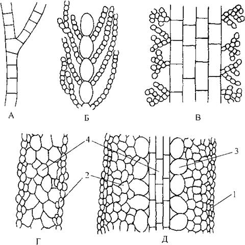
Fig. 16.3. Schematic diagram of brown algal thallus structures. A - uniaxial uniseriate thallus; B - uniaxial multiseriate; C - multiseriate; D - "simple" parenchymatous; E - "complex" parenchymatous. 1 - meristoderm, 2 - cortex, 3 - intermediate tissue, 4 — medulla.
A diverse thallus shape is characteristic of algae with parenchymatous organization. In the simplest case, such a thallus consists of only two tissue types: the cortex and the medulla. The cortex is formed by small pigmented cells that perform the photosynthetic function and give rise to reproductive organs. The medulla consists of large, predominantly colorless cells that serve conducting and storage functions.
In more complex cases, the thallus is formed by a greater number of Tissues: the meristoderm, cortex, intermediate tissue, and medulla. The meristoderm is a surface tissue that performs protective and reproductive functions. Meristoderm cells are usually small and capable of active division. The cortex is formed by larger cells with well-developed chloroplasts. The primary function of the cortex is Photosynthesis. Assimilation products accumulate in the Cells of the intermediate tissue. These cells themselves are large, typically with reduced chloroplasts. The medulla is formed by sieve tubes and tubular filaments.
The sieve tubes are elongated, with their transverse walls bearing pores penetrated by relatively thick plasmodesmata. The Cytoplasm contains a well-developed Golgi apparatus, Mitochondria, and endoplasmic reticulum. Interestingly, in some representatives, the sieve tubes possess a large lobed nucleus with a prominent nucleolus (Laminaria), whereas in others the nucleus is absent (Macrocystis). Sieve tubes perform a conducting function; special studies have established that mannitol and Amino Acids are transported through them at a rate of 0.1–0.6 meters per hour.
Often, physodes—cells filled with vesicles containing Tannins (phloroglucinol and polyphenols)—and mucilage canals with secretory cells are found within the medulla and intermediate tissue.
The growth of brown algal thalli occurs through several pathways: via Cell Division in the middle part of the thallus (intercalary growth), groups of apical cells (apical meristem), a single large apical cell, or trichothallically—through the division of a group of cells located subapically between the apical hairs and the main body of the "simple" parenchymatous thallus. The type of thallus growth is one of the diagnostic taxonomic features at the order level.
Reproduction and Life Cycles
Brown algae reproduce both asexually and sexually. Asexual reproduction can occur via thallus fragmentation, propagules, zoospores, and tetraspores.
Thallus fragmentation usually takes place during severe storms. However, in the vast majority of cases, the fragments are unable to attach to the substrate and, in a free-floating state, fail to develop into a normal new individual. Such fragments are typically washed ashore during storms and perish. Nevertheless, in some species, the cells of thallus fragments are capable of transforming into zoosporangia within a short period. Thus, fragmentation indirectly plays a role in the dispersal function.
In species of the genus Sphacellaria, Vegetative Reproduction is carried out using specialized propagules capable of attaching to the substrate and developing into a normally reproductively mature individual (Fig. 16.4).

Fig. 16.4. Organs of asexual reproduction in brown algae. 1 - unilocular sporangium; 2, 3 - successive stages of multilocular sporangium development; 4 - mature multilocular sporangium; 5, 6 - primordial (5) and mature (6) propagules in Sphacellaria (after Zinova, 1967; Petrov, 1977).
Asexual reproduction by means of zoospores is characteristic of the overwhelming majority of brown algae, with the exception of the Dictyotales and Fucales. Brown algal sporangia can be unilocular or multilocular. A unilocular sporangium represents an enlarged cell whose contents, following a series of nuclear divisions, break down into A large number of zoospores. The first nuclear division is typically reductional, and therefore the zoospores formed within unilocular sporangia possess a haploid chromosome number. Multilocular sporangia develop from a large cell in which, prior to spore formation, the protoplast is divided by thin cell walls into numerous single-nucleated chambers (locules). A single zoospore is formed in each locule (see Fig. 16.4).
In the Dictyotales, zoospores are absent; instead, non-motile tetraspores are produced, whereas in the small group of Tilopteridiales, monospores are formed.
The sexual process occurs in the form of isogamy or oogamy, while heterogamy is very rarely encountered. The gametangia of brown algae are multilocular (except in representatives of Fucales). In filamentous forms, gametangia are formed on specialized lateral branches; in heterotrichous forms, from cells of the assimilatory filaments; and in parenchymatous forms, from the cortex or meristoderm.
In species with an oogamous type of sexual reproduction, the egg cell is surrounded by a large number of spermatozoa prior to copulation. They attach to the egg membrane via their anterior flagellum. Subsequently, the flagellar membrane of one of them fuses with the oolemma, and the axoneme penetrates the protoplast, followed by the sperm nucleus. At this stage, the other spermatozoa detach and leave the egg cell. Following nuclear fusion, a zygote is formed, which grows into a new individual without a resting period.
In brown algae, The life cycle is haplo-diplontic with Morphology/12.html">ALTERNATION OF GENERATIONS (isomorphic or heteromorphic) or diplontic without alternation of generations.
In the haplo-diplontic cycle, Meiosis occurs in unilocular sporangia on the diploid sporophyte, resulting in the formation of haploid zoospores. These germinate into haploid gametophytes. Following copulation, the zygote develops directly into a new sporophyte without a resting period. If the alternation of generations is heteromorphic, the gametophytes are typically microscopic, whereas the sporophytes are macroscopic.
The alternation of generations can be regular or irregular. In regular alternation (e.g., in Laminaria), the sporophyte always produces haploid spores that germinate exclusively into gametophytes. The latter, in turn, produce gametes that are incapable of parthenogenetic development.
During an irregular alternation of generations, the sporophyte can produce both unilocular and plurilocular sporangia, yielding haploid or diploid zoospores, respectively. Haploid zoospores develop into haploid gametophytes, whereas diploid ones give rise to diploid sporophytes. The gametophyte produces gametes that either undergo copulation (with the resulting zygote developing into a sporophyte) or germinate parthenogenetically into a new gametophyte. Consequently, the sporophyte and gametophyte do not succeed each other in a regular pattern, making the alternation of generations irregular.
Taxa with a diplontic life cycle lack zoospores; meiosis occurs during gametogenesis, and the sexual process is predominantly oogamous. Oogonia and antheridia develop on specialized fertile branchlets (receptacles) housed within semi-enclosed cavities known as conceptacles. Spermatozoa produced within the antheridia of male conceptacles are subsequently released externally. In female conceptacles, the walls of the oogonia rupture, releasing the egg cells into the conceptacle cavity, where they secrete attractants that lure the spermatozoa. Following Fertilization, the zygote germinates without a resting stage, remaining within the conceptacle.
Systematics of the Division
Today, two groups of Classification systems exist for Phaeophyta: classical systems based on reproductive traits, and a synthetic system. The latter was developed using molecular data by analyzing The nucleotide sequences of various genes (including those encoding the small and large subunits of ribosomal RNA, RuBisCo, tubulin elongation factors, and Actin), and is reconciled with a complex of phenotypic traits. Importantly, the synthetic system complies with the requirements of the International Code of Botanical Nomenclature.
Classical Systems
In classical systems, the division is most commonly divided into two classes: Phaeophyceae (=Phaeozoosporosphyceae) and Cyclosporophyceae. This division is based on the type of life cycle and The ability to reproduce via spores.
Phaeophyceae (=Phaeozoosporosphyceae) comprises algae with a haplo-diplontic life cycle capable of asexual reproduction via zoospores or tetraspores. Depending on the type of alternation of generations (iso- or heteromorphic, regular or irregular), the type of sexual process, the thallus architecture, and the pattern of growth, various authors distinguish between 11 and 15 orders within the class, among which Ectocarpales, Dictyosiphonales, Cutleriales, Sphacellariales, Dictyotales, and Laminariales are considered predominant. Representative genera include Ectocarpus, Dictyosiphon, Cutleria, Sphacellaria, Dictyota, Laminaria, and Macrocystis.
Cyclosporophyceae includes algae with a diplontic life cycle that are incapable of producing spores. The class is divided into three orders: Ascoseirales, Durvilleales, and Fucales. Representative genera are Ascoseira, Durvillea, Fucus, Ascophyllum, Cystoseira, and Sargassum.
The Synthetic System
This system was formulated through collaborative research between systematists and molecular biologists, taking its definitive shape only in the final decades of the 20th century. According to this system, the division Phaeophyta comprises a single class, Phaeophyceae, and eight orders, seven of which are discussed below1.
Molecular phylogenetic clades corresponding to the ordinal level correlate with such phenotypic characteristics as the presence and STRUCTURE OF THE pyrenoid, reproductive features, the presence of zoospores, the type of sexual process, and the life cycle. The primary phenotypic traits of the orders are summarized in Table 16.1.
Ectocarpales comprises algae that, unlike other brown algae, possess a well-developed protruding-type pyrenoid. The thalli of ectocarpalean algae can exhibit filamentous, heterotrichous, or "simple" parenchymatous structure. The life cycle is diplohaplo-diplontic with an irregular alternation of generations, where the sporophytes are macro- and the gametophytes microscopic in cases of heteromorphic alternation. Molecular data indicate that Ectocarpales is the most primitive order of brown algae.
Table 16.1. Criteria for classifying brown algae into orders based on phenotypic characters
Character → Order ↓ |
Pyrenoid |
Chloroplast |
Life cycle |
Sexual process |
Asexual reproduction |
Thallus type |
Thallus growth |
Ectocarpales |
P |
Par. |
n-2n |
I, H |
zoospores |
F, H, SPT |
IG, AM |
Scytothamnales |
P |
Cent. |
n-2n |
? |
zoospores |
H |
IG, AM |
Cutleriales |
R |
Par. |
n-2n |
H |
zoospores |
SPT |
T |
Dictyothales |
R |
Par. |
n-2n |
O |
tetraspores |
SPT |
AC |
Laminariales |
R |
Par. |
n-2n |
O |
zoospores |
CPT |
IG |
Sphacellariales |
R |
Par. |
n-2n |
I, H, O |
zoospores |
H |
AC |
Fucales |
R, A |
Par. |
2n |
H, O |
absent |
CPT |
IG, AC, AM |
Abbreviations: pyrenoid: P - protruding and developed, R - rudimentary, A - absent; chloroplast: Par. - numerous parietal discoid, Cent. - solitary central lobed; life cycle: n-2n - haplo-diplontic, 2n - diplontic; sexual process: I - isogamy, H - heterogamy, O - oogamy, ? - not investigated; thalli: F - filamentous, H - heterotrichous, SPT - "simple" parenchymatous, CPT - "complex" parenchymatous; thallus growth: IG - intercalary, AM - apical meristem, T - trichothallic, AC - apical cell
An example of an alga with a highly simplified body plan is Ectocarpus (Fig. 16.5). Its sporophytes and gametophytes are morphologically indistinguishable and appear as small tufts formed of branched uniseriate filaments attached to underwater substrates by rhizoids. The ascending filaments frequently terminate in multicellular colorless hairs. Any vegetative cell of the upward-growing filament is capable of division; consequently, the upward growth of the thallus occurs intercalarily. Conversely, creeping filaments grow through the division of apical cells.
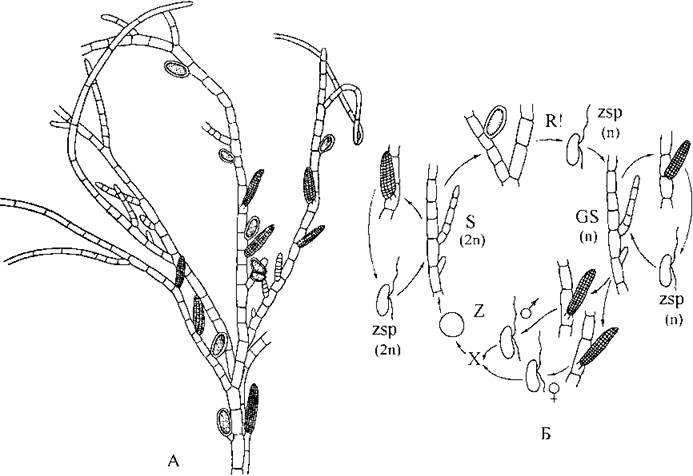
Fig. 16.5. Ectocarpus: A - sporophyte with unilocular and plurilocular zoosporangia; B - life cycle (A - Zenova, 1967; B - schematized after Moller, 1972).
The vegetative cells contain parietal ribbon-shaped chloroplasts with protruding pyrenoids typical of the order (see Fig. 16.1).
The sporophytes are diploid and produce Two Types of zoosporangia—unilocular and plurilocular—which develop at the tips of short lateral branchlets. Zoospores formed in plurilocular sporangia are always diploid and give rise to new sporophytes. Zoospores maturing in unilocular sporangia can be either diploid or haploid. Haploid spores subsequently develop into haploid gametophytes.
The gametophytes are bisexual and monoecious, bearing plurilocular gametangia that produce isogametes of both mating types. The gametes are morphologically nearly identical; however, upon release from the gametangium, the gametes acting as female ("+" gametes) secrete the pheromone ectocarpene. Male ("-" gametes) navigate along an increasing concentration gradient of ectocarpene, with a submicroscopic folded apical proboscis serving chemoreceptor functions. Following copulation, a diploid zygote is formed, which immediately begins to germinate into a new sporophyte without a resting stage.
Species of the genus Ectocarpus inhabit the littoral zone of seas across all latitudes, including the Black and Azov Seas.
A representative species with a heteromorphic alternation of generations is Dictyosiphon. The sporophytes of this alga form tufts composed of long (up to 20 cm) branched tubes, the walls of which consist of two to three outer layers of small cortical cells and an inner layer of large medullary cells. Thallus growth is apical, driven by the division of small cells within the apical meristem (Fig. 16.6: A). Unilocular zoosporangia differentiate from the medullary cells. The alternation of generations is irregular and heteromorphic. The gametophyte is microscopic, and its gametes can parthenogenetically germinate into new haploid gametophytes. Species of this genus occur in marine benthic communities of cold and temperate zones, typically attaching to rocks and mollusk shells.
Scytothamnales groups together algae that differ from all others by possessing a central lobed stellate chloroplast with a central pyrenoid. This order was established only in the late 1990s, initially based on molecular data. All scytothamnalean algae inhabit the seas of the Southern Hemisphere. A representative species is Stereocladon (Fig. 16.6: B).

Fig. 16.6. Ectocarpalean and scytosiphonalean algae. A - Dictyosiphon: 1 - general appearance of the sporophyte; 2 - Water/140.html">Anatomical Structure of the thallus; 3 - branch apex with small cells of the apical meristem. B - Stereocladon: 4 - general appearance of the thallus; 5 - cross-section through a cell (a - central lobed chloroplast, b - pyrenoid) (A - after Sauvageau, 1927; Newton, 1931; B - after Peters, Clayton, 1998).
The Cutleriales comprise marine algae in which, unlike other brown algae, the gametophytes are macroscopic, while the sporophytes are predominantly microscopic. The thalli terminate in tufts of hairs at their apices. A layer of meristematic cells is situated at the base of these hairs. The latter are capable of division and cut off segments oriented either outward or inward relative to the thallus. The outer segments drive the longitudinal growth of the hairs, whereas the inner segments differentiate into the Tissues of the main thallus body. This type of thallus growth is termed trichothallic. Representatives of this order exhibit a "simple" tissue organization, with both the sporophyte and gametophyte consistently laminar in shape. The Sexual process in cutlerialean algae is heterogamous.
In the typical representative—Cutleria—the gametophyte appears as a large, fan-shaped, upright blade that transitions at the base into a short stalk with a holdfast (Fig. 16.7). The sporophyte forms a small, microscopic blade spread across the substrate, which was long considered an independent genus, Aglaozonia. The Aglaozonia stage reproduces via haploid zoospores produced within unilocular sporangia arranged in sori. These zoospores germinate into new gametophytes.

Fig. 16.7. Cutleria: 1 - macroscopic gametophyte; 2, 3 - branch fragment with male (2) and female (3) gametangia; 4 - microscopic sporophyte (Aglaozonia stage); 5 - cross-section through the sporophyte, bearing a sorus of unilocular sporangia on its surface (after Newton, 1931; Petrov, 1977).
The Dictyotales, similar to the previous order, comprise algae with "simple" tissue organization in their thalli, which appear as intact, dichotomously, or flabellately branched blades. Alternation of generations is isomorphic. Thallus growth proceeds via the division of a single apical cell. The sexual process is oogamous. Typically, sexual organs are aggregated into groups enclosed by a common envelope, forming sori. A characteristic diagnostic feature of dictyotalean algae is the absence of zoospores; asexual reproduction occurs via non-motile tetraspores. Motile stages are represented by spermatozoa bearing a single flagellum. The most widespread genera are Dictyota, Dilophus, and Padina (Fig. 15.8).
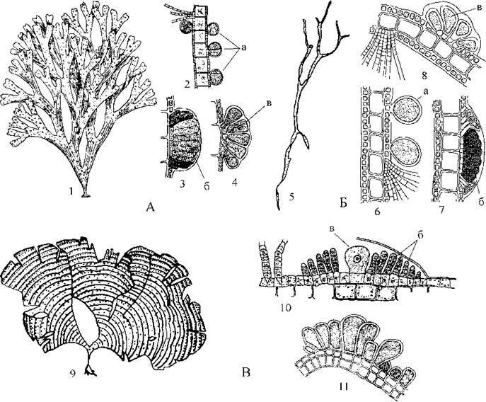
Fig. 16.8. Dictyotalean algae. A - Dictyota: 1 - general appearance of the thallus; 2-4 - cross-sections of the thallus with tetrasporangia (2), antheridial sori (3), and oogonia (4). B - Dilophus: 5 - general appearance of the thallus; 6-8 - cross-sections of the thallus with tetrasporangia (6), antheridial sori (7), and oogonia (8). C - Padina: 9 - general appearance of the thallus; 10 - cross-section of the thallus with an oogonium and antheridia; 11 - section through a tetrasporangial sorus (a - tetrasporangia; b - antheridia; c - oogonium) (after Zinova, 1967).
In Dictyota and Dilophus, the thallus blades are dichotomously branched; they are broad in the former genus, whereas they are quite narrow in the latter. The thalli of Padina appear as entire or margin-torn blades. Concentric bands formed by clusters of hairs are clearly visible On the surface of the padina thallus, and a layer of calcium carbonate is typically deposited on the lower side of the blade.
The Laminariales bring together algae whose sporophytes resemble unbranched cords or large blades with complex tissue organization. As a rule, conducting elements are present in the medulla. Thallus growth is intercalary. Alternation of generations is heteromorphic and regular, and the sexual process is oogamy. Gametophytes are microscopic, typically taking the form of short, uniseriate, branched filaments.
Laminarian algae are of considerable practical importance, as they comprise the vast majority of brown algae utilized by humans.
The best-known representative of the order is Laminaria, or kelp. In northern seas, mass species include L. saccharina and L. digitata; along the Atlantic coast of Europe, L. hyperborea; and in the Far East, L. japonica (Fig. 16.9). In the sublittoral zone of marine waters, these algae form dense beds sometimes referred to as "kelp forests."
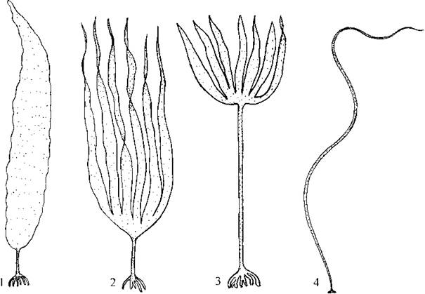
Fig. 16.9. Laminarian algae: 1 - Laminaria saccharina; 2 - L. digitata; 3 - L. hyperborea; 4 - Chorda filum (orig.).
Adult sporophytes of Laminaria reach 3–4 m in length and consist of a blade, a "stipe," and a branched holdfast system of rhizoids that anchors the alga to underwater rocky substrates. Meristematic cells are located at the transition zone between the blade and the "stipe," and their division drives the intercalary growth of the thallus.
The laminarian meristem is active during the cold seasons, with sporophyte growth occurring from January to April–May in temperate latitudes, and from February to June–August in high latitudes. Meristematic cell division is induced by short day length, a phenomenon known as the photoperiodic response. Toward the thallus apex, the intercalary meristem cuts off cells that subsequently differentiate into blade tissues (Fig. 16.10), while in the opposite direction, it produces stipe tissue cells. At the end of the growing season, the blade typically dies off and regrows the following year, whereas the "stipe" and rhizoids are perennial.

Fig. 16.10. Laminaria: A - cross-section of the blade (1 - meristoderm, 2 - cortex, 3 - storage/intermediate tissue, 4 - medulla); B - zoosporangial sorus on the meristoderm surface (5 - zoosporangium, 6 - protective elongated cells, i.e., paraphyses); C - life cycle diagram (after Petrov, 1977).
The zoosporangia of Laminaria are unilocular and develop in autumn in dense groups (sori) derived from the cortical cells of the stipe. The sori are darker in color compared to the sterile portion of the blade. Following meiotic division, each sporangium produces 32 biflagellate zoospores. Enclosed in a common mucilaginous envelope composed primarily of alginic acid, the zoospores are forcefully expelled from the blade into the external environment. The mucilaginous envelope then dissolves, and the zoospores become actively motile. Within a few minutes, they settle on a substrate and shed their flagella. Subsequently, half of the zoospores from a given sporangium develop into male gametophytes, and the other half into female gametophytes.
Male gametophytes consist of several dozen cells forming a short, profusely branched filament. The terminal cells of the lateral branchlets transform into antheridia, each producing a single spermatozoid.
Female gametophytes consist of only a few cells. Any cell of the female gametophyte can develop into an oogonium, which produces a single egg cell. Upon rupture of the oogonial wall, the egg is released to the exterior and secretes the pheromone lamoxirene to attract spermatozoa. Following copulation, a zygote is formed, which immediately begins to germinate into a new sporophyte without a resting period.
Along the coasts of the Americas, close relatives of Laminaria belonging to the genus Macrocystis act as dominants in many marine phytocoenoses. The thalli of this alga reach lengths of up to 60 m. A long, slender stipe rises at an angle from the seafloor to the surface, bearing a double row of long (up to 1–2 m) "leaf-like" blades. A pneumatocyst (float) is located at the base of each blade.
A significant proportion of laminarian algae are restricted to cold seas. For instance, the genus Chorda—a dominant of marine phytocoenoses at the lower boundary of the littoral zone—is found exclusively where the winter isotherm does not exceed +12 °C.
The Sphacellariales comprise algae with heterotrichous, multiaxial thalli that exhibit an articulate-whorled structure and appear as small, branched tufts. Alternation of generations in sphacelarialean algae is isomorphic, and the sexual process is predominantly isogamous, or more rarely hetero- or oogamous. Similar to the Dictyotales, the thalli grow via the division of an apical cell.
A typical representative, Sphacellaria (Fig. 16.11; see also Fig. 16.4), frequently occurs as an epiphyte on other macroalgal macrophytes in both cold and warm seas. The thalli of Sphacellaria are small (averaging about 1 cm in height) and differentiated into a system of upright, ascending filaments and a prostrate holdfast, sometimes accompanied by densely interwoven rhizoids. A large apical cell sits at the apex of each ascending filament, driving thallus growth through its divisions. The holdfast is capable of producing stolons, at the tips of which new holdfasts can develop, which in turn sprout new ascending filaments.

Fig. 16.11. Sphacelariales. A – apex of the thallus of Sphacellaria. B – Cladostephus: 1 – general view of the thallus; 2 – fragment of the thallus with remnants of whorled branchlets; 3 – cross-section of the thallus (after Petrov, 1977; Zinova, 1967).
The apical cell divides exclusively in the transverse direction, cutting off subapical cells. The latter are capable of longitudinal division, resulting in the formation of whorled siphons (siphonal whorls). The vast majority of cells within the whorls are incapable of subsequent divisions; however, individual cells can give rise to cells functionally similar to apical ones—namely, lateral meristematic cells. The division of these cells produces reproductive structures: gemmae (brood buds) or short lateral branches bearing unilocular sporangia (on sporophytes) or plurilocular female/male gametangia (on gametophytes). The sexual process is heterogamous, and the zygote germinates into a new sporophyte without a resting period. Gametes are also capable of parthenogenetic germination into new gametophytes, which is why the alternation of generations is considered non-established (unstable).
Another representative, Cladostephus, occurs in the benthos at shallow depths (up to 2–7 m). This alga is frequently found in storm cast-ups along the Black Sea coast. Its thalli are quite large (up to 20 cm in height), dichotomously branched, exhibit an articulate-whorled structure, and their apices are densely covered with numerous short branchlets, giving them a club-shaped appearance (Fig. 16.11).
Fucales constitute the evolutionarily youngest order of brown algae. Representatives of this order possess a complex tissue structure, predominantly oogamous (exceptionally heterogamous) sexual reproduction, and a diplontic life cycle lacking alternation of generations. In fucalean algae, sexual organs develop within specialized cavities of the cortex and medulla, known as scaphidia or conceptacles. Asexual reproduction via spores is absent in members of this order. Fucales are widespread in both cold and warm seas and typically act as dominants in littoral phytocenoses.
In cold northern seas (e.g., the White, Barents, and Okhotsk seas), the seabed exposed during low tides is often covered by a continuous carpet of Fucus and Ascophyllum thalli. Fucus thalli are large (30–70 cm high), consisting of a disc-shaped holdfast, a cylindrical stipe, and dichotomously branched blades that widen toward the apex. The stipe transitions into a central midrib within the blades. Photosynthesis is carried out primarily by the blade cells, while the stipe and midrib perform supportive and conducting functions. Near the apex, most species of the genus bear prominent, air-filled swellings known as air bladders, which keep the thalli upright in the water.
Thallus growth occurs through the division of an apical cell that cuts off cells of the subapical meristem, which subsequently give rise to all other tissues.
Sex organs develop on specialized fertile branches called receptacles. Compared to the sterile PARTS OF THE blades, these branches are thickened and have a rough surface. This roughness is due to the presence of large cavities (up to 1–3 mm in diameter) within the receptacles, called scaphidia, inside which male and female sex organs develop.
The scaphidia are filled with mucilage, and their inner surface is lined with a layer of envelope cells that give rise to short sterile filaments as well as initial reproductive cells, which form antheridia and oogonia. The scaphidia in Fucus may be bisexual or unisexual.

Fig. 16.12. Fucus. 1 – general appearance of the thallus (a – central midrib, b – air bladder, c – receptacle with scaphidia); 2 – female scaphidium with oogonia; 3 – oogonium containing eight oocytes (eggs); 4 – release of eggs from the oogonium; 5 – male scaphidium with antheridial filaments; 6 – antheridial filament with antheridia; 7 – germling; 8 – apex of the thallus (d – apical cell) (1 – orig.; 2–8 – after Petrov, 1977).
When an initial reproductive cell begins to develop into a sex organ, its nucleus undergoes meiotic division. Subsequently, the haploid cells divide mitotically and differentiate into either oogonia or antheridia. The oogonia are ellipsoidal, possessing a short unicellular stalk and eight eggs. Antheridia are formed from the apical cells of short, densely branched antheridial filaments, which represent modified paraphyses. Each antheridium produces 64 biflagellate spermatozoa.
Prior to fertilization, antheridia and oogonia are typically pushed out of the scaphidium into the external environment through an opening via mucilage pressure. Thereafter, the walls of the antheridia and oogonia dissolve, releasing the gametes. The eggs secrete the pheromone fucoserratene, which rapidly concentrates a large number of spermatozoa around each egg. Following fertilization, the zygote begins to germinate into a new thallus without a resting period.
The developmental process of antheridia and oogonia in Fucales is interpreted differently by various authors. According to classical views (Kylin, 1940; Fritsch, 1945), the sex organs in these algae are formed As a result of standard gametic reduction of specialized thallus cells, making the life cycle diplontic and devoid of an alternation of generations. Conversely, other researchers (Strasburger, 1906; van den Hoek, Mann, Jahns, 1995) suggest that the initial cells giving rise to the sex organs are homologues of unilocular sporangia, while the oogonia and antheridial filaments themselves represent microscopic haploid gametophytes. Therefore, the Life Cycle of Fucales is haplo-diplontic, featuring sporic reduction and regular heteromorphic alternation of generations. An interesting hypothesis was proposed by Yu. E. Petrov (1986), who argued that the entire layer of lining cells in the scaphidia represents a modified diploid gametophyte, and consequently, the life cycle should be regarded as diplontic, with gametic reduction and heteromorphic alternation of generations2.
Ascophyllum thalli are strap-shaped, do not widen toward the apex, and receptacles are formed not apically, but on lateral branches.
In the Black Sea, at depths of 1–4 m, marine "meadows" are formed by species of the genus Cystoseira (primarily C. barbata and C. crinita). They constitute the foundational producer link in benthic littoral phytocenoses. Cystoseira thalli resemble small bushes with more or less cylindrical, densely branched ascending shoots and short stems. In winter and autumn, chains of small air bladders develop at the branch apices. Small receptacles are also located apically.
In the littoral zone of the Atlantic and Pacific coasts, the dominant alga is Sargassum. Certain species of this genus inhabiting open Atlantic waters, unlike other brown algae, are capable of reproducing in an unattached state, forming the so-called Sargasso Sea. Sargassum thalli are bushy and differentiated into a holdfast, a stem, and main branches. Receptacles and air bladders in Sargassum develop on short branchlets in the axils of the main branches.
The primary coenose-forming Organism of the moderately cold seas of the Southern Hemisphere is Durvillea, which develops massively along the coasts of South America, Australia, and Antarctic islands. The thalli of this alga are very large (up to 10 m in height) and consist of a holdfast, a short stem, and thick tubular branches. Growth occurs through both apical and intercalary Meristems. Scaphidia are formed from several meristoderm cells of the branches that penetrate into the medulla and become enveloped by meristoderm strands originating from neighboring cells.

Fig. 16.13. Fucalean algae: 1, 2 – Ascophyllum (1 – thallus, 2 – fragment of the thallus with receptacles and an air bladder); 3, 4 – Cystoseira barbata (3 – fragment of the thallus; 4 – apex of the thallus with scaphidia); 5 – Cystoseira crinita (apex of the thallus with scaphidia); 6 – Durvillea (after Zinova, 1953, 1967; Petrov, 1977; Fritsch, 1935).
Pheromones of Brown Algae
Substances that mediate the chemotactic movement of spermatozoa toward eggs are termed pheromones. In 1963, D. Müller, while studying the reproduction of Ectocarpus siliculosus, noticed that a suspension of female gametes possessed a pleasant, sweetish odor, unlike male gametes. Oil droplets saturated with the vapor of this substance attracted male gametes. To isolate the pheromone in pure form, an apparatus was constructed to circulate clean air over the female gamete suspension, followed by the Condensation of the volatile fragrant fraction. Over the course of two years, about 100 mg of the substance under study was obtained. Chemical analysis revealed that the substance is a phenolic compound of hydrocarbon nature, which was named ectocarpene.
Subsequently, analogous studies were conducted with cultures of several species of the genus Fucus, one species of Sargassum, and the fucalean alga Pelvetia. The pheromone determining sperm chemotaxis in these species turned out to be a linear hydrocarbon named fucoserratene. Interestingly, in the presence of fucoserratene, spermatozoa did not distinguish between eggs of their "own" species and those of a "foreign" species, concentrating around any egg that secreted this pheromone or was treated with it. Recognition of the "own" egg occurred only at the Initial Stages of copulation—"foreign" spermatozoa detached from the egg, while one of the "own" successfully fused. Therefore, it was hypothesized that the egg membrane performs the recognition function in brown algae.
Today, approximately ten pheromones have been identified in brown algae, all of which are cyclic or linear hydrocarbon compounds. Some of them, notably lamoxirene isolated from Laminaria, have found application in the technological process of industrial cultivation of brown algae.
Practical Significance
Brown algae are widely used in human practical activities. Today, three main areas of their application have generally emerged: The production of alginic acid and alginates; the cultivation, harvesting, and Processing of edible species; and The Use of biomass as organic fertilizers rich in potassium and nitrogen. The primary objects of practical use are species of the genera Laminaria, Macrocystis, Fucus, Sargassum, and Cystoseira.
Alginates form The basis of the cell walls in Phaeophyta. They possess adhesive and gelling properties and are therefore used in the production of varnishes and paints, in the manufacture of textiles with specific textures, and as preservatives in the food industry. Alginates (particularly sodium alginate) also serve as a pharmacological raw material for the production of radioprotective drugs.
Edible algae are either harvested extensively or artificially cultivated on marine farms via aquaculture. The main objects of aquaculture are Laminaria saccharina, L. japonica, L. digitata, and species of the genus Macrocystis. Products made from these algae are collectively known as "kelp" or "sea cabbage". For instance, the annual harvest of Laminaria in the 1990s was around 2 million tons. Intensive breeding work is carried out on kelp, including the application of Introduction/32.html">Genetic Engineering Methods and numerous biotechnological experiments.
As organic fertilizers rich in nutrients and Trace Elements, Fucus, Cystoseira, Sargassum, and Ascophyllum are widely used. These algae are gathered along sea coasts after severe storms or in the intertidal zone during low tide.
Taxonomic Position
Brown algae belong to photoautotrophic tubulicristate stramenopiles. Based on molecular data and a complex of phenotypic traits, the division is closely related to yellow-green algae.
1 The System of the division is given according to: L.D. Druehl, C. Mayes, I.H. Tan, G.W. Saunders. Molecular and morphological phylogenesis of kelp and associated brown algae. In: Origin of Algae and their Plastids (ed. D. Bhattacharya). Springer-Verl., Wien - New York, 1997: 221-236; A. Peters, M. Clayton. Molecular and morphological investigation of three brown algal genera with stellate plastids: evidence for Scytothamnales ord. nov. (Phaeophyceae). - Phycologia (1998), 37: 106-113; F. Rousseau, B. de Reviers. Circumscription of the order Ectocarpales (Phaeophyceae): bibliographical synthesis and molecular evidence. - Cryptogamie, Algol. (1999), 20 (1): 5-18; F. Rousseau, B. de Reviers. Phylogenetic relationships within the Fucales (Phaeophyceae) based on combined partial SSU+LSU rDNA sequence data. - Eur. J. Phycol. (1999), 34: 53-64. The order Ascoseirales is not considered (see: Life of Plants, Vol. 3. Algae, Lichens. - Moscow, Prosveshchenie, 1977), representatives of which are few in number and inhabit the seas of the Southern Hemisphere.
2 The consideration of arguments "for" and "against" each of these hypotheses can be the topic of a separate seminar class. For more details, see: Petrov Yu.E. Evolution of life cycles in algae. (Komarov Readings, XXXV). - Leningrad, Nauka, 1986. - 61 p.
Last update: 07/08/2026
Editorial and Educational Adaptation: This material has been compiled based on the primary/original source text. The project team performed an editorial review, corrected technical inaccuracies, structured sections, and adapted the content for an educational format.
What was processed:
- elimination of formatting defects (OCR errors, structural breaks, corrupted characters);
- editorial organization of content;
- standardization of terminology in accordance with academic sources;
- verification of factual statements against the original source text.
All mentions of the author, publication year, and origin of the primary text have been preserved in accordance with the source.