Phycology - Kostikov I.Yu. - 2009-2013
Chapter 15. Yellow-Green Algae – Xanthophyta
The division comprises about 600 species of unicellular, multicellular, and non-cellular siphoneous Algae inhabiting fresh continental waters, soils, and aerophytic habitats. Reliable fossil remains are known from Mesozoic Triassic deposits (approximately 240 million years ago), with probable records from the Carboniferous period (300–340 million years ago). Thus, the evolutionary emergence of yellow-green algae roughly coincides with the appearance of gymnosperms. The majority of yellow-green algae are microscopic organisms distributed almost exclusively in freshwaters and soils.
Yellow-green algae belong to tubular-cristate organisms within the stramenopile phylum, and similarly to other photoautotrophic stramenopiles, possess Plastids represented by secondarily symbiotic rhodoplasts.
Morphologically, yellow-green algae strongly resemble Representatives of the green algae division. Due to Cell walls based on pectic substances and the absence of the brown pigment fucoxanthin, yellow-green algae are similar to eustigmatophytes, with which they were grouped until 1970. However, Xanthophyta differ from Eustigmatophyta in a different structural plan of the photoreceptor apparatus in monadoid Cells, the predominance of diatoxanthin-series xanthophylls over lutein-series xanthophylls, and the presence in zoospores of a connection between The Nucleus and the chloroplast Endoplasmic reticulum.
At THE MOLECULAR LEVEL, yellow-green algae proved to be more closely related to brown algae than to eustigmatophytes.
Class="center">Taxonomic CHARACTERISTICS OF THE Division
Pigments and Reserve Nutrients
The Biochemical characteristics of the division include the presence of chlorophylls a and c, and β-carotene. The dominant carotenoids are diatoxanthin-series xanthophylls, namely diatoxanthin and diadinoxanthin. Specific xanthophylls such as heteroxanthin and vaucheriaxanthin, as well as lutein-series xanthophylls (notably neoxanthin), have been detected in significantly smaller amounts in yellow-green algae. The METABOLISM/14.html">Chloroplasts of yellow-green algae are colored yellowish-green without brown tints, as the dark yellow pigment fucoxanthin, typical of many stramenopile algae, is absent.
The assimilation product is chrysolaminarin, which is always deposited extraplastidically; therefore, under optical Microscopy, the chloroplasts of yellow-green algae appear smooth and silky, without signs of granularity. Oil may accumulate in cells during Aging.
Cytological Characteristics
Cell coverings in yellow-green algae are represented by a pectic or cellulosic-pectic Cell wall, which in some representatives is impregnated with iron or manganese salts. The Cell wall can be continuous or consist of two halves overlapping each other.
In species of the genus Vaucheria, the cell wall contains Cellulose microfibrils In addition to pectin. The monadoid cells of yellow-green algae are covered only by the Plasmalemma.
Nuclear apparatus is typically eukaryotic. A distinctive feature of the nucleus is that the outer membrane of the nuclear envelope transitions into the outer membrane of the chloroplast endoplasmic reticulum, thus forming a single structural complex between the nucleus and the chloroplast in representatives of Xanthophyta (Fig. 15.1). In the space between the outer and inner membranes, microtubular parts of retronemes—three-part flagellar mastigonemes—can be formed.

Fig. 15.1. Structure of a yellow-green algal cell (using Heterococcus as an example): CW – cell wall; Chl – chloroplast, in which the outer membrane of the chloroplast endoplasmic reticulum transitions into the outer membrane of the nuclear envelope; N – nucleus; M – mitochondrion with tubular cristae; C – centrioles; Vac – vacuole (photo by A. Massalsky).
Mitosis in yellow-green algae is closed, with centrioles serving as spindle pole bodies (Fig. 15.2). Typically, the Golgi complex is located near the centrioles at the poles of the dividing nucleus.

Fig. 15.2. Mitosis in the yellow-green alga Xanthonema. Metaphase. The intact nuclear envelope, chromosome plate, and centriole are visible (photo by A. Massalsky).
In zoospores, after the period of active motility ceases During the first nuclear division, the basal bodies of the flagella perform the Functions of centrioles.
Photosynthetic apparatus. The chloroplasts in yellow-green algae are represented by secondary endosymbiotic plastids of the rhodophyte type. The number of chloroplasts per cell in yellow-green algae varies widely—from one to ten in coccoid and filamentous forms to several thousand in siphoneous representatives. Chloroplasts predominantly occupy a parietal position.
The chloroplast envelope has four membranes, with the two outer membranes forming the chloroplast endoplasmic reticulum, which transitions into the nuclear envelope. A periplastidial space is present between the outer and inner chloroplast membranes (Fig. 15.3).

Fig. 15.3. Fragment of a protoplast in a young cell of the yellow-green alga Heterococcus. The chloroplast clearly shows three-thylakoid lamellae, a girdle lamella, a genophore (at the poles of the plastid), as well as the chloroplast endoplasmic reticulum with the periplastidial space, the nuclear envelope from which vesicles with rudimentary retronemes are being pinched off, a flagellar axoneme, Mitochondria with tubular cristae, and a vacuole (photo by A. Massalsky).
In the space between the membranes of the chloroplast endoplasmic reticulum, similarly to the perinuclear space of the nucleus, the tubular parts of retronemes are formed.
Thylakoids are grouped in threes into several lamellae and are usually accompanied by a girdle lamella. Such a lamella is present in the majority of representatives of the division, although some species are known in which the plastid lacks a girdle lamella (e.g., from the genus Bumilleria). Occasionally, the plastid contains a naked pyrenoid that is not penetrated by thylakoids.
In the zoospores of many yellow-green algae, a stigma is located within the chloroplast. It is formed by a single layer of osmiophilic globules pressed against the inner surface of the chloroplast envelope membrane. The stigma is situated near the parabasal body of the short flagellum.
The chloroplast DNA-genophore is compactly concentrated at the poles of the plastid and contains 100–200 kb of nucleotide Base Pairs.
Mitochondria are single or numerous; their profiles in cross-sections feature tubular cristae. Mitochondria are often closely associated with other double-membrane Organelles, namely chloroplasts or the nucleus.
Flagellar apparatus. Monad stages in yellow-green algae are represented by zoospores and Gametes, bearing two flagella of unequal length at the anterior end of the cell or slightly laterally. The longer flagellum is tinsel and covered with mastigonemes (retronemes). The formation of mastigonemes takes place in vesicles budding off from the nuclear membrane or the chloroplast endoplasmic reticulum. The shorter flagellum is smooth and typically acts as a rudder. At its base, the short flagellum features a thickening known as the parabasal body, which functions as a photoreceptor. The stigma in Xanthophyta is located within the chloroplast and acts as a screen that shades the photoreceptor from one side. The short flagellum may be reduced, yet its basal body is retained. The transition zone of the flagellum contains a helical structure.
Three microtubular roots depart from the basal bodies of the flagella, each consisting of three to four microtubules, along with a striated microfibrillar ROOT—the rhizoplast—which connects the basal bodies to the nucleus.
Deviations from the typical structural plan are exhibited by the zoospores of the siphonean alga Vaucheria. They are large and multinucleate; the entire surface of the zoospore is evenly covered with pairs of smooth flagella of nearly equal length. It is believed that such siphonean multi-flagellate zoospores result from the fusion of numerous young biflagellate zoospores within the sporangium. Such a combined zoospore is termed a synzoospore.
The vacuolar apparatus is represented by vacuoles containing cell sap, separated from the Cytoplasm by a membrane—the tonoplast. In monad stages—zoospores and gametes—there are typically two contractile vacuoles near the basal bodies of the flagella.
Types of morphological body structures
Most yellow-green algae are coccoid, filamentous, heterotrichous, or siphonean organisms. Additionally, several species with monad and hemimonad structures have been described within the division. However, it should be noted that upon special electron microscopic and molecular-taxonomic investigations of several such representatives, all of them turned out to be algae from other divisions—primarily Chrysophyta and Dictyochophyta. Thus, the existence of algae with monad and hemimonad morphological body types within the Xanthophyta is questioned today.
Reproduction and life cycles
Yellow-green algae, with the exception of a single genus, reproduce exclusively asexually: by Cell Division, fragmentation of thalli or colonies, aplanospores, zoospores, or by means of akinetes.
The sexual process is represented solely by oogamy and is reliably known only in species of the genus Vaucheria. In these algae, The life cycle is diplontic (diplophasic), with gametic reduction and without Morphology/12.html">ALTERNATION OF GENERATIONS.
System of the division
The division includes a single class—Xanthophyceae. According to molecular data, the evolution of yellow-green algae proceeded in the direction of a transition from siphonean forms with unlimited growth to siphonean forms with limited growth, and subsequently from multinucleate to uninucleate forms (coccoid, filamentous, and heterotrichous). Uninucleate yellow-green algae, in turn, differentiated according to cell wall structure into algae with continuous walls and algae with two-valved walls. These evolutionary trends are reflected in The system of orders of the class Xanthophyceae, which comprises four main orders: Vaucheriales, Botrydiales, Mischococcales, and Tribonematales (Fig. 15.4).
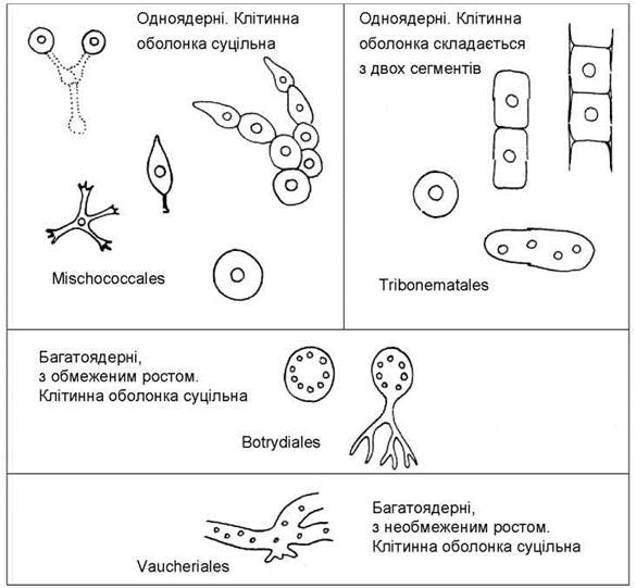
Fig. 15.4. System of Xanthophyta at the order level.
Order Vaucheriales unites species with a siphonean type of structure, possessing filamentous thalli with unlimited growth and continuous cellulosic-pectic walls (Fig. 15.5). Asexual reproduction occurs via synzoospores. All representatives of the order can reproduce sexually, with oogamy being the sexual process. Spermatozoa possess two flagella of nearly equal length, The surface of which lacks mastigonemes. The order contains a single genus, Vaucheria, which includes about 60 species. Species of this genus inhabit damp soil as well as brackish and fresh Water bodies.

Fig. 15.5. Vaucheria: 1 - general appearance of the thallus; 2 - formation and release of the synzoospore from the sporangium; 3 - fragment of the synzoospore; 4 - fragment of the thallus with sex Organs (left - antheridium, right - oogonium).
The thalli of Vaucheria appear as long, prostrate, branched tubes that lack transverse septa and are capable of unlimited growth. The tubes are attached to the substrate by means of short, branched rhizoids. In the central part of the tubes lies a vacuole with cell sap, while cytoplasm is located between the vacuole and the cell wall. Directly beneath the cell wall lies A large number of small, disc-shaped chloroplasts, and numerous small nuclei are situated between the chloroplasts and the vacuole.
During asexual reproduction, certain apical Regions of the thallus are separated by transverse septa and transform into zoosporangia. Each zoosporangium produces only a single naked synzoospore covered with pairs of nearly equal flagella. One nucleus is located beneath the basal bodies of each pair of flagella. Upon maturation of the synzoospore, the sporangial wall ruptures and the synzoospore is released; after a brief period of active movement, it settles on the substrate and grows into a new tubular siphonean thallus.
The sex organs—antheridia and oogonia—develop on the same thallus (in monoecious species) or on different thalli (in dioecious species) as lateral intercalary outgrowths which, like zoosporangia, are separated from the main part of the thallus by a transverse septum. Following septum formation, Meiosis takes place within the gametangia.
The oogonia of Vaucheria are ovoid; a single egg cell is formed within each oogonium. The antheridia have the shape of tubular hooks. A large number of spermatozoa are produced within each antheridium. Interestingly, unlike synzoospores, male gametes bear two lateral flagella of unequal length, with the longer flagellum carrying mastigonemes. Overall, the ultrastructure of Vaucheria spermatozoa closely resembles that of brown algae.
During the sexual process, spermatozoa emerge into the external environment through a rupture in the antherial wall and swim toward the oogonium. Subsequently, one of the spermatozoa penetrates the egg cell through a crack in the oogonial wall and fertilizes it. The zygote becomes enveloped in a thick wall and enters a resting stage. Upon the completion of the rest period, the zygote germinates into a new tubular siphonean thallus.
The order Botrydiales unites siphonous algae with limited growth and a continuous pectic cell wall. The thalli of botrydialeans can vary in shape, though they are never filamentous. The zoospores are biflagellate and heterokont, with the longer flagellum bearing mastigonemes (retronemes). Sometimes the shorter flagellum is reduced. A Sexual process in botrydialeans has not been reliably documented. Typical representatives of the order include the genera Botrydium and Botrydiopsis.
Species of Botrydium are adapted to life on damp soil. Their thalli consist of a system of dichotomously branched, colorless rhizoids, which anchor the alga in the soil, and an expanded, pigmented aerial portion that reaches 1–2 mm in diameter (Fig. 15.6: 1). In the aerial part, beneath the cell wall, numerous small discoid chloroplasts are located, subtended by many nuclei. In the presence of free liquid moisture, the alga reproduces via biflagellate zoospores. Under arid conditions, the protoplast contracts, pulls back into the rhizoidal region, and breaks down into uninucleate or multinucleate aplanospores. These aplanospores become enclosed in a tough, often thick, layered cell wall and enter a state of cryptobiosis. Upon the return of favorable conditions, uninucleate aplanospores germinate directly into new thalli, whereas multinucleate aplanospores transform into zoosporangia containing biflagellate zoospores.
In the genus Botrydiopsis, vegetative cells are spherical and can reach up to 0.1 mm in diameter (Fig. 15.6: 2–4). The peripheral layer of cytoplasm contains many small, parietal, discoid chloroplasts, beneath which lie up to several dozen small nuclei. The center of the cell is occupied by a vacuole containing cell sap. In the presence of free liquid moisture, the alga reproduces by means of zoospores; in its absence, uninucleate aplanospores are formed instead of zoospores. Species of this genus inhabit primarily the soils of forest and tundra phytocoenoses, as well as polar and high-mountain deserts.

Fig. 15.6. Botrydialean algae: 1 — general appearance of the thallus of Botrydium; 2–4 — Botrydiopsis (2 — vegetative cells; 3 — zoosporangium; 4 — release of zoospores).
The order Mischococcales includes uninucleate algae with continuous pectic cell walls. The more primitive mischococcales have a coccoid structure, while the more complex ones exhibit a heterotrichous Organization.
The simplest representatives of the order are species of the genus Pleurochloris (Fig. 15.7: 1–3). This genus is quite common in soils. It is characterized by solitary spherical cells resembling the aplanospores of Botrydiopsis. Species of the genus Pseudostaurastrum inhabit the plankton and possess tetrahedral cells with long appendages, which help the alga stay afloat in the water Column (Fig. 15.7: 4). In the periphytic alga Characiopsis, the cells are polar and attach to underwater substrates by means of short or long stalks formed by the cell wall (Fig. 15.7: 5).
A representative possessing a coccoid structural type and forming colonies is the genus Mischococcus. In this alga, cells are located at the tips of tree-like branched mucous tubes that attach to various underwater substrates (Fig. 15.7: 6–7).
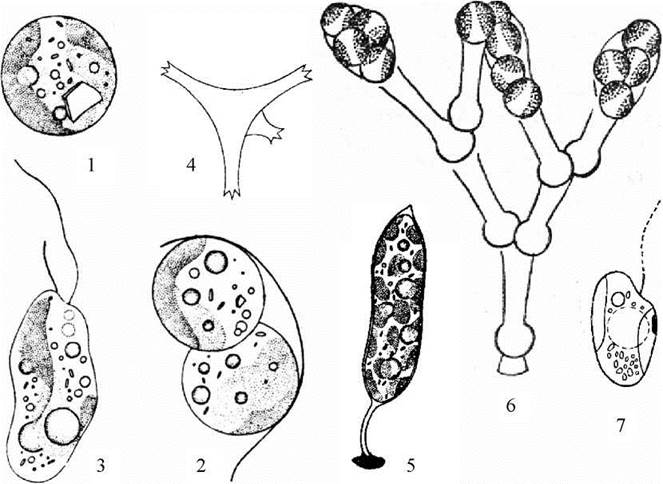
Fig. 15.7. Unicellular representatives of the order Mischococcales: 1–3 — Pleurochloris (1 — vegetative cell; 2 — release of aplanospores; 3 — zoospore); 4 — Pseudostaurastrum; 5 — Characiopsis; 6–7 — Mischococcus (6 — colony; 7 — zoospore).
Within the order Mischococcales, there are species capable of reproducing not only by zoospores and aplanospores, but also through vegetative cell division (Fig. 15.8). The Emergence of vegetative division in some mischococcalian algae led to the appearance within the order of algae with simple lamellar thalli (e.g., Chloropedia) and a numerous group of species with a heterotrichous structural type (Heterococcus). Multicellular mischococcalian algae inhabit almost exclusively terrestrial environments: predominantly in soils, and more rarely on tree bark and damp rocks.
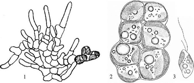
Fig. 15.8. Multicellular representatives of Mischococcales: 1 — Heterococcus; 2, 3 — Chloropedia (1, 2 — thalli, 3 — zoospore).
The order Tribonematales unites algae that possess two-valved (bipartite) cell walls. Both unicellular and filamentous organisms are represented within the order, and both groups include representatives with either uninucleate or multinucleate cells. According to molecular data, the order diverged from botrydialean algae and is sister to the Mischococcales.
Among unicellular tribonemataleans, the most widespread genus is Bumilleriopsis, the species of which inhabit soils and small, predominantly ephemeral freshwater bodies. The cells in Bumilleriopsis are elongated and sausage-shaped, covered by a cell wall consisting of two equal or unequal segments (Fig. 15.9: 1–3). The two-valved Nature of the wall is clearly visible during the reproduction of the alga—upon the release of autospores or zoospores from the mother cell, or after treating the cells with potassium hydroxide (in the latter case, the wall swells slightly, revealing the junction zone of the two-valved segments). In the parietal layer of the vegetative cell's cytoplasm, several discoid chloroplasts are located, while the center contains from one to four or eight nuclei, oil droplets, and grains of chrysolaminarin. The alga reproduces via zoospores and autospores. Sometimes, when the release of autospores from the mother cell is delayed, Bumilleriopsis forms aggregates resembling short two- or four-celled filaments.
An example of an alga that remains exclusively uninucleate in its vegetative state is the genus Diachros (Fig. 15.9: 4–5). The cells of this alga are small, spherical, and resemble the autospores of Bumilleriopsis. Reproduction occurs by means of autospores, which sometimes exhibit two contractile vacuoles. Species of this genus inhabit acidic soils and small acidic water bodies.
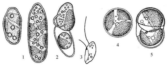
Fig. 15.9. Unicellular representatives of the order Tribonematales: 1–3 — Bumilleriopsis; 4–5 — Diachros (1, 4 — vegetative cells; 2, 5 — release of aplanospores; 3 — zoospore).
Examples of common filamentous tribonematalean algae are the genera Xanthonema and Tribonema.
Algae of the genus Xanthonema inhabit mainly soils. Their thalli appear as simple filaments formed by cylindrical cells. Each cell is covered by a two-valved cell wall (Fig. 15.10). In the contact zone between two adjacent cells, their adjoining segments are tightly or loosely connected. The cytoplasm contains from one to several chloroplasts, to the inner side of which mitochondria are tightly pressed. The center of the cell is occupied by one to four nuclei connected to the plastid via a chloroplast endoplasmic reticulum. Near each nucleus, there are one or two Golgi complexes.
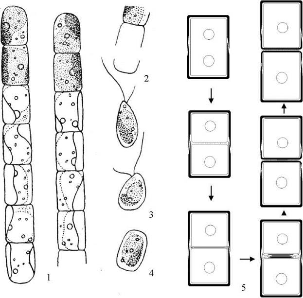
Fig. 15.10. Xanthonema: 1 — general appearance of the filaments; 2 — opening of the two-valved segments during zoospore release; 3 — zoospores; 4 — aplanospore; 5 — diagram of successive stages in the formation of cell wall segments.
Special electron microscopic studies have clarified The process of Cell wall formation. Specifically, it was established that following the completion of nuclear and protoplast division, material is secreted into the space between the plasmalemmae of the two daughter cells to form the connecting intercellular plate. Subsequently, material for the future cell wall segment is transported in vesicles derived from the Golgi complex and endoplasmic reticulum into the plate zone toward the daughter cells' plasmalemmae. This material is extruded into the space between the connecting plate and the plasmalemma, where it polymerizes to form a new cell wall segment of the daughter cell. This segment grows from the center to the periphery, simultaneously with the elongation of the daughter cell. Meanwhile, the intercellular plate decreases in size, gradually depolymerizing in a direction from the periphery toward the cell axis. If the depolymerization process stops, the remnants of the intercellular plate hold the daughter cells together. If the intercellular plate breaks down completely, the daughter cells separate from one another, resulting in thallus fragmentation.
Asexual reproduction in Xanthonema is carried out via zoospores and aplanospores. The release of zoospores and aplanospores occurs by the sliding apart of the mother cell wall segments, leaving the filaments terminating in empty half-cells that appear fork-shaped under an optical Microscope. Under unfavorable conditions, vegetative cells produce additional layers of the cell wall and transform into akinetes.
Species of the genus Tribonema inhabit primarily freshwater bodies, sometimes forming soft, light-green algal mats (frog spittle). The thalli of the alga take the form of simple, unbranched filaments that terminate in fork-like structures at their tips (Fig. 15.11). The process of cell wall formation in Tribonema is generally similar to that in Xanthonema, but an intercellular plate is not formed; instead, the material of the future cross-wall, transported in membrane vesicles, immediately polymerizes to form a single H-shaped segment between the plasmalemmae of the two daughter cells. As a result, adjacent vegetative cells are firmly connected to each other, and thallus fragmentation occurs only when the two segments of a single cell pull apart. This makes the formation of fork-like endings at the filament tips obligatory and precludes the presence of rounded filament tips.
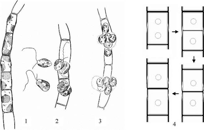
Fig. 15.11. Tribonema: 1 - vegetative filament; 2 - release of zoospores; 3 - release of aplanospores; 4 - diagram of successive stages of cell wall segment formation.
Unlike the previous genus, the vegetative cells of Tribonema are strictly uninucleate, except during the formation of reproductive cells—zoospores or aplanospores—and resting stages known as akinetes. Interestingly, zoospores in this genus can originate in several ways: from vegetative filament cells, from aplanospores, and from akinetes.
Distribution, Ecology, and Significance
Yellow-green algae inhabit soils and freshwater bodies. The most ancient group of yellow-green algae, represented by species of the genus Vaucheria, develops primarily on damp soil—such as dried-up puddles and the banks of water bodies—forming macroscopic, velvet-textured green mats. Species of the genus Botrydium grow under similar conditions. A significant portion of yellow-green algae are typical inhabitants of forest and high-altitude soils, including such genera as Botrydiopsis, Pleurochloris, Heterococcus, and Xanthonema. Terrestrial Xanthophyta are extremely sensitive to various types of anthropogenic environmental pollution, particularly smoke and gas emissions, heavy metals, herbicides, insecticides, detergents, and petroleum products. The initial symptom of such impact is the disappearance of these algae from phytocenoses.
A typical inhabitant of freshwater plankton is the genus Pseudostaurastrum, whereas Characiopsis and Tribonema are typical of the periphyton. During growth, Tribonema filaments frequently detach from the substrate, float to the surface waters, and continue Vegetative Reproduction there, forming soft green algal blooms. Other yellow-green algae occur only sporadically in aquatic environments.
Representatives of the division Xanthophyta, with the exception of Vaucheria, Botrydium, and Tribonema, almost never undergo mass development under natural conditions. Currently, there are no mass-cultivated Xanthophyta species, nor are any known to be harmful to humans.
Taxonomic Position of Xanthophyta in the Living World
Yellow-green algae are typical tubulicristate stramenopiles. Interestingly, it was in yellow-green algae that A. Massalsky and G. Lýdal first discovered tripartite flagellar hairs, later termed stramenopile mastigonemes (retronemes), which subsequently gave rise to the name "stramenopiles".
In the 1980s, transmission Electron microscopy studies conducted by D. Hibberd and G. Lýdal within the division Xanthophyta revealed a specific group of algae that was subsequently segregated into an independent division, Eustigmatophyta, in the early 1990s.
In the 1990s, research began on the phylogenetic relationships between yellow-green algae and other divisions using molecular Genetic Methods, specifically through the sequencing of the nuclear Gene encoding the 18S ribosomal RNA subunit and the chloroplast rbcL gene encoding the large subunit of ribulose-1,5-bisphosphate carboxylase. These studies confirmed the phylogenetic affinity of yellow-green algae with other stramenopile algae, as well as the hypothesis that their plastids originated from a eukaryotic ancestor within the red algae. According to molecular data, Xanthophyta proved to be more closely related to Phaeophyta than to Chrysophyta and Eustigmatophyta, contrary to what was believed in the late 1980s and early 1990s.
Taxonomic Position
Eustigmatophyte algae belong to the subkingdom Stramenopila within the kingdom Tubulicristata. Based on molecular markers, they are closely related to golden algae, from which they differ phenotypically by the specific STRUCTURE OF THE eyespot and pyrenoid, the presence of a parabasal body on the long flagellum, closed mitosis, the absence of a girdle lamella, and a specific genophore organization.
Last update: 07/08/2026
Editorial and Educational Adaptation: This material has been compiled based on the primary/original source text. The project team performed an editorial review, corrected technical inaccuracies, structured sections, and adapted the content for an educational format.
What was processed:
- elimination of formatting defects (OCR errors, structural breaks, corrupted characters);
- editorial organization of content;
- standardization of terminology in accordance with academic sources;
- verification of factual statements against the original source text.
All mentions of the author, publication year, and origin of the primary text have been preserved in accordance with the source.