Chemistry and Biology of Proteins - F. Haurowitz 1953
Albumins, globulins and other soluble proteins
Muscle proteins (contractile proteins)
Vertebrate Muscles contain approximately 15–20% protein, a large part of which can be extracted by treating freshly minced Muscle (ground with sand) with dilute salt solutions. If some time has elapsed since the animal's death, a portion of the soluble Muscle Proteins passes into an insoluble state, which affects the yield of proteins extracted by the aforementioned method. To prevent this conversion of soluble proteins into insoluble ones, it is necessary, firstly, to process the muscle immediately after sacrificing the animal and, secondly, to carry out the extraction at low temperatures.
Aqueous muscle extracts contain the following protein fractions: myogen, Myosin, globulin X, and stroma proteins. Among the listed proteins, myogen exhibits the highest solubility. Myosin precipitates when the extracts are diluted with Water and the KCl concentration in the solution drops to 0.03 M (0.22%). Upon further dilution with water (down to a KCl concentration of 0.005 M), globulin X precipitates [86]. Myosin constitutes approximately 40% of all muscle proteins; the remaining 60% of proteins are distributed roughly equally among myogen, globulin X, and stroma proteins [86].
Myogen, long considered a homogeneous protein, is actually a mixture of at least three proteins: myogens A, B, and C [87]. All three myogens have been obtained in crystalline form by fractional salting-out with ammonium sulfate at 60%, 80%, and 96% saturation [87, 88]. The average Molecular Weight of myogen, which is 81,000, decreases to 34,000 upon Treatment with urea [86]. Based on its solubility (high concentrations of ammonium sulfate are required for salting-out), myogen is classified among albumins. In myogen solutions stored at room Temperature, a portion of the myogen coagulates and turns into an insoluble compound designated as myogenfibrin. The myogen fraction contains many muscle Enzymes. For instance, phosphorylase, phosphoglucomutase, aldolase1, and glyceraldehyde phosphate dehydrogenase have been detected in this fraction [89]. These enzymes account for at least 20–25% of the total myogen content.
1 The aldolase activity of myogen was established by V. A. Engelhardt and his coworkers back in 1939 (Usp. sovrem. biol., XIV, 177, 1941). According to M. M. Zaalishvili, aldolase activity is associated with the myogen fraction that precipitates as myogen A. Myogen A contains no free sulfhydryl groups; the content of free amino groups is 0.9%, and that of carboxyl groups is 1.85% of the total protein. The ultraviolet absorption spectrum of myogen A has a maximum at 278 mμ. This maximum does not shift upon the reaction of myogen A with hexose diphosphate; consequently, the combination of the enzyme with the substrate does not occur through the groups responsible for the ultraviolet absorption spectrum shape. — Ed. Note.
Myosin, the most important muscle protein, is considered the contractile substance of muscle. Myosin can be extracted from muscle using Weber's solution (0.6 M KCl, 0.04 M NaHCO3,
0.01 M Na2CO3). Myosin solutions possess high viscosity and exhibit prominent Flow Birefringence (see p. 57) [90]. Upon standing, myosin solutions form a gel that liquefies upon shaking; this thixotropic phenomenon is observed even in 0.3% myosin solutions [90]. The flow birefringence of myosin solutions is drastically reduced by The addition of substances that lower solution viscosity, such as calcium chloride, potassium iodide, guanidine, and many others [91]. When saline solutions of myosin are extruded through a narrow orifice into distilled water, fine myosin threads are formed [92].
From all the foregoing, it appears that myosin molecules in solution have the shape of elongated filament-like particles. Physico-chemical analysis data of dry myosin support this Conclusion. Using Electron Cell/15.html">Microscopy, filaments approximately 1,000 mμ long and 5–10 mμ thick were discovered in dry myosin preparations [93]. X-Ray Diffraction Analysis of myosin showed that the identity period along the fiber axis is 726 Å, while the transverse identity period of the fiber is 200–325 Å [94]. In addition to these, transverse identity periods of 66, 42, and 33 Å were also detected; the 66 Å period is regarded as the transverse diameter of a single myosin protofibril [95]. The presence of short identity periods (equal to 2.82 Å) along the fiber axis suggests that the main peptide chains of myosin form helices around their long axis and that the side chains are tightly bound to each other [96]. The molecular weight of myosin is very high; values ranging from 600,000 to 3,900,000 have been reported for preparations obtained by various Methods [86, 97].
By combining Electrophoresis with ammonium sulfate salting-out, three different myosin fractions can be obtained, designated as a-, ß-, and y-myosin. The a-myosin fraction precipitates from solution at 23–27% ammonium sulfate saturation, and the ß-myosin fraction at 35–39% saturation [98]. Only myosin solutions exhibit flow birefringence [98].
During prolonged extraction of muscle with Weber's solution, a myosin-like protein passes into solution, possessing a much higher viscosity than the myosin obtained by short-term extraction [99]. This protein turned out to be a complex compound consisting of myosin and a second protein named Actin. To obtain actin, muscle is first extracted briefly with water, then with acetone, after which the dry residue is extracted again with water. This final extract contains actin [100]. From 1 g of fresh muscle, approximately 80 mg of myosin and 20–30 mg of actin can be obtained. Actin readily combines with myosin to form Actomyosin (myosin B). The ratio of myosin to actin in actomyosin is approximately 5:2 [101, 102]. Actomyosin is identical to the protein previously described under the name of myosin. Some researchers therefore suggest calling the actomyosin discovered by Szent-Györgyi simply myosin, and the protein that Szent-Györgyi called myosin should be termed myosin (w. r.), i.e., water-soluble myosin [102].
Actin exists in two forms: fibrous (F-actin) and globular (G-actin) [99]. Only F-actin can combine with myosin to form actomyosin; F-actin is produced from G-actin by treating the latter with potassium chloride [99, 103]. F-actin filaments can also revert to G-actin under the action of ultrasound [104, 105]1.
Actin combines exclusively with a-myosin and does not react with ß- and y-myosin [98]. Substances capable of binding free sulfhydryl groups (e.g., monoiodoacetic acid) inhibit The formation of actomyosin from its components. Based on this, it has been suggested that during actomyosin formation, actin binds to The sulfhydryl groups of myosin [106]. Actomyosin solutions, much like myosin solutions, display marked flow birefringence, whereas actin solutions lack this property [107].
Besides myosin, muscles contain 0.5% Tropomyosin—a protein very similar in properties to myosin [108]. To isolate tropomyosin, muscles are extracted sequentially with water, 50% ethyl alcohol, 97% ethyl alcohol, and diethyl ether; tropomyosin is obtained from the remaining dry powder by extraction with a 1 M potassium chloride solution. The Amino Acid Composition of tropomyosin is nearly identical to that of myosin (see Table 1). Both proteins contain unusually large amounts of Lysine—an amino acid with pronounced basic properties (about 17%). Tropomyosin yields highly viscous solutions; its molecular weight in phosphate buffer is 135,000; upon the addition of sodium chloride, tropomyosin dissociates into particles with a molecular weight of 65,000 [113]. Since both values are significantly lower than the molecular weight of myosin, it is hypothesized that myosin is a polymerization product of tropomyosin [108]. The high viscosity of tropomyosin solutions is attributed, as electron microscopy data show, to the asymmetric shape of its molecules [109], whose dissymmetry constant is f/f0 = 3 [110].
1 Data on The amino acid composition of actin have recently appeared in the literature. A paper by I. I. Ivanov and E. N. Asmolova (Biokhimiya, 15, 201, 1950) gives the following figures for individual Amino Acids (as a percentage of total nitrogen in actin): Arginine 14.2–14.6; aspartic acid 5.85; glutamic acid 9.3; Histidine 4.0; lysine 10.25; Tryptophan 1.7–1.9; phenylalanine — traces; Tyrosine 2.1; Methionine 2.4–2.95; cystine 0.81–0.83.
Expressed as a percentage of protein weight, the content of individual amino acids in actin isolated from rabbit muscle is characterized by the following figures:
|
Arginine |
7.2 |
Methionine |
4.6 |
|
Lysine |
8.55 |
6.9 |
|
|
Histidine |
2.4 |
1.7 |
|
|
Glutamic acid |
15.5 |
Valine |
2.8 |
|
Aspartic acid |
8.0 |
7.1 |
|
|
Tyrosine |
1.3 |
Hydroxyproline |
1.66 |
|
Tryptophan |
2.1 |
Leucine and isoleucine |
22.3 |
|
Cystine |
1.1 |
Phenylalanine |
0 |
Ed. Note.
Adenosine triphosphate [ATP], a mononucleotide present in muscle sap, exerts a completely specific effect on myosin and actomyosin. The addition of ATP to myosin or actomyosin solutions causes a sharp drop in the high viscosity of these solutions, and their flow birefringence disappears as well [111, 112]. This is due to the fact that upon interaction with ATP, long actomyosin filaments coil up, forming globular particles of the actomyosin-ATP complex [103, 121]. In the actomyosin-ATP complex, the ratio of actin, myosin, and ATP molecules is 1:1:1 [115]. Artificial threads of the actomyosin-ATP compound are obtained by extruding solutions of this complex through narrow orifices into distilled water. These threads contract to approximately 50–60% of their initial length when treated with a 0.01 M potassium chloride solution [112].
Cytology/cytology/93.html">ELECTRON MICROGRAPHS OF striated muscle have shown that actomyosin fibrils run uninterrupted through its isotropic and anisotropic discs [102]. These fibrils appear to be the contractile substance of resting muscle [115, 116].
In 1939, Engelhardt and Lyubimova [117] discovered that adenosine triphosphate is cleaved by myosin or a protein inseparable from myosin by existing purification methods. In this reaction, adenosine triphosphate is converted into adenosine diphosphate (ADP) [105]. Using Ultraviolet Microscopy, it was demonstrated that ADP is concentrated mainly in the isotropic discs [118]. It has recently been established that adenosine triphosphatase can be separated from myosin [119, 120]1.
Currently, most researchers believe that Muscle contraction is based on the deformation of myosin molecules. This is indicated by the decrease in birefringence during isotonic muscle contraction [83]. Birefringence in resting muscle is caused by the parallel arrangement of myosin molecules, and therefore a decrease in birefringence indicates a disruption of this oriented arrangement of myosin chains. However, X-ray diffraction analysis does not reveal any noticeable changes During muscle contraction; during contraction, the principal identity period (725 Å) remains unchanged [121]. It is possible that during contraction, myosin molecules regain their proper orientation As a result of transverse coiling [122] or supercontraction akin to that of keratin fibers [123] (see Chap. IX).
According to one existing hypothesis, the actomyosin molecule consists of myosin and actin filaments connected to each other along their lateral surfaces. Since myosin is capable of contraction while actin lacks this ability, the contraction of myosin alone must result in the bending of the entire actomyosin fiber [127] (Fig. 36). According to another view [124, 126], muscle contraction occurs as a result of end-to-end joining of small actin globules with a molecular weight of 70,000, forming actin filaments with a molecular weight of 1,500,000 that possess The ability to bind to myosin filaments [124]. Upon contraction, actomyosin dissociates back into its components [125].
1 In addition to adenosine triphosphatase activity, myosin also exhibits the ability to deaminate adenylic acid. This was established by D. L. Ferdman and I. S. Nechyporenko in 1946 (Ukr. biokhim. zhurn., 18, 105, 1946). This ability is retained by myosin even after repeated recrystallization. Following the establishment of this fact, the question arose as to whether the adenosine triphosphatase and deaminase activities of myosin preparations belong to two different proteins present in the preparations, or whether we are dealing with a single protein possessing both activities simultaneously. This question was investigated by V. A. Engelhardt and his coworkers (V. A. Engelhardt, M. N. Lyubimova, T. V. Venkstern, M. Ya. Timofeeva, Yu. V. Babskaya, DAN SSSR, XXXV, 397, 1952). The work revealed that heating myosin preparations at 53 °C for 5 minutes completely abolishes their adenosine triphosphatase activity while retaining 60–90% of the initial deaminase activity. The deaminase separated from adenosine triphosphatase is a globulin. The authors also succeeded, by precipitating myosin from dilute solutions using low concentrations of lanthanum salts, in obtaining preparations that retained adenosine triphosphatase activity but were almost devoid of deaminase activity. Of great significance are the authors' findings that only the protein possessing adenosine triphosphatase activity is capable of combining with actin, whereas the protein with deaminase activity is completely devoid of this ability. The data obtained by the authors thus reaffirm that adenosine triphosphatase activity is inseparable from the myosin fraction that plays the primary role in forming the contractile protein complex of Muscle tissue. — Ed. Note.
From the foregoing, it is evident that both The Structure of the contractile elements in striated muscle and The Mechanism of its contraction remain largely unclear. Muscle contraction begins, as is well known, within a few thousandths of a second after electrical or nerve stimulation; therefore, it is obvious that the deformation of myosin molecules can be triggered only by an extremely rapid chemical reaction. Most likely, this is an ionic reaction [128], since only ionic reactions proceed at such high speeds, whereas enzymatic reactions require considerably more time to occur.
At present, there is only scant experimental data on which to base certain assumptions concerning The Nature of the ionic reaction underlying muscle contraction. For instance, it is known that in a resting muscle, the bulk of potassium ions are in a bound, non-dialyzable form; however, immediately after muscle contraction, these ions readily pass into the dialysate. Potassium ions are presumably split off from the negatively charged carboxyl groups of myosin and other muscle proteins. A second important observation is that muscle contraction is accompanied by a slight decrease in its total volume (0.02 mm3 per 1 g of muscle) [129]. This decrease in muscle volume can be explained by the fact that The amount of free water within it diminishes due to the Hydrolysis or Hydration of proteins or other ions. The most probable assumption is that muscle contraction involves the hydration of the liberated potassium ions and carboxyl groups [129]:
Class="center">
It is possible that this Movement of water triggers the conformational changes of the myosin molecules [130].
During muscle contraction, not only potassium ions are released, but also calcium [131] and phosphate [132] ions. Since new positively and effectively negatively charged groups also appear in the peptide chains during this process, their mutual neutralization must likewise lead to the shortening of the myosin molecules [133, 134] (Fig. 37).
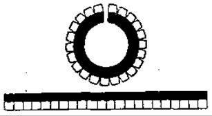
Fig. 36. Actomyosin [82].
On the other hand, it must be taken into account that divalent ions, such as calcium or phosphate ions, can form bridges between identically charged radicals of the peptide chain and thereby induce contraction [135]. This contraction is analogous to that of keratin fibers immersed in a copper-ammonia solution [136];  the minus sign in circles represents the
  groups; if the copper ions are subsequently split off by acidifying the solution, the contracted keratin fibers stretch out again [136].
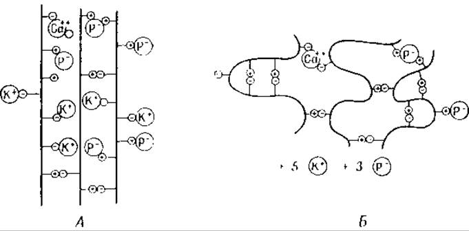
Fig. 37. Myosin molecules.
A — in a resting muscle; B — in a contracted muscle. K+ — potassium ions; P- — phosphate ions; Ca++ — Calcium Ions. The plus sign in circles represents the groups
Although we are not yet in a position to explain the MECHANISM OF ACTION of various ions on the contractile Proteins of the muscle fiber quite satisfactorily, there is nevertheless no doubt that the spatial configuration of the muscle fiber is closely related to the presence of an electrical charge on its surface. This is also supported by the fact that the pH of the muscle increases upon its passive stretch [137, 138].
None of the hypotheses proposed to explain the Mechanism of muscle contraction can satisfactorily account for the extraordinarily strong contraction of certain muscles. For instance, the muscle of the holothurian Thyone shortens upon contraction to approximately 8–9% of its initial length [84]. Such a reduction can hardly be a consequence of the fiber coiling depicted in Fig. 36 [139]. It is possible that here we are dealing with the rupture of numerous transverse ionic bonds between stretched, parallel-lying myosin fibers, which as a result transform into globular particles.
Issues related to the energetics of muscle contraction are also far from being resolved. Furthermore, we cannot even state with certainty whether muscle relaxation is a passive or an active process, although the latter assumption is more probable [140]. Assuming that the Muscle consists of rigid rod-like particles connected end-to-end by flexible links and immersed in a fluid medium, the modulus of elasticity E can be calculated using the following formula [141]:
E = 9RTW/5M,
where R is the gas constant, T is the absolute temperature, W is the weight of the elastic material per 1 ml of volume, and M is the molecular weight of an individual chain. An increase in the electrical charge On the surface of the rod-like particles leads to a decrease in the modulus of elasticity and, consequently, to muscle relaxation; conversely, a reduction in the free charge of these particles should lead to muscle contraction [142]. The relationship between the number n of charged groups and The change in the modulus of elasticity ∆E is expressed by the following equation [142]:
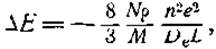
where De is the dielectric constant (~100), N is Avogadro's number, M is the molecular weight (about 106), ρ is the density, L is the average distance between the terminal charged groups (about 104 Å), n is the number of charges, and e is the magnitude of the electrical charge. If ∆E = 105 dyn/cm2, the value of n calculated using the above formula is approximately 100 [142].
During muscle contraction, chemical energy is converted into mechanical work. The thermodynamic characteristics of this reaction can be calculated if we assume that "stretched myosin" and "contracted myosin" are modifications of the same protein, and if we investigate the mutual transformations of both modifications at various temperatures (see p. 158). Measurements have shown that the changes in Free energy (∆F) amount to 7,000–8,500 cal/mol, and the thermal effect is equal to 53,500–56,000 cal/mol [143].
Last update: 06/08/2026
Editorial and Educational Adaptation: This material has been compiled based on the primary/original source text. The project team performed an editorial review, corrected technical inaccuracies, structured sections, and adapted the content for an educational format.
What was processed:
- elimination of formatting defects (OCR errors, structural breaks, corrupted characters);
- editorial organization of content;
- standardization of terminology in accordance with academic sources;
- verification of factual statements against the original source text.
All mentions of the author, publication year, and origin of the primary text have been preserved in accordance with the source.