Biochemical Engineering Fundamentals, Part 1 - Bailey J., Ollis D. 1989
The Chemical Basis of Life
Amino Acids and Proteins
Three-dimensional structure of proteins; secondary and tertiary structures
The Introduction/11.html">Secondary Structure of Proteins refers to the relative spatial arrangement of amino acid residues that are adjacent in the Amino Acid Sequence. Recall that the partial double-bond character of the amide bond accounts for its planarity. For this reason, rotation is possible around only two of every three bonds in the peptide backbone (Fig. 2.18), which limits the number of Conformations a short segment of the chain can adopt.
Class="center">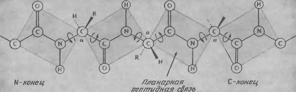
FIG. 2.18. The planar Nature of the peptide bond limits rotation about the bonds of the peptide backbone. (Reproduced from: Lehninger A. L., Biochemistry, 2nd ed., p. 128, Worth Publishers, New York, 1975; There is a Translation of an earlier edition: Lehninger A., Biochemistry. — M.: Mir, 1976.)
As previously noted (Fig. 2.14), There are two main forms of Protein secondary structure: the α-Helix and the β-structure (β-sheet). The conformation of Fibrous proteins in Hair, wool, and other structures is believed to be stabilized by Hydrogen Bonds between atoms of adjacent amino acid residues. When a protein chain folds into a helix, similar bonds can form between the —C=0 group of one residue and the —NH group of another, separated from the first by three amino acid residues. Such bonding, without significant distortion of the bonds and angles of the polypeptide chain, is possible in only one conformation, known as the α-helix. It is assumed that the Collagen molecule, the most abundant protein in higher animals, consists of three α-helical strands wound together into a single superhelix. Rigid and relatively inelastic, collagen molecules perform a mechanical (structural) function. Collagen is found in Skin, tendons, the cornea, and many other Organs; It is important in the immobilization of Enzymes and Cells, and as a component of biocompatible Materials.
Hydrogen bonds are not highly robust, so α-Helical structures are easily disrupted. Proteins can lose their helical structure In aqueous solutions due to competitive hydrogen bonding with Water molecules. If wool fibers are steamed and stretched, the wool proteins adopt a different conformation called the β-structure (β-sheet, pleated sheet). The latter is characteristic of native silk fiber proteins. The β-structure is also stabilized by hydrogen bonds, but in this case, they occur between adjacent parallel chains. This structure is characterized by flexibility and, at the same time, high tensile strength.
The properties of the secondary Structure of Fibrous proteins are utilized in the food industry. Some plant-based food products, such as soybeans, are valuable sources of Essential Amino Acids; however, they lack the consistency and texture of meat products. To impart these qualities, dissolved Globular proteins are processed into so-called "textured protein" by transforming them into structures that are closer to linear.
As a result of extremely laborious and complex experiments, the complete spatial arrangement (tertiary structure) of several proteins has been determined. For this purpose, crystals of pure protein were grown and then studied by X-ray crystallography. The scattering of X-rays by atoms produces a complex diffraction pattern containing detailed structural information about the protein at a resolution of up to 2 Å. Deciphering this information for Myoglobin (Fig. 2.19) required the calculation of 10,000 Fourier series; therefore, determining protein structures by this method is generally feasible only with The Use of high-performance computers.
As shown in Figs. 2.17,b and 2.19, the Tertiary Structure of proteins, especially globular ones, is extremely complex. In these figures, regions with helical secondary structure can be easily identified, and the Lysozyme molecule also contains segments with β-structure. How are these structures stabilized, and what purpose is served by a class of compounds whose molecules possess such a diverse architecture?
Interactions between R side groups that are far apart in the protein's amino acid sequence determine the folding or bending of the polypeptide chain to form the compact configurations typical of globular proteins. The three-dimensional structure of a protein is primarily shaped by several weak interactions, including ionic and hydrogen bonds, as well as hydrophobic interactions between nonpolar R groups (Fig. 2.20). Similar to the previously discussed lipid micelles, in many globular proteins, hydrophobic residues are concentrated in the interior of the molecule, while more hydrophilic groups are located on its surface. This conformation, sometimes referred to as the "oil drop" model, is likely the most stable in the aqueous environment natural to native proteins. (How, in your opinion, does The structure of a protein that is part of a Cell membrane differ from the one described?)
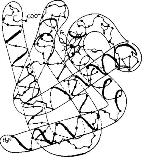
FIG. 2.19. Three-dimensional structure of myoglobin. The dots indicate the positions of the α-carbon atoms of the 121 amino acid residues in this oxygen-carrier protein. In the upper middle section of the molecule, a larger circle highlights the iron atom located at the center of the protein's single heme group. (Reproduced from: Edwards N. A., Hassall K. A., Cellular Biochemistry and Physiology, p. 57, McGraw-Hill Publishing Co., Ltd., London, 1971.)
In biochemical technology, it is extremely important from many Perspectives to understand that these weak interactions are easily disrupted by various environmental changes. In biological systems, the energy of a Hydrogen bond ranges from 3 to 7 kcal/mol, and the energy of an ionic bond is typically 5 kcal/mol; thus, even moderate heating can disrupt some of these bonds. Changes in pH, Ionic strength, physical forces, and The addition of organic substances can obviously also disrupt the native tertiary structure of proteins.
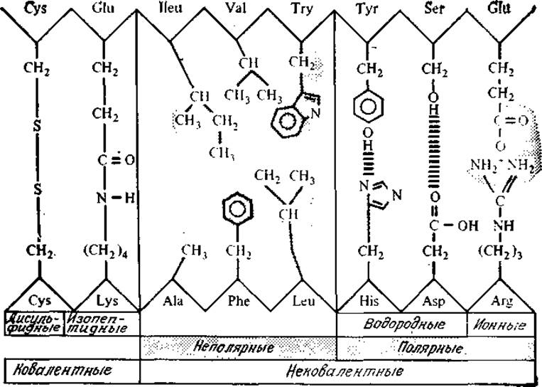
FIG. 2.20. Various types of bonds and interactions stabilizing the molecular structure of a protein. (From: Loewy A. G., Siekevitz P., Cell Structure and function. — M.: Mir, 1971, p. 245.)
Covalent bonds also play an important role in establishing stable protein conformations, most notably forming between two Cysteine residues. A disulfide bond is formed between two —SH groups by the elimination of two hydrogen atoms:

Disulfide bridges act as cross-links within a polypeptide chain, and sometimes they link two different chains; the Insulin molecule, for example, consists of two chains containing 21 and 30 amino acid residues, connected by two Disulfide Bonds; a third disulfide bridge forms an intrachain cross-link within the shorter chain (Fig. 2.21). Compared to the weak bonds mentioned above, disulfide covalent bridges are more resistant to thermal effects. At the same time, disulfide bonds are easily reduced by an excess of sulfhydryl compounds, such as β-mercaptoethanol.
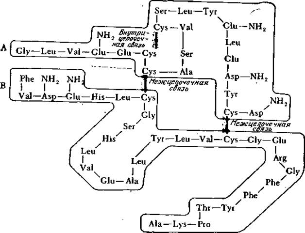
FIG. 2.21. Disulfide bridges link two peptide chains, designated as chain A (21 amino acid residues) and chain B (30 residues), in the bovine insulin molecule shown here; insulins from other animal species are structured similarly. Note the third intrachain disulfide bond between two residues of the shorter chain A. (Reproduced from: Reithel F. J., Concepts in Biochemistry, p. 237, McGraw-Hill Book Co., New York, 1967.)
Protein conformation largely determines its biological activity. Numerous experimental findings indicate that, in many cases, a protein can perform its specific function only when folded into a particular three-dimensional structure. This principle underlies the so-called "lock and key" model, which clearly illustrates the high Specificity of protein enzymes that catalyze biochemical transformations. It is well known that a given enzyme can convert only specific compounds, known as substrates. According to the lock-and-key model (Fig. 2.22), the enzyme possesses a specific site (the "lock") that is structurally complementary to the substrate molecule (the "key"); thus, only substrates with the required Spatial Structure can bind to the enzyme (and consequently undergo further catalytic transformation). Data obtained from studying the tertiary structure of proteins have not only confirmed this hypothesis but also helped to deepen our understanding of the Mechanism of Enzymatic catalysis, a topic we will explore in more detail in the next chapter. It has been repeatedly demonstrated that a direct link between the Spatial structure of Proteins and their highly specific interactions with other substances is also characteristic of permeases, Hormones, Antibodies, and other proteins.
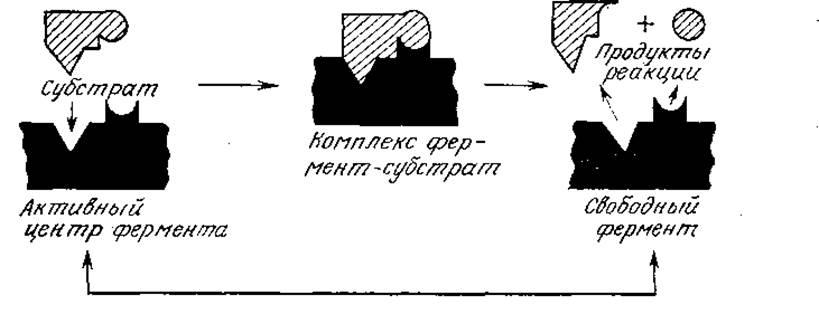
FIG. 2.22. Simplified diagram of the "lock and key" model of Enzymatic Catalysis. Here, the shape of the enzyme's catalytic Active Site (the lock) is complementary to the shape of the substrate (the key). (Reproduced from: Pelczar M. Jr., Reid R. D., Microbiology, 3rd ed., p. 158, McGraw-Hill Book Co., New York, 1972.)
If a protein is placed in conditions that differ from its normal biological environment, it may undergo structural changes, typically accompanied by a loss of its functional properties (Fig. 2.23); this process is called Denaturation. Denaturation can be triggered, for example, by relatively minor Changes in the pH and Temperature of the solution; in such cases, it is generally not accompanied by the Cleavage of covalent bonds. Upon slow cooling of a dilute solution of a denatured protein back to its physiological temperature, the reverse process, known as renaturation, often occurs, accompanied by the restoration of the protein's function. As with many other transformations, an increase in temperature promotes an increase in Entropy (disorder) and, consequently, the disruption of globular protein structures; cooling favors the interactions that stabilize the compact STRUCTURE OF THE protein and its renaturation.
The conformation adopted by a protein, which determines its properties, is characterized by a minimum of the molecule's Free energy. As of yet, this concept cannot be used to predict the structure and function of a protein based solely on its amino acid sequence. It can, however, prove useful when making informed assumptions about changes in protein properties under specific technological process conditions (which often differ significantly from the environment of the native protein) or as a result of a partial alteration in its amino acid sequence. In particular, this concept helps explain why an enzyme bound to a solid surface is less active than the same enzyme in solution.
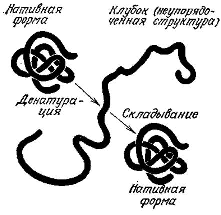
FIG. 2.23. A protein denatured by various factors can often refold into its native, biologically active conformation once these factors are removed. Such experiments demonstrate that the Primary Structure of a protein determines its secondary and tertiary structures. (Reproduced from: Lehninger A. L., Biochemistry, 2d ed., p. 62, Worth Publishers, New York, 1975; there is a Russian translation of an earlier edition: Lehninger A., Biochemistry.— M.: Mir, 1976.)
This chapter has focused heavily On the Relationship between Protein Structure and function. At the same time, it should be kept in mind that because most of the interactions and bonds stabilizing protein molecules are weak, their actual conformations can deviate significantly from a certain characteristic average conformation. Moreover, in many cases, this ability of proteins is closely linked to their biological function, much like the biological function of DNA is determined by its ability to separate into two strands under isothermal conditions within The Cell. From the perspective of a biochemical engineer, the relative ease of thermal or chemical denaturation of many proteins primarily indicates that enzymatic and cellular processes can be regulated by changing parameters such as pH, temperature, and ionic strength only within rather narrow limits. Denaturation is also an important consideration in other contexts, such as Protein Isolation (Ch. 11) and sterilizer design (Ch. 7 and 9).
Last update: 06/08/2026
Editorial and Educational Adaptation: This material has been compiled based on the primary/original source text. The project team performed an editorial review, corrected technical inaccuracies, structured sections, and adapted the content for an educational format.
What was processed:
- elimination of formatting defects (OCR errors, structural breaks, corrupted characters);
- editorial organization of content;
- standardization of terminology in accordance with academic sources;
- verification of factual statements against the original source text.
All mentions of the author, publication year, and origin of the primary text have been preserved in accordance with the source.