STRUCTURE AND PROPERTIES OF BIOMOLECULES - A. E. Zemlyakov - 2017
13. NUCLEIC ACIDS
❖ NUCLEIC ACIDS - Biopolymers responsible for storing and transmitting Genetic information in living Cells. From the perspective of macromolecular compounds, a nucleic acid is a polymer composed of NUCLEOTIDES linked by phosphodiester bonds.
Ribonucleic Acids (RNAs), which perform a wide range of biological Functions including participation in METABOLISM/35.html">Protein Biosynthesis, are distinguished from Deoxyribonucleic Acids (DNAs), which serve as the repository of genetic information in living organisms.
Nucleic acids were first isolated in 1868 by the Swiss biologist F. Miescher, who named them "nuclein". The modern term "nucleic acids" was introduced into scientific practice by the German anatomist and histologist R. Altmann in 1899.
Class="center">
The DNA of Eukaryotic cells consists of giant molecules with complex structures, with molecular weights that can reach 1010 - 1012 Da. For instance, human DNA contains 2.9 • 109 Base Pairs (bp), the DNA of the white mouse contains 2.3 • 109 bp, and the DNA of Plasmodium falciparum contains 2.3 • 107 bp.
Prokaryotic DNA is typically represented by smaller circular Chromosomes. For example, the E. coli chromosome contains 4.7 million bp. Alongside chromosomal DNA, these Bacteria also contain other DNA molecules—cyclic Plasmids (4–50 thousand bp)—which confer A number of useful and readily adaptable traits, such as resistance to specific Antibiotics.
Various types of RNA are distinguished, including Messenger RNA (mRNA), ribosomal RNA (rRNA), Transfer RNA (tRNA), viral RNA, and others. For example, in E. coli, Ribosomal RNAs containing from 100 to 3,100 nucleotides account for about 82% of the total cellular RNA mass, whereas Transfer RNAs (75–90 nucleotides) and messenger RNAs (up to 3,000 nucleotides) account for ~16% and ~2%, respectively. In recent decades, ribozymes—RNAs with catalytic activity—as well as regulatory microRNAs, have attracted considerable interest.
❖ Structure of mononucleotides. Unlike other monomers of natural polymers (Monosaccharides and Amino Acids), a nucleotide has a complex structure consisting of a carbohydrate moiety, a heterocyclic base, and a phosphoric acid residue.
✵ Heterocyclic base. Nucleic acids contain derivatives of pyrimidine, a monocyclic nitrogen-containing heterocycle—specifically cytosine, uracil (found only in RNA), and thymine (found only in DNA)—as well as derivatives of purine, a bicyclic nitrogen-containing heterocycle: adenine and guanine. Single-letter symbols are widely used to designate them.

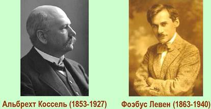
The structure of the main heterocyclic bases was elucidated between 1885 and 1901 by the German biochemist A. Kossel, recipient of the 1910 Nobel Prize in Physiology or Medicine for his body of research, which included foundational work on nucleic acids.
RNA may also contain minor nucleosides with other heterocyclic bases, including methylated and hydrogenated derivatives.

✵ Nucleoside. The type of nucleic acid is determined by The Nature of its monosaccharide component: RNA contains D-ribofuranose, whereas DNA contains 2-deoxy-D-ribofuranose. These monosaccharides were identified in nucleic acids in 1909 and 1929, respectively, by Phoebus Levene (born Fischel Levene), an American biochemist who was born and received his medical training in the Russian Empire.
The carbohydrate is linked to the heterocyclic base via a β-N-glycosidic bond involving the N1 atom in pyrimidine bases and the N9 atom in purine bases. The combination of the monosaccharide and the heterocyclic base is called a nucleoside. To distinguish them from the locants of the heterocycle, primed numbers are used for the carbon atoms of the monosaccharide.

Nucleoside names are derived from the names of their corresponding heterocyclic bases. In the case of deoxyribose, the prefix deoxy- is used, with thymidine and ribothymidine serving as exceptions.
Table 10. Names and Abbreviations of nucleosides
Base |
Nucleosides based on |
|
ribose |
2-deoxyribose |
|
cytosine |
cytidine (C) |
deoxycytidine (dC) |
uracil |
uridine (U) |
deoxyuridine (dU) |
thymine |
ribothymidine (T) |
thymidine (dT) |
adenine |
adenosine (A) |
deoxyadenosine (dA) |
guanine |
guanosine (G) |
deoxyguanosine (dG) |
Since the primary structural differences within the series of ribosides and 2-deoxyribosides lie in the nitrogen-containing heterocycle, their abbreviations are formed using the letter symbols of the heterocyclic bases with The addition of the prefix "d" in the case of the deoxysugar.
✵ Nucleotide. Phosphoric acid can esterify the primary hydroxyl group of a monosaccharide to form a nucleoside-5'-phosphate, the secondary hydroxyl group to form a nucleoside-3'-phosphate, or both hydroxyl groups simultaneously to form a nucleoside-3',5'-cyclic phosphate. Alongside monophosphates, di- and triphosphates are widespread in nature, such as nucleoside-5'-triphosphates. Examples of nucleotide structures and their abbreviated designations are given below.

❖ Biological Role of mononucleotides.
✵ Adenosine triphosphate (ATP) is a universal energy currency. The transfer of phosphoric acid residues ensures energy shuttling within biochemical systems. In The Cell, ATP (pppA) acts as the universal energy store. The Hydrolysis of the P–O bond between two phosphate groups releases a substantial amount of energy (~32 kJ/mol).

In biochemistry, covalent bonds whose hydrolysis releases more than 30 kJ/mol of energy are referred to as high-energy bonds. This released energy drives endergonic (energy-requiring) reactions. Conversely, when energy is released within the system, it is consumed in the synthesis of ATP.
ATP hydrolysis plays a critical role in powering a vast array of biochemical processes, including Muscle contraction, Photosynthesis, Bioluminescence, and the Biosynthesis of Proteins, nucleic acids, complex CARBOHYDRATES, and Lipids, among others.
A significant amount of energy is also released during the hydrolysis of ATP to adenosine monophosphate (AMP) and pyrophosphate.

In a number of biochemical processes, such as Translation, guanosine triphosphate (GTP) serves as the energy carrier.
✵ The Role of ATP in biochemical phosphorylation and adenylylation.
ATP plays a vital role in biochemical processes involving the transfer of a portion of the ATP molecule (a phosphate, diphosphate, or adenylyl group) to an acceptor molecule. These ATP-dependent reactions can proceed via phosphorylation (a), pyrophosphorylation (b), or adenylylation (c). The Mg2+ ion plays an essential role in these processes, as it is believed to enhance the reactivity of the phosphate and help direct the nucleophilic attack.

In all ATP-dependent biosynthetic processes, The formation of a covalent bond between two substrate molecules is coupled with the Cleavage of one of the pyrophosphate bonds of ATP. The first group of these reactions involves phosphotransferase (kinase) Enzymes, where a monophosphate group is transferred to the substrate and ADP is released. For example, the initial step in nucleotide biosynthesis involves the formation of ribose-5-phosphate from D-ribose.

The second group comprises enzymes that catalyze pyrophosphate-transfer reactions accompanied by the cleavage of ATP to AMP, such as in The biosynthesis of the coenzyme cocarboxylase.

Enzymes of the third group catalyze two sequential reactions involving The intermediate formation of an enzyme-bound acyl adenylate. Ultimately, ATP is cleaved into AMP and pyrophosphate. The formation of aminoacyl-tRNA serves as a classic example of such a reaction.

✵ Nucleotides as Coenzymes in carbohydrate and Lipid Biosynthesis. Uridine nucleotide coenzymes play a decisive role in Carbohydrate Metabolism. Ubiquitous in nature, uridine diphosphate sugars (UDP-sugars) feature a nucleotide-phosphate residue attached to the glycosidic center of a carbohydrate, such as D-glucose-1-uridine diphosphate (UDP-glucose).
UDP-sugars are produced through the reaction between UTP and sugar 1-phosphates, catalyzed by uridylyltransferase enzymes.

In Enzymatic Catalysis, uridine coenzymes—acting as compounds with an activated glycosidic hydroxyl group—can participate in various glycosyl-transfer reactions (glycosidation) to yield Glycosides, Oligosaccharides, and Polysaccharides.
In these processes, UDP-sugars are analogous to acyl adenylates involved in acyl-group transfer reactions. For instance, UDP-glucose facilitates the transfer of glucosyl residues to fructose to yield the disaccharide sucrose, to salicylaldehyde to form o-formylphenylglucose (the biochemical precursor of the glycoside salicin), or to the terminal glucose residue of a Glycogen molecule, leading to the elongation of the polysaccharide chain.

The second general type of UDP-sugar reactions involves chemical transformations within the sugar moiety itself. For example, UDP-glucose 4-epimerase, utilizing nicotinamide adenine dinucleotide (NAD+), catalyzes the interconversion of UDP-glucose and UDP-galactose.

Other examples of such processes include The oxidation of UDР-glucose to UDР-glucuronic acid and the decarboxylation of UDР-glucuronic acid to UDР-xylose.

Similarly to uridine nucleotides, cytidine nucleotides, such as СDР-Choline, participate in group transfer during phospholipid biosynthesis. СDР-choline itself is synthesized in a reaction between CTP and the corresponding choline phosphate. It should be noted that reactions involving cytidine nucleotides proceed with the cleavage of a pyrophosphate bond.

✵ Mononucleotides in the structure of coenzymes. A number of essential coenzymes contain an adenine moiety attached to the substrate via a pyrophosphate bridge. Examples of such compounds include coenzyme A, which participates in biological acylation processes (e.g., in FATTY ACID BIOSYNTHESIS), as well as the nicotinamide coenzyme NAD+ and flavin adenine diphosphate (FAD), which function as coenzymes in redox reactions.

✵ Nucleotide cyclophosphates as secondary messengers. 3’,5’-Cyclic phosphates of adenosine and guanosine (cAMP and cGMP) act as intracellular mediators (messengers) in biochemical reactions. Specifically, an increase in cAMP concentration leads to the activation of intracellular protein Kinases by blocking the regulatory (inhibitory) subunits of the enzyme.

Protein kinases, in turn, convert specific enzymes into an active state through phosphorylation, thereby triggering a cascade of biochemical reactions.
✵ Mononucleotide triphosphates - substrates for polymerase-catalyzed reactions. ATP and other nucleoside triphosphates serve as substrates for various DNA and RNA polymerases. The next chapter will discuss the processes of Replication and Transcription that take place with the participation of such enzymes.
❖ Structure of oligonucleotides. The phosphoric acid residue at the C3' position of one nucleotide is linked to the hydroxyl group at the C5' position of another nucleotide, and so on. Thus, nucleosides are connected to each other by a 3' —> 5' phosphodiester bond. The nucleotide with a free hydroxyl group at the C5' position is called the 5'-end, while the one with a free hydroxyl at C3' is called the 3'-end.
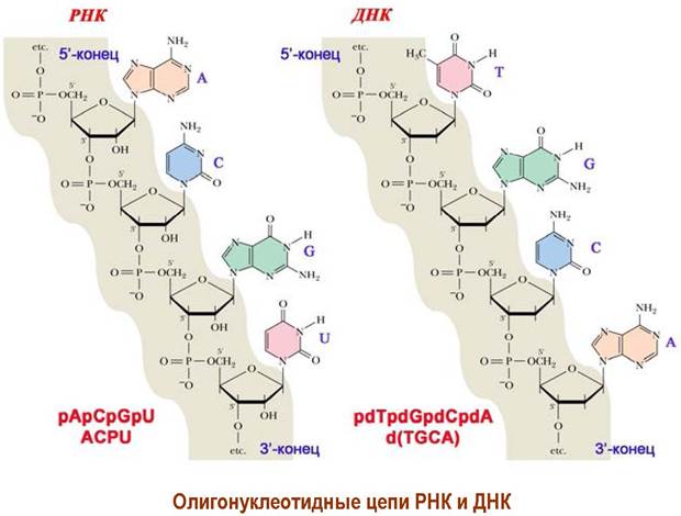
It is the Nature of the heterocyclic component that defines the nucleotide and, consequently, encodes its properties. The nucleotide sequence is as much a chemical language of living nature as the Amino Acid Sequence of proteins. Nucleotide sequences can be written using abbreviated nucleotide designations; given the commonality of the sugar-phosphate backbone in DNA and RNA, it is sufficient to list the heterocyclic bases starting from the 5'-end. For a deoxyribonucleotide sequence, the letter "d" is placed at the beginning.
❖ Introduction/11.html">Secondary structure of DNA. By the mid-20th century, the Chemical Structure of DNA had been established, and it was revealed that the length of the molecule greatly exceeds its diameter, with the heterocyclic bases located perpendicular to the molecular axis. Furthermore, it was discovered that in all DNA molecules, The amount of purine bases equals the amount of pyrimidine bases (Chargaff's rules). Based on these data, as well as a comparison of X-Ray Diffraction studies of DNA with various Spatial Models, J. Watson and F. Crick discovered the three-dimensional structure of DNA in 1953. For this discovery, they were jointly awarded the Nobel Prize in Physiology or Medicine in 1962, along with the English X-ray crystallographer M. Wilkins.

In space, the two DNA strands form a right-handed double helix, where the outer shell of the helix is formed by hydrophilic sugar-phosphate residues, while the hydrophobic heterocycles are located in the interior. The strands in The Double Helix are antiparallel—meaning the 5'-end of one strand lies adjacent to the 3'-end of the other—and complementary, with C, G, and A, T residues paired via Hydrogen Bonds.

The planes of the heterocycles are parallel to one another and perpendicular to the helix axis. Along with hydrogen bonds between complementary pairs and the Structuring EFFECT OF Water (the hydrophilic part of the molecule is surrounded by water, whereas the hydrophobic part is tucked away inside), the interaction between the π-electron systems of the heterocyclic bases in base "stacks" (stacking interactions) is the decisive factor in the self-assembly of DNA into a double helix.
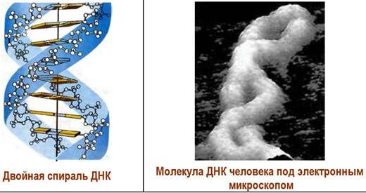
In aqueous solutions, DNA exists in the B-form, in which one turn contains 10 base pairs and has a length of 3.4 nm. The cross-sectional diameter of the molecule is ~2.0 nm. The A-form of DNA is more tightly packed—one turn is formed by 11 base pairs, whose planes are inclined by 20° compared to the B-form. The Z-form has also been identified, which, unlike the first two, is a left-handed helix with 12 base pairs per turn. Such a helix has a smaller cross-sectional diameter compared to the B-form—1.8 nm and 2.4 nm, respectively.

❖ Supercoiling of circular DNA. In many Viruses, bacteria, and Mitochondria, the double-stranded DNA molecule exists in a cyclic (circular) form. Depending on external factors (ambient pH, Ionic strength of the solution, Temperature), the nucleic acid chain can undergo additional twisting or winding around another segment. Such supercoiled DNA has been detected using Electron Microscopy.

❖ Structure of tRNA. tRNAs participate in the translation process, ensuring the targeted delivery of amino acids to the ribosomal complex. The molecular mass of such molecules is approximately 23–30 kDa. Their structure comprises 75–90 mononucleotides, of which roughly 10% are minor nucleotides, including methylated nucleotides, ribothymidine, dihydrouridine, and pseudouridine. The presence of minor nucleotides, on the one hand, protects tRNA from the action of Nucleases and, on the other hand, due to their inability to form complementary pairs, ensures the Spatial Organization of the molecule, in which single-stranded regions alternate with double-stranded ones—forming a "cloverleaf" structure.

The 3'-end of the nucleic acid chain contains the trinucleotide C-C-A-OH. The hydroxyl group of the terminal adenosine residue can be acylated by an amino acid. At the 5'-end, the guanosine residue is additionally phosphorylated. Two important regions are distinguished in the tRNA Structure: the aminoacyl (AA) site, to which The amino acid attaches, and the anticodon (AC) site, responsible for reading information from the messenger RNA. In space, the tRNA nucleic acid chain forms a three-dimensional structure resembling a three-dimensional L shape.

❖ Ribozymes. This term is formed from a combination of the concepts "ribonucleic acid" and "enzyme" and denotes the ability of RNA to catalyze biochemical processes. The catalytic activity of RNA was discovered in 1982 by the American molecular biologist T. Cech during The Study of RNA splicing, and in 1983 by the Canadian scientist S. Altman in bacterial Ribonuclease P. Prior to this, it was believed that biocatalysis was exclusive to protein molecules. For the discovery of ribozymes, T. Cech and S. Altman were awarded the Nobel Prize in Chemistry in 1989.


It was subsequently discovered that ribosome fragments also exhibit ribozyme properties during protein biosynthesis, catalyzing the elongation of the peptide chain.
❖ MicroRNAs. In the early 1990s, it was established that ribonucleic acids with a small number of nucleotides (up to 22 residues) are encoded by special DNA regions and can perform a regulatory function during translation. It is believed that such microRNAs can serve as markers in the detection of a number of dangerous diseases.

Last update: 06/08/2026
Editorial and Educational Adaptation: This material has been compiled based on the primary/original source text. The project team performed an editorial review, corrected technical inaccuracies, structured sections, and adapted the content for an educational format.
What was processed:
- elimination of formatting defects (OCR errors, structural breaks, corrupted characters);
- editorial organization of content;
- standardization of terminology in accordance with academic sources;
- verification of factual statements against the original source text.
All mentions of the author, publication year, and origin of the primary text have been preserved in accordance with the source.