STRUCTURE AND PROPERTIES OF BIOMOLECULES - A. E. Zemlyakov - 2017
14. BIOLOGICAL PROCESSES INVOLVING NUCLEIC ACIDS
Nucleic Acids drive critical biological processes in living Cells, including METABOLISM/36.html">DNA Replication and Protein Biosynthesis based on the Genetic information encoded in DNA (Transcription and Translation).
Replication
Replication is The process of DNA duplication. It simultaneously involves unwinding The Double Helix and synthesizing a complementary strand for each of the parent strands. Replicative DNA polymerases play a key role in this process, utilizing a set of mononucleoside triphosphates (pppdA, pppdG, pppdC, pppdT) to synthesize the polynucleotide chain using the original DNA strand as a template.
The incorporation of each new nucleotide is governed by The formation of stable Hydrogen Bonds with the complementary template region, ensuring high fidelity in building the duplicate DNA strand. On average, replication exhibits no more than one error per 1 billion NUCLEOTIDES.
Class="center">

Let us examine the key steps of Chromosome replication in E. coli.
❖ Unwinding of the double helix. Since single-stranded DNA acts as the template, the double helix must first be separated into individual strands. Unwinding the helix would normally cause the strands to rotate. A specialized enzyme called topoisomerase, consisting of 4 subunits with a total molecular mass of ~400 kDa, prevents this phenomenon.

Topoisomerase consists of 4 subunits with a total molecular mass of ~400 kDa. This enzyme introduces breaks into One DNA strand, passes the second strand through the break, and then reseals the nucleic acid links.

The enzyme helicase (300 kDa, 6 identical subunits) breaks the hydrogen bonds between the complementary nucleotide pairs of the DNA double helix. Working in concert with helicase, single-strand DNA-binding Proteins—which also feature a subunit Structure consisting of 4 identical protein molecules with a total molecular mass of 75.6 kDa—stabilize the separated single-stranded nucleic acid molecules. The Separation of each base pair requires the energy of Hydrolysis of two ATP molecules to ADP and phosphate.
❖ DNA polymerases. Functionally, DNA polymerase is a multienzyme complex. Its largest component is termed DNA polymerase III. DNA biosynthesis is driven by the dimeric core of this enzyme, with each half catalyzing the synthesis of a new DNA strand at a rate of up to 1000 nucleotides per second. The enzyme comprises 10 types of subunits, and its total molecular mass reaches 791.5 kDa. Its structural components are:
♦ two cores, each consisting of three subunits — α (129.9 kDa), ε (27.5 kDa), and θ (8.6 kDa) — which provide 5’ —> 3’ polymerase and 3’ —> 5’ exonuclease activity;
♦ two Ƭ subunits of 71.1 kDa each, connecting the two cores;
♦ two β proteins, each consisting of two symmetrical protein units of 40.6 kDa, which clamp the DNA strand;
♦ a γ complex composed of 5 subunits (γ - 47.5 kDa, δ - 38.7 kDa, δ’ - 36.9 kDa, χ - 16.6 kDa, Ψ - 15.2 kDa) that facilitates the movement of the multienzyme complex along the DNA strand.

✵ DNA polymerase I (molecular mass ~103 kDa) participates in replacing RNA primer segments with corresponding DNA fragments; i.e., it possesses both 5’ —> 3’ polymerase and 5’ —> 3’ exonuclease activity.
The overall rate of the process is up to 20 nucleotides per second.
❖ Template-directed synthesis.
✵ Leading strand. Polymerization begins at the 3' end and proceeds toward the 5' end. For the enzyme to initiate its work, a primer is required, which is typically a corresponding RNA segment of 10–60 nucleotides. It is synthesized by the enzyme primase (a protein with a mass of ~60 kDa). Subsequently, the DNA polymerase complex continuously elongates the second complementary strand, known as the leading strand.

✵ Lagging strand. The other double strand is synthesized in separate segments of 1,000–2,000 bp each. These are called Okazaki fragments, named after the Japanese biochemist Reiji Okazaki and his wife Tsuneko Okazaki, who first described them.
To enable the function of the second DNA polymerase III complex, which synthesizes the complementary strand exclusively in the 5'-to-3' direction, the second DNA template strand loops by 180°.
Near the site of DNA unwinding, primase associates with specific proteins, binds to the DNA template strand, and synthesizes a primer. DNA polymerase III then extends the complementary DNA strand toward the 5' end. Meanwhile, the unwinding process progresses further, a new primer attaches, a DNA segment grows from it toward the previous primer, and so forth. This results in a strand consisting of alternating RNA and DNA fragments.

To form a continuous DNA strand, a DNA Repair system comes into play, which removes the RNA primer and replaces it with DNA. In Bacteria, the removal of RNA nucleotides is carried out by the 5’ —> 3’ exonuclease activity of DNA polymerase I. Simultaneously, each excised ribonucleotide is replaced with the corresponding deoxyribonucleotide. The double-stranded DNA segment serves as a primer.
At The final stage, DNA ligase Enzymes (with a Molecular Weight of 74 kDa) join the individual polynucleotide fragments via phosphodiester bonds. Following replication, the resulting strands spontaneously recoil into helices without any Energy Expenditure or the action of additional enzymes.

Transcription
Transcription is the process of biosynthesizing RNA complementary to a specific region of DNA. The genetic information stored in DNA is not used directly for protein biosynthesis; instead, a copy—Messenger RNA (mRNA)—is transcribed from segments of the DNA strand.
A key role in transcription is played by DNA-dependent RNA polymerase (transcriptase). This enzyme was discovered in 1960 by American scientists Sam Weiss, Audrey Stevens, James Bonner, and Jerard Hurwitz.

The first enzymatic synthesis of RNA was performed by the Spanish-American biochemist S. Ochoa. In 1959, he shared the Nobel Prize in Physiology or Medicine with American scientist A. Kornberg for their pioneering work on transcription. In 2006, Arthur Kornberg's son, Roger, was awarded the Nobel Prize in Chemistry "for his studies of the molecular structure of RNA polymerase" during various Stages of Transcription.
In E. coli, transcriptase is a complex of five types of subunits (2α, β, β’, ω, and σ) with a total molecular weight of 450 kDa.
The core enzyme has a constant structure consisting of α2, β, β’, and ω. Several σ-subunits with molecular weights of 70, 32, and 28 kDa have been identified in E. coli. The first is involved in the Transcription of the majority of genes, whereas the others operate during the transcription of short or specialized genes.

Transcription consists of initiation (the starting phase), elongation (RNA chain extension), and termination (the cessation of template-directed synthesis). A large number of specialized accessory proteins, or factors (over 100 in E. coli), are involved in regulating this process.

Initiation begins when the core enzyme-σ-subunit complex binds to a specific starting region on the DNA known as the promoter, encompassing the nucleic acid. The DNA double helix unwinds locally, and a short fragment of complementary RNA (about 10 nucleotides long) is synthesized. The σ-subunit then dissociates, leaving behind the DNA-core enzyme complex, which marks the completion of the initiation phase. Thus, The primary function of the σ-factor is to recognize the promoter and ensure the successful initiation of transcription.

Next, using a pool of mononucleotide triphosphates (ATP, GTP, CTP, UTP) as substrates and one of the DNA strands as a template, the transcriptase performs template-directed Synthesis of the complementary strand. Notably, an A-U pair is synthesized instead of an A-T pair. RNA Synthesis proceeds in the 5’-to-3’ direction, meaning that RNA polymerase moves along the DNA template strand in the 3’ —> 5’ direction.
The termination of transcription occurs when RNA polymerase reaches a specific DNA sequence called a terminator, which frequently contains a series of consecutive adenosine residues. The resulting stretch of A-U Base Pairs is relatively weak, causing the newly synthesized RNA to dissociate from the DNA template. Alternatively, specialized termination factors assist in the release of the RNA.

❖ Processing. In prokaryotes, the mRNA produced during biosynthesis is immediately ready to participate in protein translation. However, The production of Ribosomal RNAs and tRNAs from precursor RNA transcripts (pre-RNA) requires a specialized maturation Procedure known as processing.
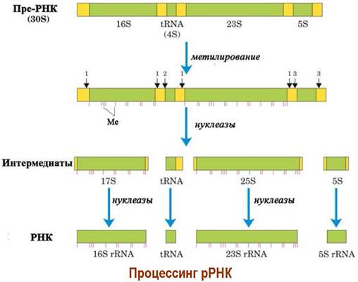
Processing involves the biochemical methylation of specific nucleotides and the removal of intervening nucleic acid segments by ribonucleases. For instance, a single 30S pre-RNA transcript in E. coli is processed to yield 23S, 16S, and 5S rRNAs, as well as several tRNAs.

tRNAs subsequently undergo processing by ribonucleases that remove unnecessary nucleotides, followed by specific modifications such as deamination, methylation, and reduction of individual nucleotides.
✵ Eukaryotic mRNA Processing. Unlike prokaryotes, eukaryotic mRNAs undergo processing. Specifically, to protect the 5’ end, 7-methylguanosine (7Me-G) is attached via a unique 5’ —> 5’ triphosphate bridge, and the 2’-hydroxyl groups of the two terminal nucleotides are methylated—a process known as "capping".

A polyadenylate fragment ranging from 100 to 250 residues is condensed at the 3’ end of the mRNA, providing protection against exonucleases and facilitating ribosome interaction. The total length of an mRNA can reach several thousand nucleotides.

❖ Splicing. Eukaryotic genes contain exons—regions encoding Protein Synthesis—as well as introns, which are non-coding DNA fragments. Consequently, transcription produces pre-mRNAs containing both exons and introns. Before translation, the Introns must be removed. This process is known as splicing, or the joining of ends.
From a chemical perspective, the 2’-hydroxyl group of an adenosine residue in the 3’ terminal region of the intron attacks the phosphate bridge at the 5’ end of the fragment to be joined. Next, the 3’-hydroxyl group of the first exon binds to the 5’-phosphate group of the second exon, releasing the lariat (cyclic) intron.

Translation is a multi-step process of Polypeptide chain synthesis based on the genetic information encoded in the mRNA nucleotide sequence. Translation takes place within a specialized intracellular structure, the ribosome, with the participation of Protein Complexes and aminoacyl-tRNAs (RNAs carrying an amino acid, see below). Specific protein-enzymes called factors are involved in all stages of translation: initiation, elongation, and termination.
❖ The ribosome is a cellular organelle that performs protein biosynthesis based on genetic information. They were first described in the 1950s by Romanian-American Cell biologist G. Palade (recipient of the 1974 Nobel Prize in Physiology or Medicine) as dense particles or granules.

In 1958, Richard Roberts proposed the term "ribosome" to replace the previously used "ribonucleoprotein particle of the microsomal fraction".
In 2009, the Nobel Prize in Chemistry "for studies of the Structure and function of the ribosome" was awarded to an international research team consisting of T. Steitz (USA), A. Yonath (Israel), and V. Ramakrishnan (UK).

Prokaryotes are characterized by 70S Ribosomes (where S is the sedimentation coefficient), whereas eukaryotes contain 80S ribosomes. For instance, a single E. coli cell contains about 15,000 ribosomes.
The 70S E. coli ribosome has a diameter of 18–20 nm and a molecular mass of ~2.5 MDa. It consists of two major subunits: the 50S subunit (~1.6 MDa) and the 30S subunit (~0.9 MDa). Each subunit has a complex nucleoprotein structure, with RNA accounting for about 60–65% of the ribosome's mass and protein structures making up the remaining 35–40%.

The small subunit consists of 21 proteins and 16S RNA, while the large subunit comprises 31 proteins and two polynucleotides: 23S RNA and 5S RNA. The protein molecules are rich in basic amino acid residues, specifically Lysine and Arginine, which facilitates their electrostatic binding to the phosphoric acid residues of the polynucleotides.
Both the large and small subunits can assemble into an intact ribosome or dissociate (see below). This process is regulated, among other factors, by the concentration of magnesium ions. In general, the small subunit is responsible for initiating translation, whereas the large subunit, which possesses the catalytic activity required to form peptide bonds, drives elongation.
The eukaryotic 80S ribosome has a mass of ~4.2 MDa and a diameter of approximately 23 nm. It is composed of 60S (~2.8 MDa) and 40S (~1.4 MDa) subunits. The large subunit contains three polynucleotides (28S, 5.8S, and 5S RNAs) and 49 proteins, while the small subunit includes an 18S RNA and 33 proteins. Ribosome synthesis in eukaryotes takes place in the nucleolus.
Despite the considerable structural similarity between eukaryotic and prokaryotic ribosomes, there are significant differences that allow certain drugs (such as Antibiotics) to preferentially bind to 70S ribosomes, thereby exerting a selective antibacterial effect.

✵ Ricin. The 80S ribosome is the target for A number of plant poisons (phytotoxins), such as ricin, which is extracted from the castor oil plant (Ricinus communis). The seeds of this plant contain up to 1 mg/g of the toxin, the lethal dose of which for humans is 10 µg/kg.
This toxin is a heterodimer consisting of an A chain (~32 kDa) with N-glycosidase activity and a B protein (~34 kDa) that Functions as a lectin. Using the lectin domain, which recognizes galactose and galactosamine residues, the toxin anchors itself to The Cell surface. Once inside the cell, the dimer is cleaved to release the toxic A component. Ricin's glycosidase targets a specific adenosine residue in the 28S RNA of the ribosomal 60S subunit, thereby disrupting the translation process.

A toxin of the same type (viscumin) has been found in the mistletoe, a poisonous parasitic plant.

❖ Genetic Code. The main part of Messenger RNA is a complementary copy of a specific DNA region and, accordingly, carries information about the Amino Acid Sequence of the synthesized protein. These nucleic acids are also referred to as informational RNA. An amino acid is encoded not by a single nucleotide, but by a sequence of three nucleotides (a codon). Out of 64 possible codon combinations, 61 correspond to standard Amino Acids (The Genetic Code). In addition, there are three so-called "stop" codons, which serve as signals to terminate biosynthesis. A number of Amino acids are encoded by multiple codons.
For establishing the principles of the genetic code in 1968, American scientists M. Nirenberg, H. Khorana, and R. Holley were awarded the Nobel Prize in Physiology or Medicine.
In 1979, deviations from the standard genetic code were discovered; for example, in plant cell Mitochondria, the codon CGG encodes The addition of Tryptophan, whereas in Yeasts of the genus Candida, the codon CUG is responsible for the incorporation of Serine.


❖ Aminoacyl-tRNA. Information from mRNA is read by tRNA, which features a complementary nucleotide sequence in its anticodon loop (see tRNA Structure). Consequently, several different tRNA molecules exist for a number of amino acids.
The process of attaching an amino acid to tRNA is catalyzed by an aminoacyl-tRNA synthetase specific to that amino acid. This enzyme drives two consecutive reactions: the synthesis of aminoacyl adenylate followed by the formation of aminoacyl-tRNA. Accordingly, it possesses two binding sites—one for The amino acid and one for the tRNA.

First, the amino acid is activated with the help of ATP. The resulting mixed anhydride of phosphoric acid and the amino acid exhibits high reactivity, enabling amino acids to attach to the hydroxyl groups of the 3’-terminal adenine residue of tRNA, thereby forming aminoacyl-tRNA.

❖ Translation in prokaryotes.
✵ Initiation. The Translation Processes in PROKARYOTES AND EUKARYOTES share common fundamental principles, yet differ significantly in their details. Let us examine the simpler translation mechanism in prokaryotes.
There is no dedicated start codon per se in the genetic code. In the 5′-terminal region of the mRNA where biosynthesis begins, near the initiating AUG codon, lies a specific group of nucleotides known as the Shine-Dalgarno sequence (e.g., AGGA or GGUUUGG). It is named after the Australian scientists John Shine and Lynne Dalgarno, who first described it. The 30S ribosomal subunit anchors to this sequence via a complementary 3′-terminal segment of its 16S RNA, thereby determining THE START OF translation.
The initiation codon for virtually all eukaryotes and the vast majority of prokaryotes is the triplet AUG. In certain prokaryotic mRNAs, however, Translation initiation may begin with other codons, such as GUG or UUG, which are also located within the mRNA initiation region.

Prior to engaging in translation, the small prokaryotic ribosomal subunit (30S) exists in a complex with initiation factor IF3, which prevents its premature association with the 50S ribosomal subunit. For this reason, this protein is referred to as the "dissociation factor." In many prokaryotes, the complex also includes the IF1 protein, which enhances the affinity of the small subunit for IF2 and IF3.
The binding of the small ribosomal subunit to mRNA can occur via two equivalent pathways. In the first pathway, the "30S subunit + IF3/IF1" block first associates with the mRNA, after which the initiator tRNA-Metf joins in a complex with initiation factor IF2 and GTP.
In the second pathway, the complex "30S subunit + IF3/IF1 + tRNA-Metf + IF2(GTP)" is formed first and subsequently anchors to the mRNA.

The initiator tRNA-Metf differs from regular tRNA-Met in that it carries an N-formylated amino acid. The anticodon of this tRNA is the sequence UAC, which is fully complementary to the AUG codon and partially complementary to the other prokaryotic initiation codons discussed above (GUG and UUG).

Next, the 50S subunit joins the "mRNA – 30S subunit – tRNA-Metf" complex, triggering a conformational change in the IF2 protein. Its enzymatic properties are activated, leading to the hydrolysis of GTP to GDP. The initiation factor IF2(GDP) then loses its affinity for tRNA and dissociates from the complex. Simultaneously, initiation factors IF3 and IF1 dissociate, and the 70S ribosomal complex is ready for action.
✵ Elongation. The 50S subunit features two tRNA binding sites: the P-site (P for peptidyl), which coordinates tRNAs carrying amidated forms (such as N-formyl-Met or growing peptide chains), and the A-site (A for aminoacyl), which binds tRNAs carrying free amino acids.

During the Formation of the initiation complex, Met-tRNAf is positioned in the P-site. The tRNA complementary to the second mRNA codon delivers the corresponding amino acid to the A-site, assisted by the GTP-containing elongation factor EF-Tu. Subsequently, GTP is hydrolyzed to GDP, inducing a conformational change in the protein and the release of this elongation factor.

In the peptide bond formation reaction, the amino group of the second amino acid acts as a nucleophile. Facilitated by a ribosomal structural domain within the large subunit that functions as the peptidyl transferase enzyme, an aminolysis of the N-formylmethionine ester bond with the nucleotide takes place. This yields a dipeptidyl-tRNA and a free tRNA.

The dipeptidyl-tRNA shifts into the P-site, dragging the mRNA along with it by precisely one trinucleotide. The deacylated initiating tRNA moves into the E-site (E for exit) and dissociates from the ribosome. This process, known as translocation, is mediated by the EF-G factor.
Next, guided by the codon sequence in the messenger RNA, aminoacyl-tRNAs are once again coordinated in the A-site to deliver subsequent amino acids, which are sequentially linked together to construct the polypeptide chain.

✵ Termination. Upon reaching a stop codon—for which no complementary tRNA exists—protein biosynthesis halts, the polypeptide chain is cleaved from the complex, and the 70S ribosome dissociates into its constituent subunits. These events are driven by release factors RF1, RF2, and RF3. The first two catalyze the release of the protein chain from the tRNA upon encountering the UAA/UAG and UAA/UGA codons, respectively, while the third promotes their action.


Translation in prokaryotes proceeds at a very high velocity, incorporating approximately 20 amino acids per second.
In many instances, transcription and translation occur concurrently. Multiple transcriptases move along the DNA strand to synthesize mRNA molecules, which in turn become densely populated with dozens of ribosomes actively executing protein biosynthesis.

✵ Antibiotics as inhibitors of ribosomal protein synthesis. The antibiotic MECHANISM OF ACTION for many drugs involves binding to specific structural sites on the prokaryotic ribosome, thereby blocking protein biosynthesis.
For instance, streptomycin, an aminoglycoside antibiotic, tightly binds to the 30S subunit, inducing alterations in the P-site and thereby preventing The entry of the initiator formyl-Met-tRNA into the ribosome, which halts the onset of protein synthesis. Tetracyclines form stable complexes within the ribosomal A-site, interfering with the binding of aminoacyl-tRNA to the 30S subunits.

The mechanism by which macrolide antibiotics, such as erythromycin, affect microorganisms is likewise linked to the disruption of ribosomal protein synthesis through binding to the 50S subunit and inhibiting the interaction between peptidyl-tRNA and the ribosomal P-site.

❖ Chromatin. The genetic material of a cell is concentrated within the Chromosomes of the Cell Nucleus. DNA molecules spanning millimeters or even centimeters in length are compacted into chromosomes measuring only a few micrometers. For example, 46 Human chromosomes with a total length of 200 µm contain 2 m of DNA. This DNA packing is achieved with the aid of small histone proteins (molecular mass 10–30 kDa), which are rich in basic amino acids (Arg and Lys), along with non-histone proteins. Together, they constitute a nucleoprotein complex known as chromatin.

Chromatin consists of discrete particles called nucleosomes, in which a stretch of DNA (~140–150 base pairs) is wrapped around a core complex of four histone pairs (two molecules each of H2A, H2B, H3, and H4). Nucleosomes are linked by stretches of free linker DNA (~50 base pairs).

A chain of six nucleosomes is anchored by histone H1 to form a single solenoidal turn. The array of solenoids is further folded into loops, which subsequently assemble into higher-order fibers (with a diameter of ~840 nm). Finally, these fibers undergo further Condensation to form chromosomes. Non-histone Proteins are essential for the formation of both solenoidal structures and higher-order fibers.

Last update: 06/08/2026
Editorial and Educational Adaptation: This material has been compiled based on the primary/original source text. The project team performed an editorial review, corrected technical inaccuracies, structured sections, and adapted the content for an educational format.
What was processed:
- elimination of formatting defects (OCR errors, structural breaks, corrupted characters);
- editorial organization of content;
- standardization of terminology in accordance with academic sources;
- verification of factual statements against the original source text.
All mentions of the author, publication year, and origin of the primary text have been preserved in accordance with the source.