Protein Chemistry - Part 2 - Selected Chapters in Special Protein Chemistry - Ashmarin, I. P. 1968
Histones
Primary and secondary structures of histones
Despite the heterogeneity of Histones, their major fractions are well characterized in terms of Amino Acid Composition, N- and C-terminal groups, and the degree of Acetylation (Tables 10, 11). All of them are characterized by a high content of the basic Amino Acids Lysine and Arginine (from 22 to 29 mol. %), a relatively high content of Alanine and Proline, the complete absence of Tryptophan, and the near-complete absence of Tyrosine, phenylalanine, and Sulfur-Containing Amino Acids (Cysteine, cystine). Recent studies have revealed only trace amounts of cysteine and cystine (about 1 mol. %) in the arginine-rich histone fraction.
Class="center">Table 10 Amino acid composition of major histone fractions from calf Thymus (in mol. %) (Johns, 1964; Phillips, 1967; Phillips a. Johns, 1965)
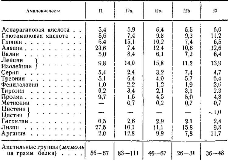
Table 11 Main Characteristics of Individual histone fractions (Butler, 1966)
|
Main characteristics |
f1 |
f2a1 |
f2a2 |
f2b |
f3 |
|
Lysine/arginine ratio |
10-13 |
<1 |
1,1-1,20 |
About 2 |
<1 |
|
Main N-terminal groups |
Acetyl |
Acetyl |
Acetyl |
Proline |
Alanine |
|
Main C-terminal groups |
Lysine |
Lysine |
Lysine |
Alanine |
|
|
High amino acid content |
Proline and alanine |
Glycine |
— |
Glutamic acid |
|
|
Molecular weight: |
|||||
|
1) sedimentation, |
9 000-15 000 |
— |
25 000—37 000 (pH 1-7) |
About 55 000 |
|
|
2) N-terminal+acetyl |
12 000-14 000 |
10 000 |
20 000 |
10 000-11 000 |
10 000 |
In addition to these features common to all histone fractions, each of them exhibits interesting differences in amino acid composition. Thus, the relative amounts of lysine and arginine vary significantly for each fraction, although the sum of these amino acids remains approximately constant. Therefore, as mentioned earlier, the lysine/arginine ratio is a strictly defined value for each fraction. For f1, it is 10–13; for f2a1, less than 1; for f2a2, 1.2; for f2b, 2; and for f3, less than 1 (see Table 11). Furthermore, each of these fractions is distinguished by an elevated content of one or two non-basic amino acids. For instance, lysine-rich histones are characterized by a high content of alanine (up to 24 mol. %) and proline (up to 10 mol. %). The f2a fraction features an increased glycine content (up to 13 mol. %), which is why it is often referred to as the glycine-rich histone, whereas the f2b fraction is marked by a high serine content (7.4 mol. %). Finally, The amino acid composition of arginine-rich histones is characterized by an elevated glutamic acid content (11 mol. %). Interestingly, in addition to standard amino acids, ε-N-methyllysine has been detected in the latter two fractions. The content of this amino acid is low (0.3–0.8 mol. %), and its presence is associated with the histone methylation process (see § 6).
The primary N-Terminal Groups of the f1 histone fraction are proline and alanine (50% and 35% of all N-terminal amino acids, respectively). For the f2a fraction, they are represented by alanine, proline, glycine, and lysine (54%, 12%, 11%, and 11%, respectively). The molecular weights of these fractions, determined from N-terminal amino acids (see Tables 8 and 11), turned out to be significantly higher than the values measured via osmotic pressure and sedimentation rate. For example, the molecular weight values for f1 found by Physical and Chemical Methods were 15,000 and 30,000, respectively, and for the f2a fraction, 15,000 and 112,000. As demonstrated by Phillips, the reason for this discrepancy was that the N-terminal groups in fractions f1 and f2a were acetylated. In the case of fractions f2a1 and f2a2, even the COMPOSITION OF THE Peptides isolated from their tryptic hydrolysates and containing the acetyl group (N-Acetyl—Ser—Gly—Arg) is known. When calculating the molecular weights of histones f1 and f2a taking into account the degree of N-terminal acetylation, results were obtained that closely matched ultracentrifugation data (see Table 11).
The N-terminal groups of fractions f2b and f3 are proline (80–82%) and alanine (95%), respectively. The relatively low heterogeneity of these histone fractions with respect to N-terminal amino acids allowed their molecular weight to be estimated at 10,000–11,000. The C-terminal groups are lysine for histones f1, f2a2, and f2b; glycine for fraction f2a1; and alanine for the arginine-rich histone f3. Thus, the entire set of C-terminal groups across the major histone fractions reduces to just Three amino acids.
Let us now turn to another feature of the Introduction/19.html">Primary Structure of histones—the distribution of basic, acidic, and non-polar amino acid residues along the polypeptide chain. The principal METHODS FOR STUDYING this are electrometric titration of histones and their enzymatic Cleavage by Trypsin. It is well established that trypsin cleaves only those bonds formed by the carboxyl groups of lysine and arginine and the amino groups of Other Amino Acids. Therefore, the analysis of peptides from tryptic hydrolysates makes it possible to evaluate the intervals (the number of non-basic amino acids) between basic residues in the original chain. For instance, the detection of the Ala—Arg peptide, which is not N-terminal, implies that the original protein must contain the sequence Lys—Ala—Arg (Arg—Ala—Arg), with a single non-polar residue positioned between two basic amino acid residues.
Studies by A number of authors have shown that Hydrolysis of fractions f2a and f3 with trypsin yields, alongside soluble peptides, a precipitate known as the "core". These precipitates, accounting for 27% of the original protein, contained nearly half as many basic Amino acids as the original histone fractions, but were much richer in dicarboxylic and non-polar amino acid residues, such as alanine, leucine, isoleucine, and valine. In the soluble hydrolysate of fraction f3, alongside free lysine and arginine, Peptides of the following composition were found:
|
Ala—Lys |
Lys—Arg |
|
Ala—Arg |
Gly—(Arg, Glu2)—Arg |
|
Asn—Lys |
Gly—(Ser, Gly, Thr)—Lys |
This indicates that a certain proportion of lysine and arginine forms doublets of the Lys—Arg, Lys—Lys, and Arg—Arg types, and that the intervals between such doublets span from 0 to 4 non-basic amino acids. Taking insoluble peptides into account, the intervals between basic amino acids extend up to 7 other residues. For fractions f2a1 and f2a2, the average intervals between basic Amino acids are 3–9 other residues. Given the relatively low content of basic amino acids in the core of the tryptic hydrolysate of fractions f3 and f2a, it can be concluded that lysine and arginine residues are irregularly distributed along the polypeptide chain and clustered closely together in specific regions. This Conclusion is in good agreement with electrometric titration data. This method demonstrated that imidazole, carboxyl, and tyrosine groups titrate at pH values close to the pK values of these groups, whereas the pK values for guanidino and ε-amino groups were significantly lower. The latter can occur only at a high concentration of positive charge, indicating that lysine and arginine residues are clustered very closely together on the polypeptide chain. The normal pK values observed for carboxyl groups indicate that these groups are not located near regions of high positive charge. Analysis of the core peptides of fraction f3 revealed a high content and frequency of dicarboxylic and non-polar amino acid residues.
However, until recently, it remained unclear whether the basic amino acid clusters are located in different Regions of the polypeptide chain of arginine-rich histones or whether they are shifted toward one of its ends. An answer to this question came from studies on the primary STRUCTURE OF THE arginine- and glycine-rich histone (fraction f2a1 or IV). Research by Bonner's group and Bush's group demonstrated that the N-terminal half of this protein molecule contains 15 of the 26 basic amino acids, with many of them gathered into clusters of 3–5 consecutive residues. On the other hand, the C-terminal portion of the molecule concentrates all aromatic amino acids, about half of the dicarboxylic amino acids, and 19 of the 32 non-polar residues. Such a distribution of residues suggests that the N-terminal part of the f2a1 histone molecule may be involved in binding to DNA, whereas the C-terminal region presumably provides the specific conformation of the histone itself and its interaction with other agents.
The lysine-rich histone fraction f1 does not form an insoluble precipitate upon tryptic hydrolysis, and one-third of the hydrolyzate peptides contain lysine as their N-terminal group. Consequently, there must be at least 7 Lys—Lys sequences per every 100 amino acid residues, which is equivalent to approximately 50% of all lysine residues. Thus, for fraction f1, the possible existence of 15 Lys—Lys sequences is postulated per protein Molecular Weight of 21,000. Furthermore, peptides containing more than one lysine residue at the N-terminus were detected in the tryptic hydrolyzate. Clearly, alongside basic amino acid doublets, fraction f1 also contains Lys—Lys—Lys triplets. According to Johns, acid degradation of fraction f1 yielded 80% fragments composed of alanine, proline, and lysine, with an average interval between basic amino acids equal to 1.7 non-basic residues. Evidently, in lysine-rich histones as well, lysine residues are distributed unevenly along the polypeptide chain and clustered into "blocks". This assumption is supported by the data of Bustin et al. on The structure of the very lysine-rich histone. These authors showed that the carboxyl-terminal half of the protein molecule contains 45 of the 61 lysine residues and about 3/4 of the proline residues. Conversely, the amino-terminal portion of the molecule is enriched in dicarboxylic amino acid residues (about 3/4 of the residues) and contains numerous hydrophobic radicals. It should also be noted that about 1/4 of the molecule, represented by two peptides, is completely devoid of proline. These two peptides are located close to the middle of the polypeptide chain within its H2N-half. Such an arrangement of amino acid residues in the histone suggests that the carboxyl-terminal region of the molecule may serve for specific interaction with DNA, whereas its amino-terminal part is likely involved in interaction with certain other agents, such as those implicated in repression and derepression processes. In the tryptic hydrolyzates of fraction f2b, a relatively small amount of insoluble peptides was found (about 10% of the original protein), containing a significant number of dicarboxylic and non-polar amino acid residues. The distances between basic amino acids here range from 3 to 6 other residues, which is very close to that observed for arginine-rich histones.
Thus, these studies demonstrate that all histone fractions are characterized by an irregular arrangement of basic amino acids, with intervals between them ranging from 0 to 9 other residues. In this respect, histones do not differ from other Proteins, such as Hemoglobin, Myoglobin, Lysozyme, and Ribonuclease, for which these intervals range from 0 to 22. However, the degree of irregularity is significantly higher in histones, since the bulk of diamino and dicarboxylic acids in them appear to be clustered in different halves of the molecule. Moreover, individual histone fractions also differ from one another in the heterogeneity of polar and non-polar residue distribution along the polypeptide chain. As electrometric titration data show, the degree of association of basic amino acids is highest in histones f3 and f2a and considerably lower in fraction f2b. As for fraction f1, it is currently difficult to assess its position among other histones regarding the degree of irregularity in basic residue placement, since the primary structure of this protein is still unknown. Nevertheless, characteristic differences in the primary structure of the f1 histone compared to other histone fractions can already be noted. These differences consist of an exceptionally high proline content, a relatively low content of dicarboxylic amino acids, and probably a relatively uniform distribution of hydrophobic residues within the polypeptide chain.
All these Features of the primary structure of individual histone fractions are reflected in the conformation of their molecules. Initial measurements performed on solutions and oriented films of these proteins demonstrated that the Secondary structure of histones is highly labile. For instance, infrared (IR) spectra of histones extracted from DNP with acid revealed only a single amide I band in the region of 1657 cm-1. However, when histones were precipitated from the extract with ethyl alcohol, along with the amide I band at 1657 cm-1, a band in the 1634 cm-1 region was also observed*. This band dominated THE SPECTRUM OF oriented films obtained from aqueous histone solutions and decreased in intensity when histones were dissolved in Ethylene chlorohydrin (a solvent favoring α-Helix formation). Optical rotatory dispersion measurements also showed that up to 60% of the protein existed in a helical state in ethylene chlorohydrin solution, whereas In aqueous solutions the proportion of the α-helix dropped to 20%. Thus, these studies demonstrated that a significant portion of native histone exists in the form of an α-helix, which readily transitions to a β-structure upon exposure to ethanol or storage in dilute aqueous solutions.
* Vibrations of the amide group give rise to a series of characteristic bands in the IR spectrum of proteins. Some of these, namely amide I (1660–1620 cm-1) and amide II (1560–1520 cm-1), are sensitive to the conformation of the polypeptide chain. The α-helix is associated with the bands at 1657 cm-1 for amide I and 1555 cm-1 for amide II, whereas the β-structure is associated with the bands at 1634 and 1530 cm-1, respectively.
Similar results were obtained in the analysis of individual histone fractions. Work by Bradbury, Murray, and Peacock proved that in aqueous solutions, the degree of helicity of all histone fractions is low, and for f1 it is equal to 0. An increase in the Ionic strength and pH of the solution increased the degree of helicity of histone fractions f2a, f2b, and f3 due to a decrease in charge and repulsion between similarly charged basic groups. However, even under these conditions, lysine-rich histones exhibited an extremely low Fraction of Helical segments (about 6%). On the other hand, an increase in optical rotatory dispersion for arginine-rich histones was observed not only upon increasing the pH, but also when it was shifted toward the acidic side. Obviously, the increase in the degree of helicity in the latter case is associated both with a reduction in the charge of carboxyl groups and with the screening effect of chloride ions on the positively charged groups of the protein.
All this indicates that the basic amino acids in histones f2a, f2b, and f3 are incorporated into those regions of the polypeptide chain that form the α-helix, and that these residues are positioned relatively close to one another. The dependence of histone helicity on The ionization of carboxyl groups suggests that acidic residues are also components of these same helical segments. Presumably, interactions between basic and acidic groups stabilize the helical regions through electrostatic attraction or salt-link formation. The low dicarboxylic amino acid content and the exceptionally high proportion of proline in lysine-rich histones explain the low degree of helicity in these proteins even at high pH and ionic strength. Finally, nuclear magnetic Resonance spectroscopy proved that the helical domains of histones f2a, f2b, and f3 also contain certain amounts of non-polar residues.
Comparing data on optical rotatory dispersion, electrometric titration, and viscometry of histones, along with the dependence of these parameters on solution pH and ionic strength, Ramm, Vorobiev et al. suggested that histone molecules possess a statistical coil conformation. At neutral pH, their polypeptide chains contain both helical and coil-like regions (Fig. 17). As already mentioned, acidic and non-polar amino acids are incorporated alongside basic residues for the first time. The coil-like regions of the polypeptide chain apparently contain A large number of basic residues and exhibit a high positive charge density at neutral pH. Consequently, these regions do not form helices in aqueous solutions and acquire an α-helical conformation only to a minor extent upon increases in solution pH and ionic strength. It is also possible that these domains are enriched in non-polar amino acids, which can act as destabilizers of the helical structure.
Thus, the degree of helicity of individual histones and the arrangement of helical and amorphous regions are determined by the distribution of charges and non-polar radicals along the polypeptide chain, i.e., by the primary structure of the protein.
At the same time, the sizes of these regions and their locations within the molecule vary among different histones. For instance, fraction f2b, where the degree of clustering of basic and non-polar residues is lower than in arginine-rich histones, apparently contains a significant number of short helical segments. In arginine-rich histones, the number of helical segments is smaller while their relative length is greater. Fraction f1 contains almost no helical domains and is characterized by an extended conformation even at high solution pH and ionic strength.
As will be shown below, these differences in the Primary and secondary structure of histone fractions are directly related to the Specific features of their interaction with DNA and the role they may play in Chromatin Organization.
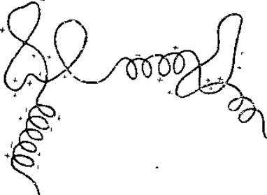
Fig. 17. Model of a histone molecule (Ramm et al., 1970).
Last update: 06/08/2026
Editorial and Educational Adaptation: This material has been compiled based on the primary/original source text. The project team performed an editorial review, corrected technical inaccuracies, structured sections, and adapted the content for an educational format.
What was processed:
- elimination of formatting defects (OCR errors, structural breaks, corrupted characters);
- editorial organization of content;
- standardization of terminology in accordance with academic sources;
- verification of factual statements against the original source text.
All mentions of the author, publication year, and origin of the primary text have been preserved in accordance with the source.