Protein Chemistry - Part 2 - Selected Topics in Special Protein Chemistry - Ashmarin, I. P. 1968
Histones
Histone-DNA Interaction
It is now well established that Histones are exclusively localized in the nuclei of differentiated Cells, where the bulk of them is associated with Chromatin. Various cytochemical techniques demonstrate a close spatial relationship between chromatin DNA and histones: all chromosome regions characterized by a high DNA content also exhibit a high concentration of histones. Analysis of isolated chromatin reveals that histones and DNA are present in approximately equal weight proportions. Macerating chromatin in a 4 M cesium chloride solution followed by centrifugation separates histones from DNA, completely disrupting the Chromosome Structure. If chromatin undergoes brief homogenization in a low Ionic strength medium (0.01 M Tris-buffer, pH 8), vigorous agitation, and centrifugation at 10,000×g, 70–80% of the chromatin DNA passes into solution. This DNA is fully bound to histones (with a histone-to-DNA mass ratio of approximately 1.35), leading to the designation of the preparation as soluble nucleohistone. Soluble nucleohistone consists of discrete DNP particles with a Molecular Weight of roughly 19∙106 and a length of 4200 Å. Given that the corresponding values for free DNA are 8∙106 and 7100 Å, it was concluded that the DNA chain in nucleohistone is folded and shortened. A second key feature of nucleohistone is that the bound histone stabilizes the DNA, increasing its melting Temperature (Tm).* For instance, the Tm of pea seedling DNA in 0.16 M sodium chloride is 70°C. However, when DNA is complexed with histones (as nucleohistone), its Tm rises to 84°C (Fig. 18). Like chromatin, nucleohistone dissociates into DNA and histones in a high ionic strength environment (1.5–2.0 M sodium chloride solution).
Thus, all these findings indicate that histones play a vital structural role in chromatin, where they are bound to DNA molecules. This binding is driven by Electrostatic Interactions between the phosphate groups of DNA and the basic groups of histones, resulting in the stabilization and conformational alteration of the DNA molecule within the resulting DNP complex.
* Nitrogenous bases in DNA are stacked like a pile of coins. Due to π–π interactions between them, this system absorbs ultraviolet light more weakly than an equivalent amount of free NUCLEOTIDES (the hypochromic effect). If a DNA solution is heated gradually, a temperature is eventually reached at which Hydrogen Bonds break, base interactions vanish, and the double-helical structure of DNA collapses. Light absorption by the system then rises to a level characteristic of an equivalent quantity of free bases (the hyperchromic effect). DNA melting occurs over a specific temperature range, which is characterized by the melting temperature (Tm) at which 1/2 of the total hyperchromicity is achieved.
Class="center">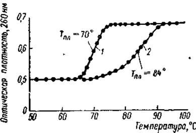
Fig. 18. Melting curves of free pea embryo DNA (1) and native nucleohistone (2) (Bonner, 1967).
What is The structure of this complex?
To address this question, we must first examine the Specificity of interaction between individual histone fractions and DNA. Work by Johns and Butler demonstrated that any of the major histone fractions can bind to bulk DNA, precipitating it in the process. Lysine-rich histones were able to precipitate twice as much DNA in a 0.14 M sodium chloride solution as the other fractions, among which few differences were observed in this regard (Fig. 19). No specific interactions driving the association of a particular histone fraction with a specific region of DNA were detected. At the same time, these and subsequent experiments showed that the interaction between DNA and histones depends on the concentration of the latter and the ionic strength of the medium. It turned out that lysine-rich histones cannot precipitate all the DNA from an aqueous solution even at a histone-to-DNA ratio of 1.6. Conversely, when the interaction took place in 0.14 M sodium chloride, complete DNA precipitation was observed at a protein-to-DNA ratio of 0.8.
On the other hand, a nearly 7-fold decrease in ionic strength (0.02 M NaCl) did not significantly affect DNA precipitation by lysine-rich histones, provided their concentration was sufficiently high (protein/DNA = 1.6). For Arginine-rich histones, the extent of DNA precipitation dropped markedly as ionic strength decreased, even at high protein concentrations in solution.
These differences in the dependence of DNA precipitation on ionic strength and protein concentration between lysine-rich and arginine-rich histones stem from variations in the number and nature of cross-links within the resulting complexes. As noted above, approximately 75% of all amino acid residues in lysine-rich histones are non-basic. Furthermore, 3/4 of all basic residues cluster in the C-terminal half of these protein molecules, forming sequences of two, three, or more lysine residues. The NH2-terminal half contains many non-polar residues and the bulk of dicarboxylic Amino Acids, which also form clusters. Consequently, the basic groups of the carboxy-terminal half can presumably sequentially neutralize the DNA phosphate groups, while the lysine-rich histone molecule itself likely resides within the major groove of the DNA. Assuming a molecular weight of 22,000 for the f1 fraction, a lysine-rich histone molecule contains about 60 basic amino acids—sufficient to sequentially neutralize the phosphate groups of roughly 30 nucleotide pairs, or about three turns of the DNA helix. Meanwhile, 75% of The amino acid residues in the f1 fraction likely take no part in this interaction, and the spatial arrangement of this protein segment relative to DNA depends on various factors.
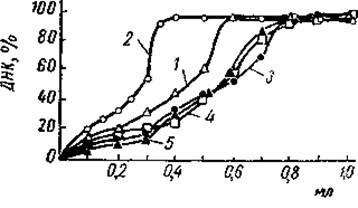
Fig. 19. Precipitation curves of DNA from solutions using total calf Thymus histones and individual histone fractions (Johns & Butler, 1964).
The ordinate represents the fraction of precipitated DNA, and the abscissa represents the volume of histone solutions added to 5 ml of DNA solution. 1 — whole histone, 2 — f1 fraction, 3 — f2a fraction, 4 — f2b fraction, 5 — f3 fraction.
Thus, at relatively low protein concentration and ionic strength, the entire lysine-rich histone molecule apparently lies within the major groove of DNA, spanning about 12 turns of The Double Helix. Under these conditions, 75% of the phosphate groups in such a "masked" DNA region remain accessible to other polycations. The f1 molecules maintain an extended conformation and are separated by free stretches of DNA. As the histone concentration in the deep groove of the DNA increases, only the C-terminal portion of the protein molecule remains there, while the remaining 75% of residues protrude outward as loops and "tails." Such protrusions have been detected via Electron Cell/15.html">Microscopy in DNP fibrils stripped of all histones except f1. These loops and protrusions are capable of interacting with other DNP Proteins and, presumably, with each other, altering their conformation and cross-linking DNA molecules into an insoluble complex.
The Nature of this binding remains unclear to this day. Many authors suggest that these loops form protein bridges between DNP particles. Alternatively, evidence indicates that insoluble DNP complexes are formed through non-covalent interactions (hydrogen, hydrophobic, electrostatic bonds) between such protrusions. The dissolution of DNP in 4 M urea, alongside the dependence of DNA precipitation on ionic strength and f1 histone concentration, strongly Supports this hypothesis. The failure of the DNA–f1 histone complex to precipitate at high histone concentrations under low ionic strength conditions is likely related to electrostatic repulsion between DNP particles due to numerous loops bearing identically charged carboxyl groups. Conversely, the precipitation of the soluble complex upon raising the ionic strength can readily be explained by the shielding of some of these groups, as well as the free phosphate groups of DNA.
Arginine-rich histones are similarly characterized by the clustering of 3/5 of their basic residues in the NH2-terminal half of the molecule, whereas all aromatic amino acids, the bulk of non-polar residues, and about 50% of acidic residues are concentrated in the C-terminal half. However, the arginine-rich histone molecule is much more compact than f1 because it contains a significant number of helical segments. As noted above, these segments are rich in basic, dicarboxylic, and non-polar residues. In such regions, each turn of the α-Helix averages about one protruding basic group, with relatively short distances between them. Consequently, these helical segments of arginine-rich histones can presumably sequentially bind a series of phosphate groups on both DNA strands, fitting snugly into the major groove of the double helix. Meanwhile, regions enriched in carboxyl or hydrophobic radicals likely form loops in the polypeptide chain that project onto the complex surface. Hydrophobic interactions between these loops may promote the Condensation and supercoiling of the DNA molecule within chromatin and DNP. At the same time, it is plausible that some surface loops are enriched in basic groups, facilitating the cross-linking of individual DNA particles. However, the size of these loops and their number in arginine-rich histone molecules are apparently small, meaning that achieving DNA precipitation requires considerably more f3 histone than f1 under otherwise identical conditions. This lower degree of cross-linking likely explains the greater sensitivity of DNA precipitation by these histones to ionic strength and protein concentration.
The features of Primary and secondary structure dictate not only the spatial relationships between individual histones and DNA, but also the strength of their binding. It is well known that adding a small amount of DNA to a large pool of mixed histones does not bind histones in the ratio observed in vivo; instead, the DNA preferentially selects those that exhibit a higher affinity for it. Arginine-rich histones bind to DNA first, while lysine-rich histones bind last. Determining the Equilibrium Constant for DNA-histone binding via equilibrium dialysis likewise demonstrated that fraction Ib has a lower affinity for DNA than fraction IV. Finally, numerous researchers have shown that fraction f1 can be extracted from DNP using 0.1 M citric acid, 0.4–0.6 M sodium chloride, or pH 1.5, whereas extracting the other fractions requires a sodium chloride concentration of 1–1.5 M and pH 0.7.
This relatively low affinity of lysine-rich histones for DNA stems from the fact that their binding is driven primarily by ionic interactions between the basic groups of the histone and the phosphate groups of DNA. According to Frierson and Kirby, however, certain lysine-rich histones also bind DNA via hydrogen bonds. In the case of arginine-rich histones, these forces are supplemented by robust hydrophobic interactions from the tails localized on the complex surface. Furthermore, non-polar amino acid radicals clustered within the helical segments of arginine histones can interact with the hydrophobic bases of DNA, further stabilizing the structure and reinforcing its bond with the histone. Finally, arginine-rich histones possess a higher number of basic amino acids per protein molecule than lysine-rich histones, and the pK of the guanidine group of arginine significantly exceeds the pK of the ε-amino group of lysine (11.6–12.6 versus 9.4–10.66, respectively). Consequently, arginine-rich histones exhibit substantially higher basicity and, As a result, a much stronger bond with DNA phosphate groups. Histone fractions f2a and f2b occupy an intermediate position in this regard, resembling arginine-rich or lysine-rich histones, respectively.
All of these data were obtained using soluble reconstituted nucleohistones. The latter were prepared from isolated histone fractions and DNA via gradient dialysis, during which the sodium chloride concentration in the medium was gradually lowered from 2 to 0.4 M. Under these conditions, weak yet noticeable gel formation occurred. However, prolonged incubation of the system in 0.4 M sodium chloride caused the DNP gel to transition into a soluble form. According to Bonner, this occurs because histone molecules, detaching from and reattaching to DNA in a medium of moderate ionic strength, eventually find the most stable conformational states that correspond to maximum complex solubility. In several properties, these complexes resembled native nucleohistones (having a histone/DNA ratio of 1.35 and a sedimentation coefficient of 26S), exhibited an elevated DNA melting temperature, and did not dissociate into their component parts upon prolonged centrifugation at 350,000×g (an acceleration sufficient to pellet DNA but not free histones). Light-scattering studies of these complex solutions revealed that nucleohistone reconstitution induces a conformational change in the DNA molecule. Specifically, the arginine-rich histone fraction f3 causes the DNA molecule to shrink, rendering its packing more compact. The lysine-enriched fraction f1 does not alter the length of the DNA molecule, which remains in the B-form within the complex. The latter is corroborated by the strong binding of actinomycin D to the DNA–f1 complex (actinomycin D is known not to bind DNA–RNA hybrids, where DNA assumes the A-form) and by minimal differences in dichroism between the native DNA and this complex.
Thus, compared to other histones, arginine-rich histones exhibit the highest affinity for DNA and induce the compaction of its molecule. These findings align well with the results of experiments investigating The Effect of individual histone fractions on DNA melting. As demonstrated by Shih and Bonner using reconstituted complexes, at a subequivalent histone-to-DNA ratio, DNA–histone IV recombinants possessed the highest melting temperature (83.7°C). For DNA–histone Ib complexes, the melting temperature was 76.3°C. The fraction of relatively lysine-rich histones occupied an intermediate position. Similar results were obtained using partially deproteinized DNP. It was found that stepwise removal of histones from DNP leaves its melting temperature virtually unchanged, provided the DNA remains bound to arginine-rich histones.
At present, explaining the exact effect of arginine-rich histones on DNA melting remains challenging. In the case of lysine-rich histones, the elevated melting temperature of the complex DNA is attributed to fraction f1 interacting with AT pairs, thereby stabilizing the structure. This stabilization can be explained as follows. It is well established that In aqueous solutions, AT pairs are the most solvated and their inter-strand bonds are weaker than those of GC pairs. The presence of regions rich in such solvated AT pairs introduces certain defects into the Introduction/20.html">DNA Structure. The attachment of Polyamines or lysine-rich histones to these regions presumably causes the dehydration of AT pairs and eliminates structural "imperfections" in the DNA. Direct confirmation of this principle comes from data by Ohba, who found that in nucleohistone—unlike deproteinized DNA—GC pairs are more sensitive to thermal Denaturation than AT pairs. He suggests that the stability of AT pairs in such a complex is conferred by the lysine residues of the histone. A similar observation was reported by Butler, who discovered that lysine-rich histones are typically associated with a DNA fraction containing abnormally high amounts of adenine.
Comparable results were obtained in experiments by G.P. Georgiev and co-workers investigating the luminescence of acriflavine complexes with DNA of varying nucleotide composition and with DNP. It was found that the quantum yield of acriflavine luminescence on DNA varies in proportion to the square of its AT-pair concentration. Consequently, the dye luminesces most intensely when adsorbed between AT–AT pairs. When acriflavine was adsorbed onto DNP, however, the luminescence quantum yield was significantly lower than on free DNA. Removing lysine-rich histones from DNP led to an increase in the luminescence quantum yield. Evidently, lysine-rich histones bind to the AT-rich regions of DNA. No such regularities have been found for arginine-rich histones.
The insights outlined above regarding the modes of interaction between DNA and individual histone fractions remain largely hypothetical, although they are supported by analyses of X-Ray Diffraction patterns, IR spectra, titration curves, and optical rotatory dispersion measurements of native deoxyribonucleoproteins. These studies indicate that lysine-rich histones form extended polypeptide chains, whereas arginine-rich and moderately lysine-rich histones feature substantial helical segments distinguished by a high content of basic, dicarboxylic, and non-polar amino acid residues. All carboxyl, imidazole, and Tyrosine groups of histones complexed with DNA remain accessible to electrometric titration. Conversely, 80% of the lysine and arginine residues are inaccessible to titration, likely because they are bound to the phosphate groups of DNA. Investigations of birefringence and dichroism in DNA, native nucleohistones, and partially deproteinized nucleohistones have shown that binding DNA to histones results in the shortening and supercoiling of the DNA molecule. This is evidenced by a decrease in the number of Base Pairs oriented perpendicularly to the molecular axis—a reduction reaching up to 60% compared to free DNA. This hypershortening of the DNA molecule is driven primarily by arginine-rich histones, since detaching a portion of these proteins from DNP triggers an increase in dichroism, Flow Birefringence, and the intrinsic viscosity coefficient.
Unfortunately, all these data still reveal little about the three-dimensional architecture of native nucleohistones. Studying this architecture is difficult because the DNP complex is non-crystalline and does not display a highly ordered structure under X-Ray Structural Analysis. Furthermore, depending on isolation conditions (the intensity of chromatin homogenization, the duration of storage in dilute saline or aqueous solutions), DNP can be obtained as a gel or as a soluble complex. The latter consists of individual DNP molecules containing double-stranded DNA with associated proteins. The structure of gel-form DNP remains unknown. It may consist of a single DNP particle folded and bent into loops, or alternatively, of individual DNP molecules. The Nature of the bonds linking different DNP fibers—or distinct Regions of the same DNP fiber—is likewise unclear. Consequently, models of DNP Organization are largely hypothetical and at first glance appear contradictory.
For instance, one of the earliest models proposed that nucleohistones form regular structures in which the polypeptide chain axis lies at an angle of 55° relative to the orientation of the DNA molecule. Since the grooves of the double helix run at an angle of roughly 58° to its axis, this model suggested that histones reside within the major groove of the DNA helix, wrapping around it like a bandage. However, this structure would require a relatively regular alternation of basic amino acid residues along the protein chain. Based on a reflection corresponding to a 37 Å period and on Infrared Spectroscopy results, Zubay proposed a lattice-like structure in which histone molecules partially occupy the major grooves of DNA while crossing its strands at an angle of 55–60° to the double-helix axis (Fig. 20). Finally, A number of authors suggest that the Tertiary Structure of nucleohistone arises from a system of histone bridges linking separate DNA molecules or distinct regions thereof.
In this arrangement, the helical segments of the histone chain may reside within the major grooves of the DNA helix, whereas the amorphous regions occupy the spaces between its molecules.
The existence of such histone bridges is supported by studies on Various Forms of DNP. As previously mentioned, depending on the chromatin Treatment, DNP can be obtained in either a soluble or a gel-like form. For a long time, it was believed that there were fundamental differences between them and that only one of them represented truly native DNP. For instance, Zubay and Doty considered only the finely dispersed soluble material to be native, whereas Itzhaki, Frederic, and other authors argued that the soluble form was a degradation product of the DNP gel. This Conclusion was based on the observation that the soluble DNP form contained less protein and lower molecular weight DNA. However, it was already shown at the time that both fractions contained DNP particles with similar parameters, and that the DNA and Amino Acid Composition of the histones were identical in both. This suggested that a high concentration of histones is necessary for The formation of the DNP gel network.
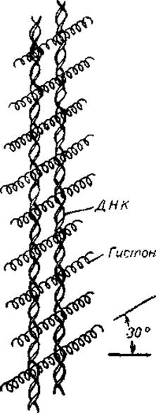
Fig. 20. Model of the nucleohistone, according to which histones form bridges between DNA molecules (Zubay, 1964).
Recent studies have shown that the differences between the soluble and gel forms are primarily related to Changes in the organizational level of DNP. Upon intensive fragmentation of chromatin or dilution of the DNP gel, the latter breaks down into separate nucleohistone components of varying size and structural complexity, which share a number of properties with the DNP gel (chemical composition, UV absorption spectra, melting profiles, birefringence, and dichroism). However, they differ in having a lower elastic modulus, a slightly lower protein-to-DNA ratio, and a lower DNA molecular weight. Apparently, the fragmentation of the DNP gel involves not only the loss of some protein but also the rupture of certain DNA strands. Similar components can be obtained by extracting a portion of the lysine-rich histones from the DNP gel; reversible association in this case restores DNP with its original properties. It is likely that these nucleohistone components are assembled into a three-dimensional gel network via cross-linking protein bonds, a role played by a fraction of the lysine-rich histones.
At the same time, work by G. P. Georgiev and co-workers, as well as by Chalkley and Hnilica, demonstrated that the destruction of the gel network can also be achieved without removing the lysine-rich histones. To do this, it is sufficient to treat the DNP gel with 4 M urea at low ionic strength. Under these conditions, DNP solubilization occurs without protein loss or any DNA degradation. It is known that urea disrupts Hydrogen bonds and affects hydrophobic interactions. Therefore, it can be assumed that bonds of this type are responsible for cross-linking between individual DNP molecules (or separate regions of a single molecule). Since, at the same time, the existence of the DNP gel is impossible without the involvement of lysine-rich histones, it can be hypothesized that the formation of such bonds proceeds with their direct participation. In other words, individual DNP particles are linked into a supramolecular gel structure not by the lysine-rich histone chains themselves, but through hydrogen bonds and hydrophobic interactions of these chains. Which of these models corresponds to reality is currently difficult to say. However, regardless of the nature of the bonds between DNP molecules (or regions), one thing is certain: lysine-rich histones play a decisive role in the Formation of the gel network.
Not only lysine-rich histones, but also other proteins may play a specific role in DNP organization. It is known that the elastic modulus of reconstituted gel-like DNPs is lower than that of native DNP gels. Upon dissociation and subsequent reassociation of native DNPs, gels with identical properties can be obtained. Presumably, histones alone are not yet sufficient to form the native DNP gel lattice. Complete removal of histones from DNP and Analysis of the remaining DNA showed that the latter is firmly bound to residual acidic proteins. These proteins contain a significant number of SH groups and disulfide bridges, which link DNA molecules and form a loose, three-dimensional fibrillar network. Such complexes of DNA and residual acidic proteins exhibit relatively high viscosity and elastic modulus, resembling native DNP gels. The Cleavage of —S—S— bonds leads to the complete loss of gel properties in these complexes. Obviously, residual acidic proteins are the component that, alongside lysine-rich histones, is essential for building the gel lattice. It is also possible that interactions of hydrophobic loops of arginine-rich histones play a role in the formation of DNP gels. In DNP, the bulk of the charged basic groups of histones are shielded by the phosphate groups of DNA. The remaining polar groups are no longer sufficient to shield non-polar radicals, whose interaction may facilitate the formation of DNP gels.
Cross-linking histone bridges and acidic proteins also play a significant role in chromatin organization. It is known that chromatin in The Nucleus exists in two forms: as dense condensed clumps and as diffuse regions consisting of freely extended fibers with a diameter of about 100–150 Å. Although condensed chromatin contains the bulk of the nuclear DNA, only diffuse chromatin is active in supporting RNA Synthesis. Work by Littau, Allfrey, Mirsky, and other researchers has shown that selective removal of lysine-rich histones from dense chromatin or mitotic Chromosomes leads to the disappearance of condensed clumps and the appearance of a fibril network. The removal of arginine-rich histones has no such effect. If all histones are removed from mitotic chromosomes, they consist of a loose network of fibrils. The addition of lysine-rich histones to such chromosomes results in the formation of dense chromatin. Arginine-rich histones, although bound by such histone-depleted chromosomes, leave them in the same loose state. All this indicates that lysine-rich histones can form links between individual DNP threads of chromatin, converting it into a condensed state.
However, this does not mean that the difference between condensed and diffuse chromatin is due to quantitative differences in lysine-rich histone content. Recent studies have shown that the histone-to-DNA ratio is virtually identical in condensed and diffuse chromatin. Electrophoretic analysis of individual histone fractions from both types of chromatin also revealed no differences in histone spectra. Obviously, the quantity and types of histones are practically identical in these chromatin fractions, and the transition from diffuse to condensed chromatin depends on a number of other factors. Such factors likely include acidic proteins, Phospholipids, and chromatin RNA. As demonstrated in the works of Bonner, Wang, Comings, Frenster, and other researchers, diffuse chromatin contains significantly more RNA and phospholipids, four times more phosphoprotein phosphorus, and twice as much acidic protein as condensed chromatin. Chromatin acidic proteins make up approximately 20% of its mass, consist of at least four fractions, exhibit a high turnover rate, and contain up to 0.14% phosphorus and up to 3.6% RNA. They are capable of forming stable complexes with both histones and DNA. Apparently, an increase in the amount and phosphorylation level of acidic proteins is the primary factor preventing the formation of lysine-rich histone bridges. It is also possible that acidic proteins, being bound to DNA, can directly act as regulatory proteins. Interestingly, the content of these proteins in sea urchin embryo chromatin increases during the transition from blastula to larva.
The data presented above merely indicate The Role of individual proteins in chromatin organization. However, they are still largely hypothetical and incomplete, failing to provide a clear picture of the "architecture" of this complex chromosomal substance. At the same time, they provide no insight into the arrangement of individual types of histone molecules along DNA strands in chromatin. Meanwhile, the question of whether each DNA molecule receives a full set of various histone types or binds only specific types of histones is of fundamental interest. Electrophoretic analysis of native DNPs, as well as the stepwise removal of histones from them followed by The Study of partially deproteinized DNP properties, have shown that the localization of histones along DNA strands in chromatin is complex. Alongside regions where DNA is bound to a single type of histone, there are chromatin areas where almost all types of histone molecules are associated with DNA. Direct electron microscopic observations confirm this conclusion. Using native chromatin fibrils from which all histones except the lysine-rich ones had been removed, Bonner and co-workers were able to demonstrate that subfractions of these proteins are located adjacent to one another and form protrusions on the DNA. These protrusions are regularly spaced relative to the chromosomal DNA at intervals of 805 Å. All other histone fractions are located within these intervals, with each histone type apparently covering a DNA segment of no more than 40–50 Å in length.
Thus, the arrangement of histones relative to chromatin DNA is characterized by a certain degree of specificity, despite the fact that their interaction appears to be purely ionic. How can this contradiction be explained? As will be shown below, the specificity of histone–DNA interaction and the specific regularity of histone arrangement relative to DNA in chromatin are ensured by an important component—vector RNA. By binding several histone molecules, this RNA can locate its complementary operator Gene and ensure the interaction of specific histones with the corresponding structural gene. In the case of lysine-rich histones, the base COMPOSITION OF THE DNA also plays a significant role.
Last update: 06/08/2026
Editorial and Educational Adaptation: This material has been compiled based on the primary/original source text. The project team performed an editorial review, corrected technical inaccuracies, structured sections, and adapted the content for an educational format.
What was processed:
- elimination of formatting defects (OCR errors, structural breaks, corrupted characters);
- editorial organization of content;
- standardization of terminology in accordance with academic sources;
- verification of factual statements against the original source text.
All mentions of the author, publication year, and origin of the primary text have been preserved in accordance with the source.