Protein Chemistry – Part 2 – Selected Chapters in Special Protein Chemistry - Ashmarin, I. P. 1968
Histones
Genetic Function of Histones
The putative Functions of Histones are quite diverse and can be divided into three main groups. These include structural functions (such as “cementing” DNA molecules together, stabilizing DNA, forming the chromosomal Skeleton, etc.), genetic functions (repression of Gene function, i.e., RNA Synthesis, blocking or stimulating DNA Synthesis, and functioning as ribonucleases), and several others (such as inhibitory effects on Cells and alterations in mitochondrial activity). Among these, the genetic functions of histones—and particularly the modulation of gene action—appear to be of the greatest interest.
The idea that histones might play a role in genome regulation was first suggested back in 1943 by the Stedmans, based on the observation that The amount of DNA in various Cell nuclei did not vary nearly as much as the proportion of histones and acidic Proteins (“chromosomin”). This hypothesis was later formulated into the concept that histones block those regions of DNA that are inactive in a given cell type. The “unmasked” regions of DNA remain active and determine the Specific characteristics of these cells.
This hypothesis was subsequently supported by the work of numerous investigators. For instance, Allfrey and Mirsky demonstrated that up to 80% of DNA can be removed from Thymus nuclei by DNase without significantly altering The rate of RNA synthesis. Clearly, the major portion of nuclear DNA does not participate in this process and remains in a repressed state. Similar results were obtained in experiments where Chromatin isolated from rat Liver cells was used as a template for RNA synthesis. As shown by Huang, Bonner, and other researchers, The activity of chromatin in supporting DNA-dependent RNA synthesis is only 1/5 of that of DNA prepared from the same chromatin (Fig. 21). Moreover, this transcriptional restriction is specific in nature. The latter was convincingly proven by Paul and Gilmour through RNA competitive Hybridization experiments. It turned out that RNA transcribed in vitro from nuclear DNA hybridizes with 40—50% of deproteinized DNA, whereas RNA synthesized on chromatin hybridizes with only 5—10% of free DNA. Obviously, only a fraction of chromosomal DNA is read by RNA polymerase. On the other hand, populations of RNA molecules,
generated from chromatins of various Tissues, show almost no competition with each other upon hybridization with denatured DNA and contain different base sequences. This indicates the differentiated activity of The Cell's hereditary material.
While chromatin itself was active in RNA synthesis in the presence of RNA polymerase and RNA precursors (GTP, CTP, UTP, and C14-ATP), the nucleohistone component of chromatin (the DNA—histone complex) lacked template activity. This also indicated that DNA exists in chromatin in two forms. The latter Conclusion is supported by direct experimental data as well. Thus, chromatin melting is a two-step process: the first step has a Tm corresponding to that of pure DNA, whereas the second has a Tm corresponding to nucleohistone (Fig. 22). In addition, pea seedling chromatin was successfully separated into two fractions, one of which (10—20% of chromatin DNA) was depleted in histone (DNA/histone ratio of 0.4) and actively supported RNA synthesis in the presence of its precursors and RNA polymerase. Finally, it was shown that the DNA-histone complex, in which the phosphate groups of the nucleic acid are completely neutralized by the basic residues of the proteins, cannot serve as a template for the RNA polymerase reaction.
Thus, all these data indicate that a portion of DNA is present in chromatin in a form capable of supporting RNA synthesis, whereas the other and larger portion (80—90%) is in the form of nucleohistone and is inactive in RNA generation. Obviously, this inactivity of the nucleohistone complex is due to the presence of the histone component. A number of experimental findings confirm this conclusion.
Class="center">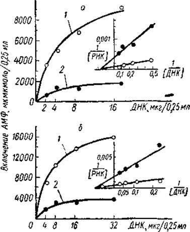
Fig. 21. Comparison of the template activity of rat liver chromatin and deproteinized DNA from the same chromatin (Marushige a. Bonner, 1966).
a — E. coli RNA polymerase content 30 mg, KM — 5.3 μg, 0.25 ml; b — E. coli RNA polymerase content 60 mg, KM = 10.5 μg/0.25 ml. 1 — DNA; 2 — chromatin.

Fig. 22. Melting curve of purified pea embryo chromatin (Bonner, 1967).
Selective removal of Lysine-rich histones (but not other proteins) from chromatin using a 0.4 M sodium perchlorate (NaClO4) solution dramatically increased its template activity. That this Treatment of chromatin activates previously repressed genes was proven by immunochemical Analysis of the synthesized proteins. Using histone-depleted pea seedling chromatin, E. coli RNA polymerase, four riboside triphosphates, and a ribosomal protein-synthesizing system, Huang and Bonner achieved the synthesis of proteins, among which pea seed globulin was identified. Under normal conditions, this protein was synthesized only in a system containing chromatin from developing cotyledons. Chromatin from pea seedlings could not serve as a template for the Synthesis of the necessary mRNA. Consequently, in seedling chromatin, the gene responsible for seed globulin production is repressed by lysine-rich histones. The latter constitute a significant portion of the total seedling histones and therefore presumably suppress the activity of many other genes as well.
Similar results were obtained by G. P. Georgiev and co-workers with Nucleoproteins from ascites cell nuclei. When lysine-rich histones were removed from them using a 0.6 M sodium chloride solution, a significant increase in the template activity of the nucleoproteins was observed, accompanied by an increase in the degree of hybridization of newly synthesized RNAs with the DNA of these complexes. Obviously, such nucleoproteins differed not only in their level of template activity, but also qualitatively, yielding RNA of a different composition.
The fact that histones, upon complexing with DNA, alter its ability to be transcribed by RNA polymerase has also been demonstrated by other studies. It is known that nucleohistones can be reconstructed from DNA and histones by co-dissolving them in a medium of high Ionic strength (1—2.5 M sodium chloride) followed by its reduction via dialysis. Such nucleohistones were reconstituted by Huang and Bonner, and by Barr and Butler, from pea seedling or calf thymus DNA and four histone fractions from the same sources. These complexes were tested for their activity in supporting DNA-dependent RNA synthesis. It was found that reconstituted nucleohistones exhibit varying capacities to support such synthesis. For example, nucleohistone containing lysine-rich histones practically lacked template activity, whereas complexes including Arginine-rich histones retained up to 66% of control activity. Histone fractions relatively rich in lysine and arginine occupied an intermediate position. Similar results were obtained in experiments by Hnilica et al. and A. G. Bukrinskaya et al., who studied RNA synthesis in Novikoff ascites tumor nucleoli and the incorporation of C14-uridine into chicken fibroblasts. Here too, lysine-rich histones inhibited RNA synthesis much more effectively than arginine-rich histones.
The inhibitory effect of histones and their individual fractions on RNA synthesis was also demonstrated in experiments by Allfrey and Mirsky on whole calf thymus nuclei. Along with suppressing the RNA polymerase reaction, histones inhibited a number of other nuclear processes, including ATP synthesis. Wishing to prove that histones truly alter the template activity of DNA rather than inhibiting RNA synthesis by blocking energy processes, the authors performed a series of experiments involving the removal of nuclear histones with Trypsin. To limit the enzyme's action and preserve nuclear Structure, a specific trypsin inhibitor, soybean trypsin inhibitor, was added to the system. Under these conditions, a 70% increase in RNA synthesis was observed, with the predominant type of newly synthesized RNA no longer being A—U-type, but rather G—C-type. All this indicated that histone removal activates previously repressed DNA regions with a different average base composition.
However, in one respect, the data of Allfrey and Mirsky differed significantly from the results of Huang and Bonner obtained on reconstituted nucleohistones. They showed that the most effective inhibitors of RNA synthesis in thymus nuclei are not the lysine-rich, but the arginine-rich histones (Fig. 23). The latter not only suppressed the genetic activity of Chromosomes more strongly, but also altered their Morphology. As demonstrated on lampbrush chromosomes of newt oocytes, even brief exposure to arginine-rich histones led to the retraction of chromosomal loops, which are sites of active RNA synthesis. In this respect, arginine-rich histones resembled actinomycin D.
Similar results were obtained by a number of other researchers. For instance, in Skalka's experiments, equal suppression of RNA synthesis in a system containing DNA, four riboside triphosphates, and E. coli RNA polymerase required five times more lysine-rich thymus histones than arginine-rich histones.
Thus, all these studies indicate that histones are precisely the inhibitors of DNA template activity. It is true that in some experiments, DNA-dependent RNA synthesis was most effectively inhibited by lysine-rich histones, while in others, by arginine-rich histones. It is possible that these discrepancies are due to unequal histone concentrations in the model systems used. It is known that the template activity of chromatin can be largely determined by its physical state, and that RNA synthesis proceeds much more intensely in diffuse (soluble) chromatin than in condensed (gel) chromatin. Since all these experiments were carried out in low ionic strength solutions, at a histone/DNA ratio of 0.8—1.5, the most active DNA repressors could be lysine-rich histones. Recall that the latter precipitate DNA most completely at the specified concentrations. At a greater excess of protein, arginine-rich histones might become the primary repressors, because the f1 fraction under these conditions could cause re-solubilization of the DNP complex and make it accessible for METABOLISM/31.html">Transcription. This assumption is supported by data from Jones and Forrester, who showed that in low ionic strength solutions, lysine-rich histones cause the dissolution of the DNA—f1 histone complex if the protein/DNA ratio exceeds 1.6.
On the other hand, the suppression of the RNA polymerase reaction by histones may also depend on the Structural Features of the histones themselves. This is indicated by model experiments on RNA synthesis in a system containing nuclear RNA polymerase. It turned out that under these conditions, the polymerase reaction was most strongly inhibited by both arginine-rich thymus histones and lysine-rich pea seedling histones. According to Allfrey and Mirsky, differences in the inhibitory action of individual histones on RNA synthesis are determined by such factors as surface charge distribution, degree of coiling, secondary structural changes, and degree of aggregation. In other words, the inhibitory effect of individual histones on the polymerase reaction may depend on the Specific features of their Primary and secondary structure. Substantial confirmation of this proposition is provided by a wealth of data showing that modification of histone structure through Acetylation, phosphorylation, or The formation of disulfide cross-links significantly alters their ability to suppress RNA synthesis (see § 7). It is also possible that the differing inhibitory effects of individual histone fractions may stem from their varying affinity for RNA polymerase itself.
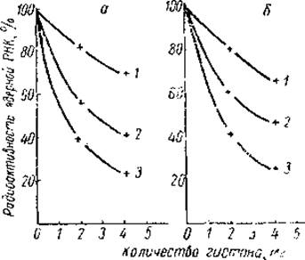
Fig. 23. Effect of histones on the incorporation of 8-C14-guanosine (a) and 8-C14-adenosine (ff) added to calf thymus nuclear preparations (Allfrey et al., 1963).
1 — fraction f1, 2 — total histones, 3 — fraction f3.
Finally, differences in the inhibitory action of individual histones may presumably be determined not only by the CHARACTERISTICS OF THE histones themselves, but also by The base sequence order in DNA. Some data regarding the enhanced affinity of lysine-rich histones for AT Base Pairs of DNA have already been cited above. Work by L. P. Ananieva, G. P. Georgiev, and co-workers has shown that the chromosomes of higher organisms exhibit a non-uniform recurrence of DNA nucleotide sequences. The restriction of transcription from DNA in DNP is specific in nature — transcription is restricted primarily on repetitive DNA nucleotide sequences. Treatment of DNP with 0.6 M sodium chloride, i.e., the removal of lysine-rich histones, leads to an enhancement of RNA synthesis precisely on these sequences. From this, it was concluded that lysine-rich histones are responsible for restricting transcription from repetitive DNA sequences, which presumably are enriched in AT base pairs. In this regard, data indicating that repetitive DNA sequences are topographically associated with compact chromatin, and that lysine-rich histones play a special role in organizing The structure of the latter, are of particular interest (see § 5).
The Role of DNA nitrogenous bases in their interaction with histones is also supported by experiments studying The Effect of polycations on transcription. Thus, Skalka, Gurvich, and Vidholm demonstrated that the histone-mediated suppression of the RNA polymerase reaction is proportional to the AT base content in the DNA template (the higher the A+T/G+C ratio, the lower the nucleotide incorporation into RNA at the same histone concentration in the system). The Nature of the synthesized RNA (the A+U/G+C base ratio and the frequency of neighboring nucleotide combinations) depended on both the type of inhibitor histone and the thymine and adenine content in the DNA. For example, when DNA with an A+T/G+C ratio of 1.8 was used as a template, the A+U/G+C ratio in the synthesized RNA was 3.15. Upon addition of lysine-rich histones to the system, this ratio decreased to 2.08, whereas arginine-rich histones increased it to 4.5—5.3. A significant reduction of AU pairs in the synthesized RNA upon reaction suppression by the f1 histone indicates an increased affinity of the latter for DNA AT pairs.
Thus, existing data indicate that in cell chromatin, DNA exists in two forms, one of which is inactive in supporting RNA synthesis (80—90% of total DNA). The suppression of DNA template activity is caused by histones, whose positive groups electrostatically interact with DNA. Different types of histones exert unequal effects not only on the intensity of DNA-dependent RNA synthesis, but also on the type of synthesized RNA, presumably by suppressing the activity of different genes. At the same time, the Repression of the RNA polymerase reaction depends not only on the histone type, but also on secondary Changes in the histone occurring during the modification of its primary and secondary structures. A certain role in this process may also be played by the histone/DNA ratio, as well as the ionic strength of the medium.
It is likely that the binding of lysine-rich histones is determined by the nitrogenous bases of DNA and occurs predominantly at regions enriched in AT base pairs. However, the selectivity of the interaction between Other types of histone and specific DNA regions (the Specificity of repression) has not yet been proven. Therefore, one of the fundamental questions in histone biochemistry is how the effect of histones on DNA is combined with specificity. To resolve this question, it is first necessary to determine whether histones themselves possess tissue and species specificity.
Last update: 06/08/2026
Editorial and Educational Adaptation: This material has been compiled based on the primary/original source text. The project team performed an editorial review, corrected technical inaccuracies, structured sections, and adapted the content for an educational format.
What was processed:
- elimination of formatting defects (OCR errors, structural breaks, corrupted characters);
- editorial organization of content;
- standardization of terminology in accordance with academic sources;
- verification of factual statements against the original source text.
All mentions of the author, publication year, and origin of the primary text have been preserved in accordance with the source.