Vertebrate Zoology - V. M. Konstantinov 2011
Chordates
Gnathostomes
Class Chondrichthyes - Subclass Elasmobranchii
The subclass Elasmobranchii comprises two primary groups: sharks (Selachomorpha) and rays (Batomorpha), which are generally classified at the superorder rank. They are characterized by the presence of placoid scales. Gill openings typically number 5 to 7 pairs, with each opening independently on the body surface. Spiracles are present. Due to the extension of the anterior end of the HEAD, known as the rostrum, the Mouth is positioned on the ventral side of the head as a transverse slit. The Skull is most commonly hyostylic, and more rarely amphistylic. The terminal section of the intestine is the cloaca.
To better understand the Structural Organization of elasmobranchs, let us examine the Anatomy of the shark.
Structural Features of Elasmobranchs
(using the shark as an example)
External Morphology. The body shape of most sharks is elongated and spindle-like. The anterior part of the head features a nasal projection, the rostrum. Eyes are well developed. Gill slits are visible along the sides of the head, usually numbering 5 on each side, though only a few modern species have 6 or 7. Two openings located behind the eyes lead into the Pharynx; these are the spiracles, which represent vestigial gill slits originally situated between the 3rd (mandibular) and 4th (hyoid) arches. The cloacal opening is located on the ventral surface of the body near the Base of the tail. The caudal fin is heterocercal, with THE Vertebral Column extending into its larger upper lobe.
Paired appendages—the pectoral and pelvic fins—are positioned horizontally. In males, the inner margins of the pelvic fins are modified into finger-like appendages called claspers, which function as copulatory Organs.
The integument consists of the epidermis and the cutis (or corium). The epidermis is stratified, containing numerous glandular Cells that secrete mucus onto the Skin surface. The corium is dense and fibrous. The skin is covered with placoid scales; each scale consists of a flat basal plate embedded in the fibrous layer of the dermis and a posteriorly directed spine resting upon it. The scale develops within the corium and is composed of osteodentin, a bone-like tissue structurally similar to the dentin of other vertebrate Teeth. Externally, the spine is covered by a thin layer of enamel, a derivative of specialized cells located in the basal layer of the epidermis. These scales cover the entire body and extend along the margins of the mouth onto the jaws, where they enlarge and function as teeth. This similarity in Cytology/cytology/67.html">Development and Structure highlights the Homology between teeth and placoid scales.
Skeleton. The endoskeleton of sharks remains cartilaginous throughout life, although calcium salts are deposited in certain regions.
The Axial Skeleton comprises the Vertebral Column and the neurocranium. The spine is divided into two regions: trunk and tail. The vertebrae are fully formed, consisting of a centrum and two arches. The centra are deeply concave at both anterior and posterior ends (amphicoelous vertebrae). The notochord persists within the cavities between adjacent centra and through a narrow canal running through the center of the vertebral bodies. The Spinal Cord runs through the neural canal formed by the dorsal arches.
Short Ribs articulate with the vertebrae, restricting the body cavity dorsally and laterally.
The neurocranium (Fig. 28) consists of the braincase, sense capsule cartilages, and the rostrum. The sense capsules are paired and firmly fused to the basal plate of the cranium. The cartilaginous roof of the braincase is incomplete, leaving a large opening in its anterior region—the anterior fontanelle—which is covered by a Connective Tissue membrane. The occipital region of the skull forms through the incorporation of the first vertebra into the cranium. The rostrum is composed of three rod-like cartilages extending from the anterior part of the skull.
Class="center">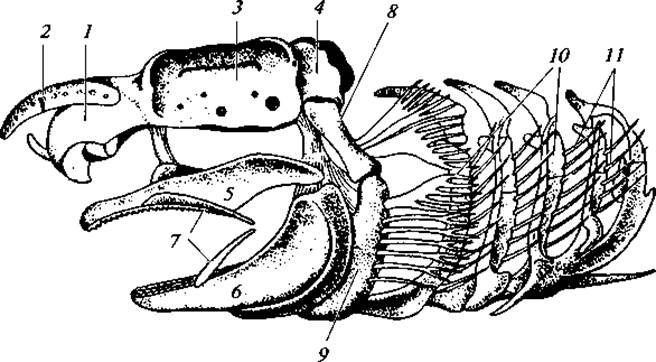
Fig. 28. SKULL OF A shark:
1 — olfactory capsule; 2 — nasal process (rostrum); 3 — Orbit; 4 — occipital capsule; 5 — palatoquadrate Cartilage (upper jaw); 6 — Meckel's cartilage (lower jaw); 7 — labial cartilages; 8 — hyomandibula; 9 — hyoid; 10 — branchial arches; 11 — gill rays
The visceral skeleton undergoes significant modification. Its arches are not connected to one another by horizontal cartilages, a condition that allowed for functional specialization, notably The formation of jaws. The visceral skeleton includes the mandibular arch, hyoid arch, branchial arches, and two pairs of labial cartilages.
The mandibular arch is composed of paired cartilages. The upper pair, termed the palatoquadrate cartilages, Functions as the upper jaws, while the lower pair forms Meckel's cartilages, which serve as the lower jaws. In most sharks, the palatoquadrate cartilage articulates with the neurocranium only anteriorly. Its posterior region lacks direct attachment to The Skull and is instead suspended from it via the upper element of the hyoid arch, the hyomandibular cartilage (hyostyly).
Positioned anterior to the mandibular arch are two pairs of small labial cartilages. These represent vestigial remnants of the First and Second visceral arches of ancestral primitive vertebrates, making the functional jaws of modern sharks the third visceral arch.
The hyoid (fourth) arch consists of paired hyomandibular cartilages, paired hyoid cartilages, and an unpaired connecting element called the basibranchial (or copula). The hyomandibular cartilage articulates with the cranium, the hyoid, and the mandibular arch. The copula connects the left and right hyoid components.
The branchial arches, which typically number five, are composed of four paired cartilages and one unpaired ventral cartilage situated along the midline of the body floor, joining the left and right sides. Cartilaginous gill rays project along the posterior margin of the branchial arches, supporting the interbranchial septa.
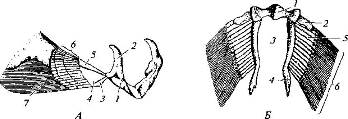
Fig. 29. Skeleton of the girdles and paired fins of a male shark:
A — Pectoral Girdle and pectoral fin: 1 — coracoid region; 2 — scapular region; 3, 4, 5 — basals; 6 — radials; 7 — ceratotrichia; B — Pelvic Girdle and pelvic fins: 1 — pelvic girdle proper; 2, 3 — basals; 4 — copulatory cartilage; 5 — radials; 6 — ceratotrichia
The skeleton of the paired appendages includes the girdle, embedded within the body musculature to provide structural support, and the skeleton of the free appendage (Fig. 29).
The pectoral girdle is represented by a cartilaginous arch lying freely within the Muscle mass, with its apex directed ventrally. Projections situated midway along each half (left and right) of the arch serve as the articulation points for the free limb skeleton. The Regions of the girdle dorsal to this projection are termed the scapular regions, while those ventral to it are the coracoid regions.
The skeleton of the free pectoral appendage comprises three distinct zones. The base of the fin contains three basal cartilages that articulate with the girdle. Attached to the basas are rod-like radial cartilages arranged in several rows, a structural arrangement known as a uniserial fin. Finally, long, slender elastoidin fibers (ceratotrichia) of epidermal origin extend outward from the radials.
The pelvic girdle (of the ventral fins) consists of an unpaired cartilage located within the musculature transversely across the body just anterior to the cloaca. The skeleton of the pelvic fin itself contains only one or two basalia. Radials attach to their outer margin.
The skeleton of the unpaired fins consists of radials and elastoidin fibers.
Digestive System. Movable jaws bordering the mouth opening typically bear rather large teeth. The Oral Cavity leads into the pharynx, which is pierced by gill slits. The aforementioned spiracles, representing rudimentary gill slits, also open into the pharynx. A short Esophagus opens into an arc-shaped Stomach, from which a short Small Intestine extends. The Pancreas lies within its mesentery. The Large Intestine has a considerable diameter and is equipped with a spiral valve. In sharks, the spiral valve serves the same function as in cyclostomes, but its structure features steeper folds, resulting in a greater number of turns. The trilobed Liver is provided with a Gallbladder, the Bile duct of which empties into the initial section of the small intestine. The digestive tract terminates in the cloaca.
As we can see, a general morphological feature of the digestive tract in cartilaginous Fishes that indicates its evolutionary advancement is a greater degree of division into distinct sections and a general elongation of the entire tract, associated with the formation of bends in the digestive tube.
Respiratory system. Each gill slit opens internally into the pharynx and externally on the body surface independently. The gill openings are separated from one another by broad interbranchial septa, within the thickness of which lie cartilaginous gill arches. Gill filaments are situated on the anterior and posterior walls of the gill slits, forming hemibranchs. Respiration is characterized by a countercurrent flow of Blood and Water.
Circulatory system. The Heart of cartilaginous fishes is two-chambered, consisting of an atrium and a ventricle (Fig. 30). A broad, thin-walled venous sinus, into which venous blood flows, is adjacent to the atrium. An arterial cone (conus arteriosus) is adjacent to the terminal part (along the blood flow) of the ventricle; it is morphologically part of the ventricle, although externally it appears as the Origin of the ventral aorta. The belonging of the arterial cone to the heart is evidenced by the presence within it (as in the other PARTS OF THE heart) of striated musculature. Blood pressure in the ventral aorta reaches 7–45 mm Hg.
The ventral aorta originates from the arterial cone; four pairs of branchial Arteries extend from it through the gills. Blood enters the gill filaments via afferent branchial arteries; oxygenated blood is carried away from the gills by efferent branchial arteries. The latter empty into an unpaired longitudinal vessel—the ROOT of the aorta—which pours blood into the main arterial trunk, the dorsal aorta. It lies beneath the spinal column and supplies blood to the Internal Organs. The carotid arteries, carrying arterial blood to the head, branch off anteriorly from the first pair of efferent arteries. At the level of the pectoral girdle, paired subclavian arteries branch off from the dorsal aorta, supplying blood to the pectoral fins. Venous blood from the head collects into paired anterior cardinal Veins. Venous blood from the tail collects into the caudal vein, which divides into two renal portal veins. After passing through the Kidneys, blood from the trunk collects into paired posterior cardinal veins, which merge at the heart level with the anterior cardinal VEINS OF THE corresponding side, forming paired Cuvierian ducts that then empty into the venous sinus. Blood from the intestine enters the subintestinal vein, which forms a hepatic portal system in the liver. From the liver, blood flows via the hepatic vein into the venous sinus.
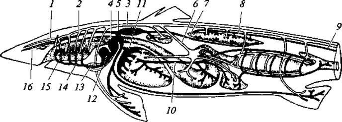
Fig. 30. CIRCULATORY SYSTEM OF the spiny dogfish (Squalus acanthias): 1 — carotid artery; 2 — epibranchial efferent artery; 3 — dorsal aorta; 4 — venous sinus; 5 — Cuvierian duct; 6 — celiacomesenteric artery; 7 — posterior cardinal vein; 8 — renal portal vein; 9 — caudal vein; 10 — HEPATIC PORTAL VEIN; 11 — hepatic sinus; 12 — atrium; 13 — ventricle with conus arteriosus; 14 — ventral aorta; 15 — afferent branchial artery; 16 — anterior cardinal vein
The Spleen, a hemopoietic organ, lies in the body cavity near The Stomach. Similar to the kidneys, it produces formed elements of the blood, such as erythrocytes, leukocytes, and others.
Nervous system. The Brain is relatively large. All its major divisions are well developed: the Forebrain, Diencephalon, Midbrain, Cerebellum, and Medulla Oblongata. Nervous Tissue is present on the floor, sides, and roof of the forebrain. The cerebellum is enlarged compared to that of lampreys.
Sense Organs. The paired olfactory sacs lie within cartilaginous capsules and communicate with the external environment through nostrils. Sharks possess extremely sensitive olfactory organs capable of detecting the scent of blood at a distance of about 0.5 km from the source. The paired eyes have a typical piscine structure: the cornea is flat, the lens is spherical, and eyelids are absent. A few species possess a nictitating membrane capable of pulling the Eyeball from its anteroventral margin toward the dorsal side. In most sharks and rays, cones are absent in the retina; therefore, they presumably do not distinguish colors, which is also reflected in the overall camouflage coloration of these fishes. The Organ of Hearing and Equilibrium is represented by the Inner ear—the membranous labyrinth. There are three semicircular canals. The fish perceives the direction of water currents through the mechanoreceptive lateral-line system, which is a system of canals located along the head and as lateral lines along the sides of the body. In most fishes, it is a canal lying within the skin and communicating with the external environment through closely spaced pores. In primitive sharks (Chlamydoselachus), the lateral line is an open dorsal groove.
Excretory system. The primary kidneys, or mesonephros, serve as the organs of excretion. Liquid metabolic wastes are eliminated both from the body cavity (via nephrostomes) and from the blood (via Malpighian corpuscles). Paired Wolffian ducts, which empty into the cloaca, function as their excretory ducts. Ammonia is excreted through the gills.
Reproductive System. The Ovaries in most sharks are paired. The paired oviducts (Müllerian ducts) are not connected to the ovaries, but open into the body cavity via funnels in the immediate vicinity of them. Shell glands are located in the upper section of the oviducts. The widened lower sections of the oviducts open into the cloaca. The genital and urinary tracts in females are separate throughout their length. Alongside oviposition, ovoviviparity is characteristic of sharks, during which a yolk Placenta develops (see Fig. 34).
The paired Testes of males are connected by a system of tubules to the anterior sections of the kidneys. These tubules pass through the renal tissue and empty into the Wolffian ducts, which serve males not only as Ureters but also as vas deferens. The anterior sections of the kidneys lack an excretory function and serve as epididymides (testicular appendages). Only the posterior sections of the kidneys function as excretory organs.
The Wolffian ducts are expanded in their lower section to form thin-walled Seminal Vesicles. Both Wolffian ducts empty into a urogenital sinus, which opens into the cloaca. Sharks exhibit various modes of reproduction, ranging from egg-laying to ovoviviparity and even the formation of a yolk placenta.
Last update: 13/08/2026
Editorial and Educational Adaptation: This material has been compiled based on the primary/original source text. The project team performed an editorial review, corrected technical inaccuracies, structured sections, and adapted the content for an educational format.
What was processed:
- elimination of formatting defects (OCR errors, structural breaks, corrupted characters);
- editorial organization of content;
- standardization of terminology in accordance with academic sources;
- verification of factual statements against the original source text.
All mentions of the author, publication year, and origin of the primary text have been preserved in accordance with the source.