GENERAL MICROBIOLOGY - T.P. Pyrog - 2004
5. STRUCTURE OF THE MICROBIAL CELL
5.2. MICROBIAL CELL MEMBRANES
5.2.1. Cytoplasmic Membrane
The Cell of any Organism contains various membranes that differ morphologically and functionally. Directly beneath The Cell wall lies the cytoplasmic membrane (Plasma Membrane, Plasmalemma).
This membrane covers the Cytoplasm and plays a vital role in the cell's life, serving as much more than just a structural morphological component. The membrane acts as the osmotic barrier of the organism, regulating the internal Osmotic Pressure of the cell. It mediates the selective transport of nutrients
from the environment into the cell and the export of Metabolic waste products. The membrane is the site where certain cellular structures are synthesized, notably the cell wall and the capsule. A number of Enzymes are localized within or associated with the membrane (Electron Transport and Oxidative Phosphorylation enzymes, which in eukaryotes are found in Mitochondria, are localized inside or On the surface of The Plasma Membrane in Bacteria; Components of the Electron Transport Chain — the Respiratory Chain — are found exclusively in membranes). The entire photosynthetic apparatus of purple bacteria is housed within the membrane. It is also highly likely that the METABOLISM/36.html">DNA Replication center is localized on the membrane.
Membranes can be isolated by subjecting protoplasts, obtained using Lysozyme, to osmotic Shock. The membrane is rich in Lipids, especially Phospholipids (Table 5.2). Comprising only 8-15 % of the cell's dry matter, membranes contain 70-90 % of all its lipids.
Class="center">Table 5.2.
Composition of membranes in certain bacteria
Components |
Content, % of dry membrane mass |
|
Micrococcus luteus |
Purple bacteria |
|
Lipids: |
28-37 |
40-50 |
neutral |
9 |
10-20 |
phospholipids |
28 |
30 |
50 |
50 |
|
Hexoses |
15-20 |
5-30 |
The Structure of microbial, plant, and animal cell membranes is remarkably similar, which provides grounds for THE CONCEPT OF a universal "unit membrane".
One of the classic early models of Membrane Structure was the Danielli–Davson–Robertson model (Fig. 5.5). The membrane consists of a lipid bilayer sandwiched between thin protein layers. The inner layer of the membrane is composed of lipids, which, as is well known, possess a polar (hydrophilic) end capable of ionization, and a non-polar (hydrophobic) end that is chemically a hydrocarbon chain. The lipids in the membrane are oriented with their hydrocarbon ends facing each other and their polar ends facing outward, thus forming a bilayer (bimolecular layer). A monomolecular layer of non-lipid nature, typically protein, is juxtaposed with the polar ends.
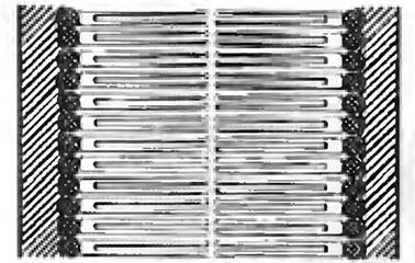
Fig. 5.5. Danielli–Davson–Robertson membrane model.
The diagram shows two layers of lipids: the Water-insoluble terminal groups (Fatty acids) point inward toward each other, while the water-soluble groups point outward (middle part of the diagram). The Lipid Bilayer is located between two protein layers (shaded strips)
The hypothesis that proteins are present in the membrane was first proposed by British scientists J.F. Danielli and H. Davson in 1935 to account for the low surface tension of cell membranes. Since a high surface tension would be expected at an oil-water interface, these researchers concluded that the Hydrophobicity of the lipid components must be counterbalanced by some hydrophilic protein. In 1959, based on the work of American scientist J.D. Robertson (Electron Microscopy revealed two electron-dense membrane layers separated by a less dense region), the unit membrane hypothesis was formulated. The presence of the electron-dense outer membrane layers was attributed to proteins associated with the hydrophilic surfaces formed by the lipid molecules. However, this model was contradicted by the results of freeze-fracture Electron Microscopy of membrane preparations, which demonstrated that protein molecules are not only located on the membrane surface but some also span it entirely. The fluid-mosaic model of membrane structure was proposed in 1972 by G. Nicolson and S.J. Singer (Fig. 5.6). According to this model, which is universally accepted today, Proteins can be envisioned as icebergs floating in a lipid sea.
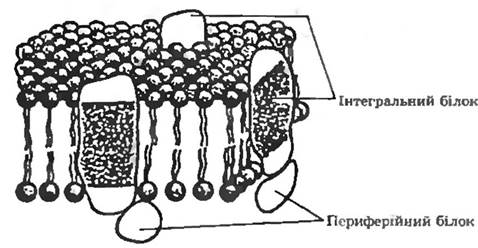
Fig. 5.6. Fluid-mosaic model of the plasma membrane by Nicolson and Singer.
Integral proteins are embedded in the lipid bilayer. Peripheral proteins are located on the membrane surface
Membrane Proteins. There are two Types of Membrane proteins: integral and peripheral (see Fig. 5.6).
Peripheral proteins are easily washed out of the membrane by mild detergents or even distilled water, as they are bound to the membrane surface. In contrast, integral proteins span the thickness of the membrane completely. As a rule, integral proteins exist in complexes with lipids. These Two Types of proteins differ in the arrangement of their hydrophobic amino acid residues. The surface of peripheral proteins is hydrophilic (these proteins are water-soluble), with hydrophobic amino acid residues buried inside the protein globule. In integral proteins, hydrophobic residues are localized on the surface, ensuring maximal interaction with the non-polar environment inside the membrane. However, in some integral proteins, polar groups are also located on the surface, interacting with the polar groups of lipids and with peripheral proteins.
According to their biological Functions, membrane proteins are broadly divided into three groups:
those possessing enzymatic activity;
those that specifically bind various substances required by the cell, i.e., proteins with receptor function (permeases);
structural, but they remain chemically understudied. It is known that all of them are poorly soluble in water due to the presence of large hydrophobic regions. This facilitates The formation of stable structures with lipids—Lipoproteins.
Membrane CARBOHYDRATES. There are few free carbohydrates in cell membranes; most carbohydrate residues are part of Glycolipids and Glycoproteins. The same Monosaccharides have been identified in their composition: galactose, glucose, N-acetylglucosamine, N-acetylgalactosamine, fucose, mannose, and xylose.
Membrane Lipids. Lipids are mainly represented by Phospholipids and glycolipids. Among phospholipids, phosphatidylglycerol and phosphatidylethanolamine are most commonly found in bacteria. Phosphatidylcholine and phosphatidylinositol are less common. Phospholipids contain phosphorus in their molecule, bound by two ester bonds. Phospholipids share one common component—glycerol, to which two long-chain Fatty acids are linked by ester bonds, along with a phosphorus-containing compound. The presence of two nonpolar fatty acid residues within membrane structures is their characteristic feature. In bacterial cultures, lipids, including phospholipids, predominantly contain saturated fatty acids (although unsaturated ones are also present). Branched-Chain Fatty Acids are also present in membranes (especially in sarcinae and micrococci).
The absence of sterols in bacteria is considered to be one of the features that distinguish bacterial lipids from the lipids of other microorganisms.
In addition to glycerol-containing lipids, Ethylene glycol is present in some compounds. Such lipids are called diol lipids. At high concentrations, diol lipids disrupt membranes. However, in very limited quantities, they only modify membrane properties, for example, by increasing permeability to small ions and molecules. Apparently, Cells exploit this property. For instance, during periods of rapid growth, they intensively synthesize diol lipids. When growth slows down, the synthesis of diol lipids ceases.
Glycolipids are carbohydrate derivatives of lipids.
The structure of membranes also includes divalent Metal Ions. It is assumed that they form chelate complexes with phospholipids, thereby imparting the necessary stability to the membrane through greater compactness.
Last update: 13/08/2026
Editorial and Educational Adaptation: This material has been compiled based on the primary/original source text. The project team performed an editorial review, corrected technical inaccuracies, structured sections, and adapted the content for an educational format.
What was processed:
- elimination of formatting defects (OCR errors, structural breaks, corrupted characters);
- editorial organization of content;
- standardization of terminology in accordance with academic sources;
- verification of factual statements against the original source text.
All mentions of the author, publication year, and origin of the primary text have been preserved in accordance with the source.