Pediatric Medical Genetics - S.I. Smiian 2003
Hereditary blood disorders
Hereditary diseases occupy a leading place in The Structure of pediatric hematological pathology. Recent advances in genetics have led to the refinement of diagnostic Methods, which has improved the quality of medical care for these children. Hereditary Disorders of the Blood system are characterized by impaired hematopoiesis at various levels (Hemoglobin synthesis, clotting factors, erythro-, leuko-, and thrombocytopoiesis). In particular, hereditary thrombocytopathies are diagnosed in 60-80 % of children presenting with vascular-platelet bleeding. According to the WHO, there are over 100 million people worldwide with structural hemoglobin abnormalities, and about 300 thousand with erythrocyte glucose-6-phosphate dehydrogenase deficiency.
The Classification of hereditary blood disorders distinguishes the following groups:
I. Hereditary hypo- and aplastic anemias:
— with impaired hematopoiesis;
— with impaired leukopoiesis;
— with impaired thrombocytopoiesis.
II. Hereditary hemolytic anemias:
— with impaired erythrocyte Membrane Structure;
— with erythrocyte enzyme deficiency.
III. Disorders of hemoglobin synthesis:
— hemoglobinopathies;
— hemoglobin disorders with increased oxygen affinity;
— methemoglobinemias;
— thalassemias.
IV. Hereditary thrombocytopathies:
— primary;
— secondary.
V. With isolated Impairment of the intrinsic mechanism of thrombokinase formation:
— deficiency of clotting factors (hypofibrinogenemia, hemophilia A, B, von Willebrand disease);
— deficiency of kallikrein-kinin system components.
VI. With isolated impairment of the extrinsic mechanism of thrombokinase formation.
VII. With impaired extrinsic and intrinsic mechanisms of thrombokinase and prothrombin formation.
VIII. With disorders of The final stage of coagulation.
Among the causes, three main groups of factors are distinguished:
1) exogenous factors; 2) endogenous factors; 3) Gene Mutations (incomplete deletion, deletion of one or more clones in different Regions of the gene, duplication, single Amino Acid Substitutions).
It is worth focusing in detail on the most common forms of hereditary blood disorders.
Fanconi anemia is a hereditary disorder characterized by generalized impairment of hematopoiesis, congenital developmental anomalies, and impaired repair capabilities of the body.
The prevalence of Fanconi anemia is significantly higher in children than in adults. It affects boys more frequently.
The inheritance pattern is autosomal recessive with variable penetrance and genetic heterogeneity.
Etiology. 20% of children with Fanconi anemia were born to consanguineous marriages. It has been established with high probability that the cause lies in chromosomal abnormalities such as chromatid breaks, rearrangements, and translocations. The mutated genes responsible for the reduced repair capabilities of the body are located on Chromosomes 22 and 20.
The Pathogenesis of this pathology involves the suppression of all three hematopoietic lineages (erythroid, myeloid, and megakaryocytic), fatty infiltration, and Bone Marrow hypocellularity. The hematopoietic defect occurs at the stem cell level. Hematopoietic Cells exhibit prolonged maturation times. An increased level of fetal hemoglobin is characteristic, and the lifespan of red Blood Cells is reduced by 2.5-3 times compared to normal.
Clinical presentation. During the first years of life, symptoms are virtually minimal, providing no grounds for diagnosing anemia. The anemic syndrome typically manifests at 6-8 years of age. The leading symptoms include failure to thrive (delayed physical development). Children are born with low birth weight, and subsequent growth slows down, with the delay in height and weight being proportional.
Characteristic features include numerous skeletal anomalies: microcephaly, absent or hypoplastic thumbs, reduced number of Carpal Bones, polydactyly, Syndactyly, clinodactyly, delayed appearance of ossification centers, congenital hip dislocation, Clubfoot, Scoliosis, Kyphosis, mandibular hypoplasia, and anomalies of rib and vertebral development.
An important diagnostic criterion is the pigmentation of the Skin and mucous membranes, which is predominantly diffuse and most pronounced in the axillary and inguinal regions, on the neck, abdomen, and genitalia. Hyperpigmentation of the skin is accompanied by dryness and peeling.
The Clinical presentation of Fanconi anemia is further characterized by various Congenital Malformations of The Cardiovascular system, respiratory tract, digestive tract, Reproductive System (Hypogenitalism, cryptorchidism, hypospadias, Atrophy of the cervix and Vagina, bicornuate Uterus, Ovarian Cysts), Thyroid Gland, eyes (microphthalmia, strabismus, epicanthus), and Nervous system (Hydrocephalus). In summary, Fanconi anemia occurs against a Background of severe endocrine insufficiency combined with congenital anomalies and skin pigmentation. Hematological changes develop in children after one year of age, coinciding with the appearance of skin manifestations (bleeding, petechiae, ecchymoses).
Early symptoms of bone marrow hypoplasia include pallor, headache, weakness, dizziness, loss of appetite, abdominal pain, and hemorrhages. They most commonly appear following an acute infection.
Hematological parameters at this stage are characterized by normochromic anemia, anisocytosis, poikilocytosis, decreased reticulocyte count, leukopenia, and thrombocytopenia. Thrombocytopenia, as an early symptom of hematopoietic hypoplasia, subsequently reaches its peak as the disease progresses.
Sternal aspiration reveals hypocellularity with a relative predominance of erythropoiesis, subsequently progressing to bone marrow hypoplasia. The classic hematological triad (normochromic anemia, reticulocytopenia, and thrombocytopenia) in Fanconi anemia develops after 5 years of age.
Treatment of Fanconi anemia consists of conservative supportive care and radical therapy. The latter involves bone marrow transplantation followed by immunosuppressive therapy. However, in most cases, this is not feasible, not only due to the high cost of the Procedure but also because of the difficulty in finding an "ideal" donor. Conservative treatment is carried out with a combination of glucocorticoids (2-3 mg/kg/day) and androgens (testosterone propionate 1-2 mg/kg/day). Severe anemia requires periodic blood transfusions to maintain hemoglobin levels within 80-100 g/l.
Prognosis. If bone marrow transplantation is not possible, children die rapidly. From the onset of classic hematological changes, the average life expectancy is about 6 years. The cause of death in Fanconi anemia is most commonly superimposed opportunistic infections, due to severe neutropenia and immune deficiency.
Hereditary microspherocytosis (Minkowski-Chauffard hemolytic anemia)
Hemolytic anemias are disorders characterized by a shortened lifespan of red blood cells, clinically presenting with periodic jaundice of varying degrees due to indirect bilirubin, Splenomegaly, and long-term skeletal changes.
The prevalence of hereditary microspherocytosis is 2-3 cases per 10 000 population. The inheritance pattern is autosomal dominant with incomplete penetrance.
Etiology. The gene for hereditary microspherocytosis is mapped to the short arm of chromosome 8. However, in 25 % of cases, the pathology is sporadic, caused by de novo mutations.
The pathogenesis of the disease lies in a red blood cell defect caused by a Qualitative and quantitative deficiency of structural Membrane Proteins (spectrin and ankyrin). The membrane defect leads to an increased influx of sodium, and subsequently Water, into the erythrocyte, activation of Glycolysis, and generation of ATP required for the cation pump, as well as increased METABOLISM of surface Lipids, spherocytosis, and the loss of the erythrocytes' ability to deform while circulating. In the splenic microvasculature, erythrocyte rigidity leads to increased intracellular osmotic pressure and osmotic lysis. The phagocytic activity of the Spleen in hereditary microspherocytosis drives its remodeling from a "graveyard" into a "slaughterhouse" for red blood cells.
The clinical presentation of hereditary microspherocytosis typically manifests in preschool and early school-age children. However, characteristic symptoms can be observed from the first months of life; the earlier they appear, the more severe the clinical course. The classic triad consists of the following symptoms: jaundice, anemia, and splenomegaly. Jaundice is caused by indirect Hyperbilirubinemia, with normal stool color. Anemia is accompanied by reticulocytosis.
Among the first symptoms to appear are decreased activity in children, pale skin, loss of appetite, headache, and dizziness. Stigmata of dysembryogenesis are frequently detected: high-arched (gothic) palate, saddle Nose, heterochromia iridis, microphthalmia, syndactyly, malformed auricles, and dental anomalies. Children with hereditary microspherocytosis are diagnosed with Congenital Heart defects, most commonly ventricular septal defect, more frequently than others. With a prolonged course of anemia, these patients are diagnosed with cholelithiasis.
Diagnostic criteria for hereditary microspherocytosis include the following laboratory parameters: anemia, reticulocytosis (greater than 5 %), decreased erythrocyte diameter, presence of microspherocytes, altered osmotic fragility of erythrocytes, with decreased minimum and increased maximum resistance.
Exacerbation of the patients' condition can be caused by a hemolytic or aplastic crisis. A hemolytic crisis is triggered by infectious diseases, chemical compounds, or stress. Clinically, it manifests as a deterioration in the child's general condition, fever, sudden pallor and jaundice (lemon-yellow skin tone), dizziness, headache, nausea, vomiting, loss of appetite, and pain in the left upper quadrant of the abdomen. Palpation reveals splenomegaly and splenic tenderness.
A hypoplastic crisis is caused by parvovirus B19 infection, and 10–14 days prior, the child experienced signs of an acute respiratory viral infection with migratory joint pain. Clinically, a hypoplastic crisis manifests as headache, weakness, lethargy, tachycardia, and tachypnea (hemic Hypoxia), with worsening anemia in the peripheral blood and a decreased reticulocyte count. The course of the crisis is favorable, lasting up to 2 weeks, and does not require treatment adjustment (except in cases where There is a real need for supportive blood transfusion).
Treatment of Hereditary microspherocytosis is limited to packed red blood cell transfusions when hemoglobin drops below 5060 g/l and clinical signs of hemic hypoxia are present. Splenectomy is the most effective method, resulting in the resolution of jaundice; although the baseline functional and morphological abnormalities of erythrocytes persist after splenectomy, the red and WHITE BLOOD CELL counts approach age-specific norms, and erythrocyte lifespan increases. To enhance the efficacy of splenectomy, the surgical intervention is recommended for children older than 5 years, with antipneumococcal vaccination administered prior to surgery and bicillin prophylaxis for 6 months postoperatively (to prevent severe infections).
The prognosis is favorable provided that a timely splenectomy is performed. If one parent has hereditary microspherocytosis, the risk of having an affected child is 50 %.
von Willebrand disease is a hereditary disorder characterized by increased bleeding tendency combined with prolonged bleeding time, low factor VIII levels, and decreased platelet adhesion (angiohemophilia).
Prevalence. No data on the frequency of this pathology could be found in the available medical literature, but there are reports that women are affected more frequently. The inheritance pattern is autosomal dominant. The disease is caused by a deficiency of von Willebrand factor, which is synthesized under the control of an autosomal gene on chromosome 12.
Pathogenesis. von Willebrand factor is a carrier protein for factor VIII, which is essential for platelet adhesion to the myofibrils of the vascular wall. During the adhesion phase, it binds platelet glycoprotein Ib to the subendothelial structures of the damaged vessel wall, and during the aggregation phase, it binds to glycoprotein IIb/IIIa on the platelet surface. The bulk of the factor is synthesized in specific granules of the vascular endothelium.
Quantitative and qualitative characteristics of von Willebrand factor allow for the classification of Three types of the disease: Type I — partial quantitative deficiency (70-80 %); Type II — qualitative deficiency of von Willebrand factor with the absence of large multimers (10-12 % of patients); Type III — complete qualitative deficiency of von Willebrand factor VIII (3-5 % of patients).
Clinically, the hemorrhagic syndrome, which manifests in early childhood, comes to the fore. Massive bleeding of various locations is observed even with minor wounds or injuries. In severe cases, when the factor VIII level is less than 5 % of normal, the clinical presentation is identical to that of hemophilia. At higher factor levels, bleeding is of the vascular-platelet type. Recurrent massive cutaneous hemorrhages, epistaxis, uterine, gastrointestinal bleeding, and postoperative bleeding occur, which can last for several days and lead to The Development of posthemorrhagic anemia.
Diagnostic Criteria for the disease include altered laboratory parameters: a sharp increase in bleeding time; low platelet adhesion to Glass beads; low blood factor VIII levels; and low ristocetin-induced platelet aggregation.
Treatment. In mild to moderate cases of type I von Willebrand disease, when the factor VIII level is above 5 %, antidiuretic hormone is used. The prognosis for life is favorable.
Coagulation disorders in childhood are mostly hereditary. They are based on a genetically determined defect in one of the clotting factors. Isolated deficiency of a single factor is most common, while combined deficiencies of different clotting factors (IX + VII, VIII + V, VII + II, VIII + IX) are rarer.
In the Diagnosis of hereditary coagulopathies, a correctly and thoroughly gathered medical history is of great importance, providing grounds to suspect the hereditary Nature of the disease — the onset of hemorrhagic manifestations at an early age, especially when the child's physical activity increases (1-2 years), and the presence of hemorrhagic diathesis in relatives. Family history and determining the inheritance pattern help in the diagnosis of coagulopathy. Subsequently, Differential diagnosis is based on laboratory test results. Finding a prolonged Thrombin time and decreased plasma fibrinogen levels points toward a hereditary coagulopathy: a-, hypo-, or dysfibrinogenemia.
One representative of hereditary coagulopathies is hemophilia A. The disease was first described by the Arab physician from Cordoba, Al-Zahrawi, in 1100. Depending on the clotting factor deficiency, hemophilia B (caused by factor IX deficiency) and hemophilia C (factor XI deficiency) are distinguished.
Hemophilia A is caused by a deficiency of factor VIII — antihemophilic globulin. In this case, the level of the VIII:C component is sharply reduced, while The activity of VIII-vWF and VIII-Ag remains unchanged. Inheritance pattern: X-linked recessive. Prevalence: occurs with a frequency of 47-100 cases per 1 million inhabitants.
Pathogenesis. The hemophilia gene is located on the X chromosome — Xh. Women have an XX karyotype. Therefore, female carriers, being heterozygous carriers of the trait (XXh), show no signs of the disease (the normal X chromosome compensates for the deficiency). In contrast, the male sex chromosome set is represented by X and Y chromosomes (XY). If a male receives the pathological gene (XhY), the clinical manifestations of hemophilia are present, as the male Y chromosome does not contain the gene responsible for factor VIII synthesis and therefore cannot compensate for the existing deficiency.
According to the laws of X-linked recessive inheritance, a marriage between a male with hemophilia and a healthy female results in healthy sons, as well as daughters who are hemophilia carriers (Fig. 24).
Class="center">
Fig. 24. Marriage of a man with hemophilia and a healthy woman.
If a healthy man and a female hemophilia carrier marry, healthy and hemophilic sons, as well as healthy daughters and female hemophilia carriers, are born with equal frequency (Fig. 25).

Fig. 25. Marriage of a healthy man and a female hemophilia carrier.
However, a marriage between a man with hemophilia and a female carrier is entirely possible. Under such circumstances, the number of affected children increases. Specifically, among males, affected and healthy sons occur with equal frequency; and among females, affected individuals and hemophilia carriers occur with equal frequency (Fig. 26).
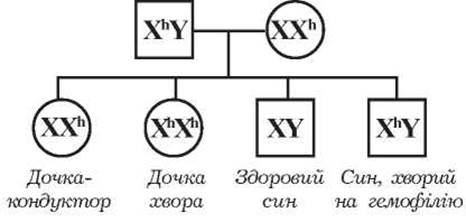
Fig. 26. Marriage of a man with hemophilia and a female carrier.
Recently, the number of hemophilia patients and carriers in the population has increased. At the same time, an increase in hereditary forms of the disease has been noted. This is explained by a significant reduction in childhood mortality from hemophilia due to the increased efficacy of modern drug therapy. Hemophilia patients live to middle age, marry, and have children, among whom daughters transmit the disease. In this regard, The Need for MEDICAL Genetic Counseling for the diagnosis of heterozygous carriers of the abnormal gene is growing.
The clinical presentation of hemophilia is characterized by recurrent bleeding episodes (mucosal bleeding, prolonged bleeding following trauma, joint hemorrhages, hematomas, and Hematuria). Hemorrhagic manifestations evolve with age. Although children are born with hemophilia, most do not exhibit specific symptoms during the neonatal period. However, bleeding from the umbilical stump can occur. Subsequently, teething can trigger bleeding, as can prolonged bleeding from a cut or torn lingual frenulum. In most cases, the first signs of hemophilia appear at the end of the first year and during the second or third years of life. There is a correlation between the onset of the first symptoms and the severity of the disease. The severity of clinical manifestations in hemophilia is determined by the level of factor VIII in the blood. Based on severity, the following forms of hemophilia are distinguished: a) factor VIII level below 1 % — severe; b) 1 to 5 % — moderate; v) 5 to 10 % — mild; g) above 10 % — latent.
In addition to the aforementioned symptoms, it should be noted that bleeding from the oral and nasal mucosa carries a risk of asphyxia due to the accumulation of blood and clots in the upper Airways. Sometimes, hemophilia is diagnosed during injections or vaccination. As children become more physically active, their risk of injury increases. During this period, they fall frequently, leading to an increased incidence of nosebleeds and the appearance of hematomas on the face and trunk. In the event of skin integrity disruption, bleeding can last for several hours to several days, which is disproportionate to the depth of the injury. Soft tissue hemorrhages can be subcutaneous, intramuscular, or intermuscular. Hematomas commonly develop in the shoulder joints, chest, abdomen, and neck.
After three years of age, physical activity increases, and the most Typical symptoms of hemophilia A emerge: joint hemorrhages (hemarthroses). Hemorrhages predominantly occur in the knees, elbows, and ankles, and somewhat less frequently in the shoulders, hips, and small JOINTS OF THE hands and feet.
Clinically, hemarthrosis is characterized by rapid joint Swelling and pain; the overlying skin is tense, erythematous, and warm to the Touch, with pain increasing sharply during joint movement. As the blood resorbs, mild symptoms of systemic toxicity, fever, leukocytosis, and an elevated ESR may occur. With recurrent joint hemorrhages, the blood is not completely resorbed. Gradually, a proliferative process develops, leading to joint deformity.
In early childhood, bleeding from the Urinary Tract or gastrointestinal tract is rare. Occasionally, hemorrhages occur in the abdominal Organs, Peritoneum, or retroperitoneal tissue. Intracranial or meningeal hemorrhages are rarely diagnosed, but they carry a high risk of severe neurological damage or death.
Treatment. The primary principle is replacement therapy. Overall, the management plan is aimed at: 1) controlling bleeding using systemic and local agents; 2) treating the consequences of bleeding; 3) managing complications (inhibitor forms, secondary rheumatoid syndrome); 4) preventing bleeding episodes; 5) genetic counseling and Prevention in families with hemophilia patients.
In the event of prolonged bleeding, treatment should be administered as follows:
1. Administer type-specific cryoprecipitate intravenously as a bolus (15-20 U/kg body weight as a single dose), followed by subsequent administrations every 4-6 hours until the bleeding stops.
2. For large hemarthroses — joint aspiration to remove blood.
3. Immobilization of the affected joint for 5 days.
4. In cases of severe post-hemorrhagic anemia — transfusion of washed red blood cells at a dose of 10-15 mL/kg of body weight.
5. ε-Aminocaproic acid 0,2 g/kg of body weight orally every 4 hours.
6. Non-specific hemostatic agents: hemophobin, hemostatic sponge.
1. Classification of hereditary blood disorders.
2. What are the main groups of hereditary hemolytic anemias?
3. Name the etiological factors in hereditary blood disorders.
4. What is the inheritance pattern of Fanconi anemia?
5. What are the pathogenetic pathways of Fanconi anemia?
6. Clinical manifestations of Fanconi anemia.
7. Hematological criteria for Fanconi anemia.
8. Principles of treatment for Fanconi anemia.
9. Etiopathogenesis of hereditary microspherocytosis.
10. Clinical presentation of hereditary microspherocytosis.
11. Clinical and paraclinical criteria of hemolytic crisis.
12. Clinical and paraclinical criteria for hypoplastic crisis.
13. Treatment of hereditary spherocytosis.
14. Etiopathogenesis of von Willebrand disease.
15. Clinical manifestations of von Willebrand disease.
16. Paraclinical manifestations of von Willebrand disease.
17. Treatment of von Willebrand disease.
18. Etiopathogenesis of hemophilia.
19. Possible marriages in hemophilia (draw a pedigree).
20. Early clinical manifestations of hemophilia.
21. Paraclinical manifestations of hemophilia.
22. Principles of treatment for hemophilia.
Last update: 11/08/2026
Editorial and Educational Adaptation: This material has been compiled based on the primary/original source text. The project team performed an editorial review, corrected technical inaccuracies, structured sections, and adapted the content for an educational format.
What was processed:
- elimination of formatting defects (OCR errors, structural breaks, corrupted characters);
- editorial organization of content;
- standardization of terminology in accordance with academic sources;
- verification of factual statements against the original source text.
All mentions of the author, publication year, and origin of the primary text have been preserved in accordance with the source.