Operative Surgery and Topographic Anatomy - 2016
Content Module No. 1. Introduction to Topographic Anatomy and Operative Surgery. Topographic Anatomy and Operative Surgery of the Head and Neck Regions
Topic 5. Topographic Anatomy of the Neck. General Overview. Boundaries of the Neck. Division into Regions. Fasciae and Interfascial Spaces. Medial Triangle of the Neck. Suprahyoid Region, Submandibular and Submental Triangles. Submandibular Salivary Gland. Pirogov's Triangle. Infrahyoid Region. Carotid Triangle Region. Vessels and Nerves. Region of the Sternocleidomastoid Muscle. Incisions for Neck Phlegmon. Exposure of the Internal Jugular Vein, External and Common Carotid Arteries. Vagosympathetic Blockade According to A.V. Vishnevsky and M.N. Burdenko
1. Relevance of the topic: vital Organs (carotid Arteries, internal jugular Veins, Vagus nerve, Trachea, etc.) are located in the neck region. Their injury can lead to severe emergency conditions and requires prompt action from the physician, demanding precise knowledge of TOPOGRAPHIC AND ANATOMICAL relationships.
2. Specific objectives:
1. Explain the topographic relationships of anatomical structures within the neck regions.
2. Explain The Structure of the cervical fasciae and interfascial spaces.
3. Explain the pathways of spread of abscesses and phlegmons in the neck, and the incisions used for phlegmons.
4. Explain how to ligate the common, external carotid, and lingual arteries.
5. Explain how to perform vagosympathetic blockade using the Methods of A.V. Vishnevsky and M.N. Burdenko.
3. Tasks for independent work in preparation for the Class.
3.1. List of basic terms, parameters, and characteristics that the student must master when preparing for the class:
Term |
Definition |
1. Cervical vagosympathetic blockade according to A.V. Vishnevsky 2. Cervical vagosympathetic blockade according to M.N. Burdenko |
1. A type of local anesthesia performed to prevent pleuropulmonary Shock in chest injuries and during complex surgeries on the thoracic and Abdominal cavity organs 2. Cervical vagosympathetic blockade classified as an invasive Procedure, since the Skin, subcutaneous tissue, and deeper Tissues are dissected along the anterior margin of the m. sternocleidomastoideus. |
3.2. Theoretical questions for the class:
1. BOUNDARIES OF THE neck, division into regions.
2. Triangles of the neck.
3. Fasciae of the neck.
4. Cellular spaces of the neck.
5. Submandibular triangle.
6. N.I. Pirogov's triangle.
7. Carotid triangle.
8. Topography of the main neurovascular bundle of the neck.
9. Differences between the external and internal carotid arteries.
10. Branches of the External Carotid Artery in the carotid triangle.
11. Vagosympathetic blockade according to A.V. Vishnevsky and M.N. Burdenko.
3.3. Practical skills acquired during the class:
1. Performance of incisions in purulent processes of the neck.
2. Isolation of the external and internal carotid arteries.
3. Execution of vagosympathetic block according to A.V. Vishnevsky.
4. Topic outline
On an embalmed cadaver, students identify the boundaries of the neck and its individual regions, demonstrating the boundaries of each cervical triangle. They pay attention to variations in neck shape, sex- and age-related differences, and study the cervical fasciae.
Cervical fasciae
According to V.N. Shevkunenko's Classification of fasciae with modifications, 5 cervical fasciae are currently distinguished: I — superficial fascia; II — deep fascia (investing layer), forming the sheath for the trapezius and sternocleidomastoid Muscles, as well as the capsule of the Cytology/practical/98.html">Submandibular salivary gland; III — omoclavicular fascia (pretracheal layer), forming the sheath for the Infrahyoid muscles; IV fascia — endocervical fascia. It consists of 2 layers — parietal and visceral. The former forms the sheath for the neurovascular bundle of the neck (Internal jugular vein, common carotid artery, vagus nerve). The previsceral space is located between the layers of the fourth fascia, and the prevertebral space lies posterior to the fifth fascia. V fascia — prevertebral fascia (Fig. 26).

Fig. 26. Fasciae and fascial spaces of the neck on a sagittal section:
1 - fascia superficial; 2 - lamina superficial fasciae cervicalis; 3 - lamina pretrachealis fasciae cervicalis; 4 - fascia endocervicalis; 5 - lamina prevertebralis fasciae cervicalis; 6 - spatium prevertebrale; 7 - spatium retroviscerale; 8 - spatium pretracheale; 9 - spatium interaponevroticum suprasternale.
Students should be made aware of the presence of closed and communicating fascial spaces, and consequently, the differences in the spread of abscesses and hematomas in the neck.
The instructor emphasizes the execution of standard incisions for neck phlegmons.
Phlegmon of the floor of the Mouth is opened via an incision in the submental triangle between the anterior bellies of the right and left digastric muscles, along the raphe of the mylohyoid Muscle. Submandibular phlegmon is incised in the submandibular triangle 2–3 cm below and parallel to the edge of the lower jaw, anterior to its angle (Fig. 27).

Fig. 27. Surgical approaches to the neck:
1 - parallel to the edge of the Mandible; 2 - hyoid collar approach to the Pharynx; 3 - collar approach to the superior thyroid artery; 4 - median longitudinal; 5 - along the anterior edge of the sternocleidomastoid muscle; 6 - collar approach to The Thyroid Gland; 7 - parallel to the upper edge of the clavicle.
Retrobulbar/retropharyngeal abscess drainage is performed through the Oral Cavity via an incision in the posterior pharyngeal wall (in children). In adults, an extraoral approach is more commonly used — via an incision along the posterior-lateral border of the sternocleidomastoid muscle. Opening of the fascial Sheath of the cervical neurovascular bundle is also performed using the de Quervain incision in the lower neck and the Küttner incision (for phlegmon in the upper portion of the vascular sheath).
Attention is drawn to the potential spread of phlegmons into the anterior and posterior Mediastinum, the risk of Hemorrhage due to vascular wall necrosis, as well as thrombophlebitis of major veins (Fig. 28).
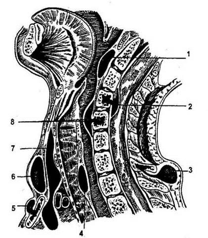
Fig. 28. Schematic representation of neck abscesses:
1 - retropharyngeal abscess; 2 - intraspinal (epidural) abscess; 3 - abscess in the posterior region of the neck originating from a vertebra; 4 - abscess between the trachea and Esophagus; 5 - pre-sternal abscess; 6 - abscess in the spatium interaponevroticum suprasternale; 7 - deep abscess in the spatium praeviscerale extending into the anterior mediastinum; 8 - abscess between the vertebra and esophagus (resulting from vertebral pathology).
The median triangle of the neck is defined, bordered by the lower edge of the mandible and the anteromedial borders of the sternocleidomastoid muscles. It contains two medial triangles of the neck.
Medial triangle of the neck
It is bounded medially by the midline of the neck, superiorly and laterally by the edge of the mandible, and anteromedially by the edge of the sternocleidomastoid muscle. A horizontal plane passing through the greater horns of the Hyoid bone divides the anterior region of the neck into the suprahyoid and infrahyoid areas.
Suprahyoid Region of the Neck
It houses the submandibular and submental triangles. The submandibular triangle is bounded superiorly by the Base of the mandible and laterally by the anterior and posterior bellies of the digastric muscle.
Contents of the submandibular triangle. Layer-by-layer preparation of the suprahyoid region. A skin flap is outlined within an area 3 cm wide, with its base facing outwards (towards the sternocleidomastoid muscle). Regions of the area. Skin and its structural features. Subcutaneous adipose tissue and its structural features (age- and sex-related). Superficial Vessels and nerves. Superficial fascia and its connection with the platysma. The Second layer of the Cervical fascia. Its course within the submandibular triangle. Capsule of the submandibular salivary gland. The gland has an irregular shape, consists of 10-12 lobules, and features an anterior process. Its excretory duct lies between the mylohyoid and hyoglossus muscles and opens onto the sublingual caruncle alongside the duct of the sublingual gland.
The glandular capsule is formed by the splitting of the investing (second) layer of the deep cervical fascia. The facial artery and facial vein are located within the bed of the submandibular salivary gland. Submandibular Lymph Nodes are situated above, within the thickness of, and beneath the superficial lamina of the second cervical fascia. Using a blunt hook, the gland is retracted superiorly (toward the mandible), and students begin dissecting the deep layers of the region. This is the Location of Pirogov's triangle. It is bounded superiorly by the Hypoglossal nerve, inferiorly by the tendon of the digastric muscle, and anteriorly by the free margin of the mylohyoid muscle. Its floor is formed by the hyoglossus muscle, upon which the lingual vein lies. The lingual artery is situated deep to this muscle.
Position of the patient and their HEAD during the surgery: the patient lies supine, with the head hyperextended and turned to the side opposite to the surgical intervention. Attention is drawn to the LOCATION OF THE lingual artery and vein, which lie in different anatomical planes (see above). Students apply a ligature to the lingual artery.
Infrahyoid Region
Boundaries. The instructor points out the Practical significance of the carotid tubercle for temporarily arresting hemorrhage from the carotid artery. The Significance of the carotid triangle in neck surgeries.
Layer-by-layer dissection of the region. The flap is outlined within the area, stopping one fingerbreadth short of the midline of the neck. The base of the flap is located near the anterior border of the sternocleidomastoid muscle.
Carotid Triangle of the Neck
Bounded superiorly by the posterior belly of the digastric muscle, laterally by the sternocleidomastoid muscle, and inferiorly (medially) by the superior belly of the omohyoid muscle. This triangle contains the neurovascular bundle of the neck, enveloped by the parietal layer of the fourth cervical fascia. It consists of the common carotid artery (which bifurcates at the level of the upper border of the thyroid Cartilage into the external and internal carotid arteries), the internal jugular vein (lying most superficially and laterally), and the vagus nerve (positioned between the artery and vein, near their posterior surfaces). THE POSITION OF the common carotid artery is crucial for endovascular interventions on cerebral vessels. The external carotid artery lies medially, whereas the internal artery lies laterally at its origin. The latter gives off no branches in the neck, whereas the external carotid artery branches into the superior thyroid, superior laryngeal, ascending pharyngeal, lingual, facial, and other arteries. The terminal branch of the external carotid artery is the superficial temporal artery, the pulse of which can be palpated anterior to the tragus of the ear. Ligation of the external carotid artery abolishes the pulse in the superficial temporal artery.
Region of the Sternocleidomastoid Muscle
Following an oral review, students study the layer-by-layer topography of this region through dissection. Here, a flap 3 cm wide should be positioned in the lower PARTS OF THE region. The base of the flap must be located near the posterior border of the sternocleidomastoid muscle. Anteriorly, it should be placed 1 fingerbreadth from the midline of the neck. During dissection, the instructor draws attention to Specific features of the area: the arrangement of the superficial fascia with the platysma muscle situated between its leaves, the specific layout and variations of the superficial veins, and the localization of the main branches of the Cervical plexus. After incising the second fascia using a Kocher probe, students enter the plane between the fascia and the muscle, noting that the interfascial cellular space of the sternocleidomastoid muscle is closed.
While dissecting, students use blunt hooks to retract the muscle and reveal the neurovascular bundle of the neck beneath the deep lamina of the second cervical fascia, surrounded by the parietal layer of the fourth cervical fascia (the carotid sheath). Attention is directed to the specific relationships within the neurovascular bundle at different levels of the region: the upper, middle, and lower thirds of the sternocleidomastoid muscle.
During dissection, several deep lymph nodes should be isolated and the pathways of lymph drainage accounted for.
On the specimen, students identify the bifurcation of the common carotid artery into the external and internal carotid arteries, as well as the cervical branch of the hypoglossal nerve. They determine the positions of the internal jugular vein and the vagus nerve.
At the beginning of the first hour of the class, while questioning the students, the instructor emphasizes the Anatomical and physiological rationale for surgical interventions on neck organs, incisions for neck phlegmons, surgical approaches to cervical organs, and the indications and technique of Vashnevsky's vagosympathetic block. The indications and technique for exposing the carotid arteries and the External Jugular Vein are also reviewed.
Since the topic of the session is dedicated to the fundamentals of neck surgery, the instructor highlights the correct execution of surgical approaches and techniques.
Vagosympathetic Block According to A.V. Vyshnevsky
Attention is drawn to the position of the corpse on the operating table, the injection site, and the direction and depth of needle insertion. Students draw 20 ml of a colored solution into a Record-type syringe and, targeting the injection point (the intersection of the external jugular vein with the posterior border of the sternocleidomastoid muscle), advance the needle toward the bone. To verify the correct execution of Vyshnevsky's vagosympathetic block, students should be prompted to displace the tissues along the previously incised edge of the sternocleidomastoid muscle. If they observe that the needle lies posterior to the common carotid artery and the vagus nerve, it confirms that the injected novocaine solution will infiltrate and block the area containing the vagus and sympathetic nerves (Fig. 29).

Fig. 29. Technique of cervical vagosympathetic block according to Vyshnevsky.
Forty to fifty milliliters of a 0.25% novocaine solution are injected into the cellular tissue located posterior to the sheath of the neurovascular bundle. Indicators of a successfully performed block include facial and scleral hyperemia, along with Claude Bernard-Horner syndrome (myosis, narrowing of the palpebral fissure, and enophthalmos).
When performing the block using M.N. Burdenko's method, the neurovascular bundle is exposed, and 2 cm of a 2% novocaine solution is administered endoneurally.
Exposure of the External and Common Carotid Arteries
During the exposure of the carotid arteries, the instructor assigns the following tasks to the students:
1. To be able to position the patient correctly on the operating table.
2. To be able to properly outline the projection line for surgical access to both the external and common carotid arteries.
3. To be able to navigate through the layered tissue dissection.
4. To be able to identify the differences between the external and internal carotid arteries.
When exposing the arteries, the patient lies in a supine position with a bolster placed under the shoulder girdle; the head is tilted back and turned to the side opposite to the surgical intervention. The incision is made along the anterior margin of the sternocleidomastoid muscle. The skin, subcutaneous tissue, first fascia with the platysma, and second fascia forming the sheath for the sternocleidomastoid muscle are sequentially incised. The muscle is retracted laterally, and the posterior wall of its sheath along with the parietal layer of the fourth fascia are incised. The neurovascular bundle is then exposed. As noted above, the arrangement of the structures within the bundle from lateral to medial and deep is: vein, nerve, artery.
When exposing the external carotid artery, it is necessary to differentiate it from the Internal Carotid Artery (see above). If required, the ligature should be placed above the Water/144.html">Origin of the superior thyroid artery.
During the exposure of the carotid arteries, the instructor draws attention to the necessity of preserving the superior cervical loop (Fig. 30).
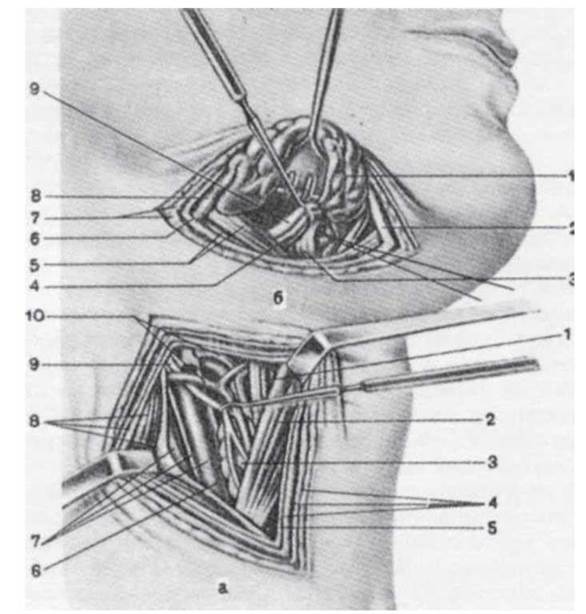
Fig. 30. Exposure of the common carotid (a) and lingual (b) arteries.
a: 1 - thyrohyoid muscle; 2 - superior belly of the omohyoid muscle; 3 - thyroid gland; 4 - superficial (first) cervical fascia and platysma; 5 - investing (second) cervical fascia; 6 - neurovascular sheath formed by the deep (fourth) cervical fascia; 7 - common carotid artery, vagus nerve, and internal jugular vein; 8 - sternocleidomastoid muscle and its sheath formed by the investing (second) cervical fascia; 9 - superior ROOT of the cervical loop (descending branch of the hypoglossal nerve - XII cranial nerve); 10 - superior thyroid artery and vein;
b: 1 - submandibular salivary gland; 2 - investing (second) cervical fascia; 3 - hyoglossus muscle; 4 - lingual vein; 5 - posterior belly of the digastric muscle and stylohyoid muscle; 6 - facial vein; 7 - superficial (first) cervical fascia and platysma; 8 - retromandibular vein; 9 - hypoglossal nerve (XII cranial nerve); the lingual artery is looped on a ligature.
5. Self-Assessment Materials
A. Self-Assessment Tasks
Test No. 1
During the primary surgical debridement of a neck wound, the surgeon identified damage to a muscle attached to the hyoid bone by its intermediate tendon. Which muscle is injured?
a. Omohyoid
b. Mylohyoid
c. Digastric
d. Hyoglossus
e. Sternohyoid
Test No. 2
During the ligation of the lingual artery within Pirogov's triangle, the surgeon injured a nerve, resulting in impaired motor Innervation of the Tongue. Which nerve was injured?
a. Lingual
b. Recurrent laryngeal
c. Glossopharyngeal
d. Hypoglossal
e. Vagus
Test No. 3
A physician is performing Palpation in the left lateral triangle of the neck. Which structure forms the inferior boundary of this triangle?
a. Inferior belly of the omohyoid muscle
b. Posterior belly of the digastric muscle
c. Hyoid bone
d. Jugular notch of the Sternum
e. Clavicle
Test No. 4
A patient has an inflammatory process spreading downward from the previceral space of the neck. Where has it extended?
a. Parapharyngeal space
b. Retropharyngeal space
c. Anterior mediastinum
d. Posterior mediastinum
e. Preperitoneal tissue
Test No. 5
A patient has an injury to the external carotid artery. Against the carotid tubercle of which cervical vertebra can this artery be compressed to temporarily stop the hemorrhage?
a. 2nd
b. 3rd
c. 4th
d. 5th
e. 6th
Test No. 6
A surgeon is performing an approach to the common carotid artery. In which direction must the sternocleidomastoid muscle be retracted?
a. Inward and forward
b. Outward and backward
c. Inward and backward
d. Outward and forward
e. Upward
Test No. 7
A surgeon mistakenly ligated the internal carotid artery instead of the external carotid artery in the carotid triangle. How does the ligated artery differ from the external carotid?
a. Has no branches
b. Has branches
c. Located more medially
d. Located more superficially
e. Located more anteriorly
Test No. 8
To ligate the lingual artery, the surgeon identified Pirogov's triangle in the wound. Which structure forms the anterior wall of this triangle?
a. Lingual nerve
b. Hypoglossal nerve
c. Hyoglossus nerve
d. Intermediate tendon of the digastric muscle
e. Mylohyoid muscle
B. Self-Assessment Tasks
Task 1. During dissection in the submandibular triangle, a student found M.I. Pirogov's triangle, where the lingual vein is clearly visible, but failed to locate the lingual artery. How can this artery be found?
Task 2. During a mandibular resection, the surgeon first exposed the bifurcation of the common carotid artery within the carotid triangle. How to ensure that the branch chosen for temporary ligation is the external carotid artery?
Task 3. During class, a student claimed that There are five fascial layers in the carotid triangle of the neck. Was the student's answer correct?
Task 4. During surgery, the surgeon transected the external jugular vein. Attempting to stop the bleeding, the surgeon clamped the peripheral end of the vein. Was the bleeding stopped correctly? What is your management strategy?
Task 5. To ligate the external carotid artery, the surgeon exposed its segment from the bifurcation of the common carotid artery to THE ORIGIN OF the superior thyroid artery and performed the ligation. Did the surgeon choose the correct ligation site?
Task 6. During a vagosympathetic block, a patient developed facial hyperemia. Is this symptom sufficient to evaluate the correctness of the performed block?
Task 7. A patient consulted a surgeon complaining of a pulsating mass in the neck and impaired Vision. Near the anterior border of the sternocleidomastoid region, slightly above the clavicle, a pulsating mass is palpable. A systolic murmur is auscultated over the mass. The face on the side of the lesion is hyperemic, and there is miosis, narrowing of the palpebral fissure, and enophthalmos. The surgeon diagnosed an Aneurysm of the common carotid artery. What is the relationship between the arterial aneurysm and Claude Bernard-Horner syndrome?
Main
1. Operative Surgery and Topographic Anatomy; edited by M.S. Skrypnykov. — K.: Vyshcha Shkola, 2000. — P. 209-230.
2. Operative Surgery and Topographic Anatomy; edited by M.P. Kovalsky. — K.: Medytsyna, 2010. — P. 105-122.
Supplementary
1. Kovanov V.V. Operativnaya khirurgiya i topograficheskaya anatomiya / V.V. Kovanov. — M., 2001. — P. 108-129
2. Operative Surgery and Topographic Anatomy; edited by K.I. Kulchytsky. — K.: Vyshcha Shkola, 1994. — P. 91-106.
3. Operativnaya khirurgiya i topograficheskaya anatomiya; edited by G.E. Ostroverkhov. — Rostov-on-Don, 1998. — P. 373-401.
4. Elizarovsky S.I. Operativnaya khirurgiya i topograficheskaya anatomiya / S.I. Elizarovsky, R.N. Kalashnikov. — M., 1979.
5. Matyushin I.F. Vvedenie v kurs operativnoy khirurgii i topograficheskoy anatomii / I.F. Matyushin. — Gorky, 1976.
6. Tomashuk I.P. Rukovodstvo po operativnoy tekhnike dlya nachinayushchikh khirurgov / I.P. Tomashuk, I.I. Tomashuk. — K.: Iz-vo Evropeyskogo universiteta, 2001. — 860 p.
7. Frauchi V.Kh. Kurs topograficheskoy anatomii i operativnoy khirurgii / V.Kh. Frauchi. — M., 1976.
Last update: 10/08/2026
Editorial and Educational Adaptation: This material has been compiled based on the primary/original source text. The project team performed an editorial review, corrected technical inaccuracies, structured sections, and adapted the content for an educational format.
What was processed:
- elimination of formatting defects (OCR errors, structural breaks, corrupted characters);
- editorial organization of content;
- standardization of terminology in accordance with academic sources;
- verification of factual statements against the original source text.
All mentions of the author, publication year, and origin of the primary text have been preserved in accordance with the source.