Operative Surgery and Topographic Anatomy - 2016
Content Module No. 1. Introduction to Topographic Anatomy and Operative Surgery. Topographic Anatomy and Operative Surgery of the Head and Neck Regions
Topic 6. Topographic Anatomy of the Neck. Neck Organs: Cervical Parts of the Trachea and Esophagus, Blood Supply and Innervation. Branches of the Cervical Plexus. Lateral Triangle of the Neck. Scalenus Anticus, Interscalene, and Scalenovertebral Fascial Spaces. Tracheostomy. Complications and Errors in Tracheostomy. Intubation. Surgery on the Cervical Esophagus. Subtotal Subfascial Thyroid Resection According to O.B. Nikolaiev
1. Relevance of the Topic
The Scope of surgical interventions on the Trachea, Thyroid Gland, and cervical Esophagus is quite extensive, and performing them is impossible without a deep understanding of the TOPOGRAPHIC AND ANATOMICAL features of these Organs and their relationships with adjacent structures.
2. Specific Objectives
1. Analyze the Topography of the neck organs (Larynx, trachea, Pharynx, esophagus, thyroid, and Parathyroid glands).
2. Explain the technique of layered dissection of the studied neck regions.
3. Explain how to perform tracheostomy and surgeries on the cervical esophagus.
4. Explain the administration of anesthesia during subtotal subfascial thyroid resection.
3. Tasks for Independent Work in Preparation for the Class.
3.1. List of key terms, parameters, and characteristics that the student must master in preparation for the class:
Term |
Definition |
1. Thyroid resection 2. Strumectomy 3. Tracheotomy |
1. Partial removal of The Thyroid Gland 2. Removal of a goiter. 3. Surgical opening of the trachea to provide immediate airflow into the Lungs |
3.2. Theoretical questions for the class:
1. BOUNDARIES OF THE lateral cervical triangle and its division into the omotrapezoid and omoclavicular triangles.
2. Layers of the lateral triangle of the neck.
3. Fascial spaces of the lateral triangle of the neck.
4. Topography of the trachea.
5. Topography of the cervical part of the esophagus.
6. Branches of the Cervical plexus.
7. Topography of the cervical segment of the phrenic nerve.
8. Technique of tracheostomy.
9. Errors and Complications during tracheostomy.
10. Features of the surgical approach to the cervical esophagus.
11. Surgeries on the thyroid gland.
3.3. Practical skills acquired during the class:
1. Dissect the lateral triangle of the neck.
2. Perform a tracheostomy.
4. Topic contents:
At the beginning of the class, the instructor quizzes students on the topography of the cervical organs. Special attention is paid to the skeletotopy of the cervical trachea, its relation to the thyroid gland, esophagus, and the neurovascular bundle of the neck. Blood supply and Innervation of the trachea. Using anatomical specimens and illustrations, students examine the topographical features of the cervical esophagus, its relationship with the cervical trachea, the neurovascular bundle of the neck, and the recurrent laryngeal nerve, emphasizing the Practical significance of topographical differences between the right and left recurrent laryngeal nerves. Blood supply and innervation of the cervical esophagus. Within the lateral triangle of the neck—bounded by the trapezius Muscle, the posterior border of the sternocleidomastoid muscle, and the clavicle—two smaller triangles are distinguished: the omotrapezoid and omoclavicular triangles. The boundary between them is the inferior belly of the omohyoid muscle.
On a formalin-fixed neck specimen, students study the topographical features of the cervical trachea and esophagus. Through dissection, they examine the topography of the right and left recurrent laryngeal nerves relative to the esophagus.
By retracting the neurovascular bundle laterally with blunt hooks, students expose the fifth layer of the Cervical fascia, incise it, and isolate the Sympathetic trunk along with its superior and middle ganglia. In the lower region, students dissect the venous angle, the initial cervical segment of the Subclavian Artery with its branches, the Vagus nerve, the sympathetic trunk, and the cervicothoracic ganglion.
Above the clavicle, following the exposure of the second fascial layer, the third fascial layer covering the omohyoid muscle is revealed. The instructor points out that this fascia is absent in the region of the omotrapezoid triangle. After incising and retracting the inferior belly of the omohyoid muscle, students isolate the fifth fascial layer and the scalene Muscles. During dissection, it is essential to study the position, boundaries, and Contents of the scalenovertebral triangle, as well as the interscalene space. The topography of the phrenic nerve and the subclavian vein is examined. Attention is drawn to The formation of the phrenic nerve and the risks associated with injury to the subclavian vein.
Tracheostomy
Incision of the trachea (tracheotomy) or creation of a stoma (tracheostomy) are life-saving emergency Procedures. These interventions are routinely performed in intensive care units, maxillofacial surgery, and otolaryngology departments.
Tracheotomy is classified into superior and inferior procedures, depending on whether the trachea is incised above or below the ISTHMUS OF THE thyroid gland. Inferior tracheotomy is typically the Procedure of choice (and exclusively used in children) (Fig. 31).

Fig. 31. Superior tracheostomy:
a — incision of the Skin, subcutaneous tissue, and platysma with the superficial (first) layer of the cervical fascia; b — linea alba colli; c — incised linea alba exposing the arch of the cricoid Cartilage, the initial tracheal rings, and the thyroid isthmus; d — thyroid isthmus retracted inferiorly, with the trachea fixed by sharp single-hook retractors and opened via a longitudinal incision; e — insertion of the tracheostomy cannula (with its flange oriented in the sagittal plane); f — cannula fully inserted (with its flange oriented in the frontal plane).
Indications include laryngeal tumors, acute and inflammatory conditions, foreign body airway obstruction, and tracheal trauma. A tracheostomy stoma is established when a patient requires prolonged mechanical ventilation.
The patient is placed in the supine position with a roll placed beneath the shoulder girdle. The HEAD is hyperextended and maintained in the midline position. Anesthesia is local, or general in young children. Performing tracheostomy under general anesthesia with an endotracheal tube in place allows for unhurried surgery under optimal pulmonary ventilation while preventing blood aspiration into the trachea.
The surgeon incises the skin, subcutaneous tissue, superficial and deep cervical fasciae, and the third fascial layer covering the Infrahyoid muscles, using a grooved probe, and laterally retracts the muscles with blunt hooks. The thyroid isthmus is visible in the wound. After incising the ligament fixing the thyroid isthmus to the cricoid cartilage, the isthmus is retracted superiorly using a blunt hook. The trachea is stabilized with a sharp single-hook Grünfeld retractor. Following tracheal fixation, the resident surgeon incises 2–3 tracheal rings. A tracheal dilator is inserted to widen the opening, and a Luer tracheostomy cannula is introduced. During insertion, the cannula flange is initially positioned in the sagittal plane and then rotated into the frontal plane as the cannula is advanced (Figs. 32, 33, 34).
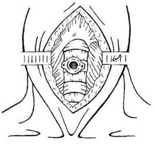
Fig. 32. Insertion of the cannula into the tracheal lumen

Fig. 33. Björk tracheostomy and conicotomy:
Tracheostomy — surgical creation of an opening (stoma) in the trachea (1);
Conicotomy — surgical creation of an opening (stoma) in the larynx via incision of the cricothyroid membrane (2).
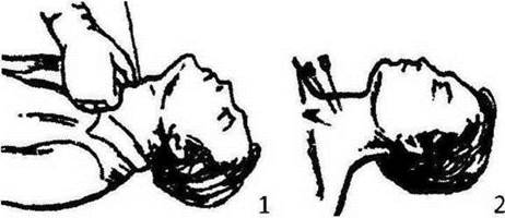
Fig. 34. Performing conicocentesis:
Conicotomy is a surgical procedure to create a percutaneous opening in the larynx by puncturing the cricothyroid membrane with 2-3 needles.
Dissection of the Cervical Esophagus
The surgical approach is performed from the left side. The Tissues are dissected layer by layer. Using a finger, students identify the spine and trachea. Attention is drawn to the risk of injuring the neurovascular bundle of the neck, the recurrent laryngeal nerve, and the inferior thyroid artery during this procedure. Deep within the wound, students locate the esophagus by its characteristic features: it has a dark red color and longitudinally oriented muscle fibers. Temporary ligatures are placed on the exposed esophagus, after which it is incised and catheterized. The operation concludes by suturing the esophageal wound in a transverse direction and closing the surgical incision while placing a drain in the lower angle of the wound (Fig. 35, 36).
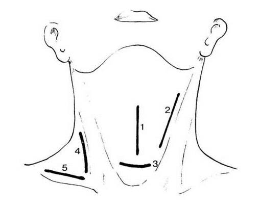
Fig. 35. Incisions for draining purulent-inflammatory processes in the neck:
1 - for Phlegmon of the pretracheal space; 2 - for adenophlegmon of the sternocleidomastoid muscle; 3 - for phlegmon of the suprasternal interfascial space; 4 - for phlegmon of the retropharyngeal (retrovisceral) space; 5 - for phlegmon of the lateral triangle of the neck.
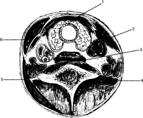
Fig. 36. Phlegmons of the neck:
1 - prevertebral (previsceral) space; 2 — perivascular space; 3 - retrovisceral space; 4 - posterior region of the neck; 5 — trapezius muscle bed; 6 - sternocleidomastoid muscle bed.
Operations on the Thyroid Gland (Oral Structure/133.html">Discussion)
The instructor emphasizes that the thyroid gland is a frequent site of surgical intervention (endemic goiter, DIFFUSE TOXIC GOITER (Graves' disease), malignant tumors) and outlines the steps of subtotal subfascial thyroidectomy. The Importance of preserving a portion of the gland (approx. 5.0 g) on each side of the lower pole of the lateral lobes—near the entry point of the inferior thyroid artery and the adjacent parathyroid glands—is stressed, along with exercising careful handling of the recurrent laryngeal nerve (Fig. 37).
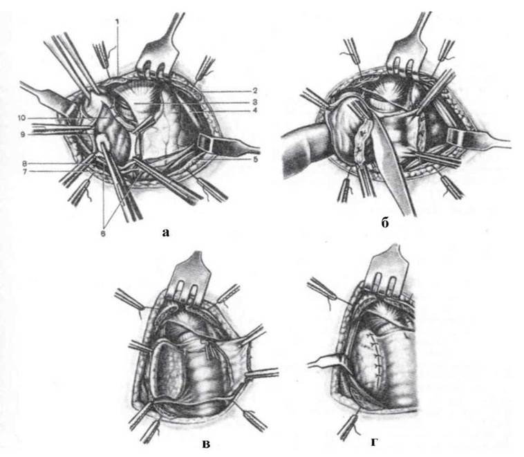
Fig. 37. Technique of thyroidectomy:
a - the right lobe of the thyroid gland is delivered into the wound, its outer capsule is incised and pushed back to the resection line of the lateral lobe; Blood Vessels are clamped: 1, 2, 5, 7 - ends of the severed sternocleidomastoid muscles; 3, 6 - edges of the incised parietal layer of the deep cervical fascia (fourth fascia); 4, 8 - sternocleidomastoid muscles; 9 - external fascial capsule of the thyroid gland, formed by the visceral layer of the deep cervical fascia; 10 - true capsule of the right thyroid lobe; b - resection of the right thyroid lobe fixed in the wound on a finger; c - initiation of catgut suturing along the edges of the fascial capsule of the right lobe; d - sutures applied to the capsule.
5. Self-Assessment Materials
A. Self-Assessment Tasks
Test No. 1
A victim presents with a Combined Trauma to the trachea and the thyroid isthmus. At the level of which tracheal rings is the isthmus typically located?
a. 1-2
в. 2-3
c. 3-4
d. 4-5
e. 5-6
Test No. 2
As an emergency measure for a patient with mechanical asphyxia, a conicotomy was performed. What procedure did this entail?
a. Tracheotomy
b. Tracheostomy creation
c. Cricothyroidotomy (incision)
d. Cricothyroid puncture
e. Laryngostomy creation
Test No. 3
A surgeon is performing an inferior tracheostomy. In which direction should the trachea be incised relative to the isthmus of the thyroid gland?
a. Upward
b. Downward
c. Outward
d. Inward
e. Forward
Test No. 4
A surgeon is performing a subtotal subfascial thyroid resection. During the procedure, they fail to exercise due caution while working in the hazardous (posteromedial) zone of the gland. Which extracapsular structures are at risk of injury?
a. Recurrent laryngeal nerves
b. Vagus nerves
c. Common carotid Arteries
d. Internal jugular Veins
e. Parathyroid glands
Test No. 5
As an emergency measure for mechanical asphyxia, a physician performed a stab incision through the cricothyroid membrane. To locate the puncture site, the physician palpated two laryngeal cartilages beforehand. Below which of these cartilages was the puncture made?
a. Thyroid
b. Cricoid
c. Arytenoid
d. Styloid
e. of the corniculate cartilage
Test No. 6
A surgeon has incised the first tracheal ring and inserted a tracheostomy cannula. Against which laryngeal cartilage will the cannula press in this case?
a. Thyroid
b. Cricoid
c. Arytenoid
d. Styloid
e. Corniculate
Test No. 7
During a thyroid gland resection, the physician mistakenly removed all parathyroid glands. What is the anatomical arrangement of these glands?
a. Left, middle, right
b. Superior, middle, inferior
c. Anterior, middle, posterior
d. Anterior and posterior
e. Superior and inferior
Test No. 8
A surgeon has performed a tracheostomy. What did he do to the patient's trachea in this procedure?
a. Incised it
b. Sutured it
c. Resected it
d. Created a fistula
e. Stabilized it
B. Self-Assessment Problems
Problem 1. A patient with a traumatic laceration of the larynx was admitted to the surgical department of a regional hospital. Emergency tracheostomy had already been performed at the district hospital. Upon examination, signs of progressive asphyxia are present, the soft Tissues of the neck are edematous, and crepitation is palpated. What error was made during the tracheostomy? How can it be corrected?
Problem 2. Following the incision of the tracheal rings and insertion of the cannula, the patient's signs of asphyxia did not resolve. What was the surgeon's mistake? How could it have been prevented?
Task 3. During a tracheostomy, while making a transverse incision of the soft tissues strictly along the midline, the surgeon injured a vessel directly on the trachea. Massive bleeding hinders the subsequent steps of the operation. Which vessel could the surgeon have injured, and how should they proceed — ignore the bleeding and open the trachea, or stop the bleeding first and then incise the trachea?
Task 4. To expose the esophagus, a student decided to use an approach along the posterior border of the right sternocleidomastoid muscle. Is the student's tactic correct?
Task 5. During a tracheostomy, the surgeon entered the tracheal lumen to the full depth of the scalpel blade. After incising the tracheal rings, a gaping wound of the posterior tracheal wall is visible. What error was made by the surgeon, and how can it be prevented?
Task 6. While performing a strumectomy, the surgeon detached the lobe of the gland from the trachea along its entire length. The patient developed hoarseness. What is the cause of the hoarseness, and what was the surgeon's mistake?
Basic
1. Operative Surgery and Topographic Anatomy; ed. by M.S. Skrypnikov. — K.: Vyshcha Shkola, 2000. — P. 234-249.
2. Operative Surgery and Topographic Anatomy; ed. by M.P. Kovalsky. — K.: Medytsyna, 2010. — P. 123-138.
Additional
1. Kovanov V.V. Operative Surgery and Topographic Anatomy / V.V. Kovanov. — M., 2001. — P. 286-297.
2. Operative Surgery and Topographic Anatomy; ed. by K.I. Kulchytsky. — K.: Vyshcha Shkola, 1994. — P. 108-120.
3. Operative Surgery and Topographic Anatomy; ed. by G.E. Ostroverkhov. — Rostov-on-Don, 1998. — P. 406-426.
4. Elizarovsky S.I. Operative Surgery and Topographic Anatomy / S.I. Elizarovsky, R.N. Kalashnikov. — M., 1979.
5. Matyushin I.F. Introduction to the Course of Operative Surgery and Topographic Anatomy / I.F. Matyushin. — Gorky, 1976.
6. Tomashuk I.P. Guide to Surgical Technique for Beginning Surgeons / I.P. Tomashuk, I.I. Tomashuk. — K.: European University Publishing House, 2001. — 860 p.
7. Frauchi V.Kh. Course of Topographic Anatomy and Operative Surgery / V.Kh. Frauchi. — M., 1976.
8. Trutnev V.N. Tracheostomy / V.N. Trutnev. — M., 1962.
Last update: 10/08/2026
Editorial and Educational Adaptation: This material has been compiled based on the primary/original source text. The project team performed an editorial review, corrected technical inaccuracies, structured sections, and adapted the content for an educational format.
What was processed:
- elimination of formatting defects (OCR errors, structural breaks, corrupted characters);
- editorial organization of content;
- standardization of terminology in accordance with academic sources;
- verification of factual statements against the original source text.
All mentions of the author, publication year, and origin of the primary text have been preserved in accordance with the source.