Operative Surgery and Topographic Anatomy - 2016
Content Module No. 2. Topographic Anatomy and Operative Surgery of the Regions and Organs of the Thoracic Cavity
Topic 7. Topographic Anatomy of the Thoracic Wall. Soft Tissue Layers. The Mammary (Breast) Gland: Blood Supply, Innervation, Lymphatic Drainage. Intercostal Spaces (Arteries, Veins, Nerves). The Pleura. Operations on the Mammary Gland. Pleural Puncture. Rib Resection
1. Relevance of the topic: chest trauma, common Diseases of the mammary gland, resection of damaged Ribs as well as healthy ones to gain surgical access to the thoracic Organs require thorough knowledge of the Topographic anatomy of the chest, as well as the mastery of various surgical approaches and techniques.
2. Specific objectives:
1. Explain surgical approaches and techniques for interventions on the chest wall and mammary gland.
2. Analyze the Pathways of the spread of inflammatory processes within the Thorax.
3. Explain the Topography of the Pleura, its recesses, and the projection of these structures onto the chest wall.
4. Explain how to perform a pleural puncture.
5. Explain how to make incisions for mammary gland abscesses.
6. Explain how to perform a rib resection.
7. Explain how to carry out primary surgical debridement of penetrating chest wall wounds.
3. Tasks for independent work during preparation for the Class.
3.1. List of basic terms, parameters, and characteristics that the student must master when preparing for the class:
Term |
Definition |
1. Mastectomy 2. Rib resection 3. Pleural puncture |
1. Removal of the mammary gland. 2. Removal of a portion of a rib. 3. Puncture of the pleura for diagnostic or therapeutic purposes. |
3.2. Theoretical questions for the class:
1. BOUNDARIES OF THE thorax, division into regions.
2. Conventional landmarks for determining the projection of thoracic cavity organs.
3. Anterosuperior region of the thorax.
4. Topography of the mammary gland and pathways of lymphatic drainage. Its Blood supply and innervation.
5. Topography of the intercostal spaces.
6. Boundaries of the pleura.
7. Pleural recesses and their topography.
8. Incisions for mastitis. Radical mastectomy.
9. Pleural puncture.
10. Rib resection.
11. Methods for closing an open pneumothorax.
3.3. Practical skills acquired during the class:
1. Perform incisions for mastitis.
2. Perform pleural puncture on a cadaver.
3. Perform rib resection.
4. Perform pneumothorax closure.
4. Topic Content
During the questioning, the instructor tests students' knowledge of musculoskeletal landmarks and their projection onto the chest wall, Muscle layers, The Structure of fascial sheaths and fascial spaces of the chest, the topography of blood Vessels and nerves, as well as the Location OF THE regional Lymph Nodes of the mammary gland. During the practical session, the instructor analyzes topographical specimens with students, drawing attention to important topographo-anatomical relationships of organs and neurovascular bundles.
Mammary gland. It is located on the anterior chest wall from the 3rd to the 6th rib, reaching the parasternal line medially and the anterior axillary line laterally. It consists of 15-20 lobes. A lactiferous duct extends radially from each lobe toward the nipple. These ducts open onto the nipple through 8-10 openings, having previously formed dilatations known as lactiferous sinuses. The gland is surrounded by a capsule formed by the superficial fascia. The capsule anchors the gland to the clavicle and the deep layers of the chest wall by forming suspensory ligaments of the breast. Blood supply to the gland is provided by the intercostal, internal thoracic, and lateral thoracic Arteries. The gland is innervated by Branches of the intercostal, supraclavicular, and anterior Thoracic nerves; Lymphatic vessels form deep and superficial plexuses. Anastomoses between lymphatic vessels are well developed. Lymph nodes that collect lymph from various PARTS OF THE mammary gland are considered regional. Lymph drainage from the lateral quadrants occurs into the axillary lymph nodes, from the upper quadrants into the subclavicular and supraclavicular lymph nodes, and from the inner quadrants into the parasternal lymph nodes. Students' attention is drawn to the pathways of metastasis spread in malignant tumors of the mammary gland and the necessity of mandatory wide excision of the axillary, subscapular, and subclavicular lymph nodes during radical surgery for breast Cancer (Fig. 38, 39).
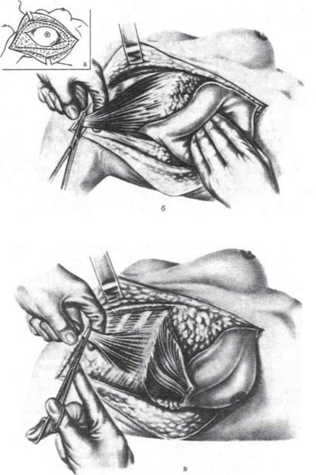
Fig. 38. Radical mastectomy.
a - elliptical Skin incision with undermining of the subcutaneous adipose tissue; the dashed line indicates the incision line of the superficial pectoral fascia; b - transection of the pectoralis major tendon; c - pectoralis major retracted inferiorly, pectoralis minor tendon transected with scissors.
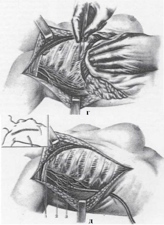
Fig. 39. Radical mastectomy (continued).
d - adipose tissue with lymph nodes along the axillary vessels and in the axillary fossa removed; Muscles and mammary gland detached from the chest wall; e - view of the wound after mammary gland removal; a drain is inserted into the wound: 1 - a. et v.axillares; 2 - a.thoracica lateralis; 3 - n.thoracicus longus.
Surgeries for mastitis and subpectoral phlegmons. The instructor emphasizes The Need for conservative yet sufficiently deep incisions (radial for single abscesses and inframammary semicircular ones for retromammary abscesses and deep-seated abscesses of the lower part of the breast).
Incisions for subpectoral phlegmons. The incisions along the lower-outer margin of the pectoralis major muscle (opening the spaces anterior and posterior to the clavipectoral fascia) are reviewed theoretically. The instructor discusses segmental resection of the mammary gland and radical mastectomy, focusing on the removal of regional lymph nodes (Fig. 40).

Fig. 40. Various types of purulent mastitis and corresponding incisions: a - types of mastitis:
1 - retromammary; 2 - interstitial; 3 - subareolar; 4 - antemammary; 5 - parenchymal; b - incisions: 1, 2 - radial; 3 - inframammary.
Intercostal spaces. Layers, topography of the intercostal neurovascular bundle. Topography of the internal thoracic artery, its relation to the endothoracic fascia.
Rib resection. Indications. Anesthesia. Patient position during surgery. Students perform subperiosteal resection of the 7th and 8th ribs. Attention is drawn to the differences in the technique of rib resection depending on the indications (removal of injured ribs, creating access to thoracic cavity organs, plastic surgeries (thoracoplasty, etc.) (Fig. 41, 42).
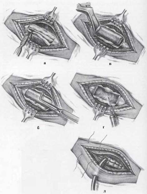
Fig. 41. Rib resection.
a – Separation of the periosteum on the outer surface of the rib using a curved periosteal elevator; b – stripping of the periosteum from the inner surface of the rib using a Doyen raspatory; c – transection of the resected rib segment using rib shears; d – diagnostic thoracentesis through the periosteal bed of the resected rib; e – wound closure following rib resection and pleural cavity drainage.

Fig. 42. Closure of the chest wall wound using transmural sutures encompassing adjacent ribs (a). Diagram of the suture placement technique (b).
Technique for open pneumothorax closure. Indications. Anesthesia. Patient positioning during surgery. A penetrating wound involving rib and pleural injury is simulated on a cadaver. Students perform wound debridement and close the open pneumothorax. All methods of pneumothorax closure are reviewed orally: pleuromuscular sutures, musculocutaneous flaps,
pneumopexy, diaphragmopexy.
Pleura. Students review the boundaries and recesses of the pleura, paying special attention to the cupola of the pleura, which extends 2–3 cm above the clavicle. When studying the pleural recesses, it should be emphasized that during expiration, the costodiaphragmatic recess reaches the 9th rib, which justifies choosing this site for thoracentesis. The anterior costomediastinal recess is located anteriorly. The posterior costomediastinal recesses lie near THE Vertebral Column, corresponding to the transition from the costal to the mediastinal pleura. The phrenicomediastinal (diaphragmo-mediastinal) recesses are small potential spaces in the pleural cavity where the diaphragmatic pleura transitions into the mediastinal pleura.
Thoracentesis (pleural puncture). Indications, anesthesia, patient positioning during surgery. Students perform thoracentesis in the eighth and ninth intercostal spaces between the scapular and mid-axillary lines, along the upper border of the rib (Fig. 43).
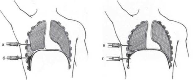
Fig. 43. Pleural cavity puncture and potential complications.
a – the needle has passed into the pleural cavity above the effusion; b – the needle has entered an adhesion between the pleural layers of the costodiaphragmatic recess; c – the needle has passed above the effusion directly into the lung parenchyma; d – the needle has passed through the lower part of the costodiaphragmatic recess into the Abdominal cavity.
5. Self-Assessment Materials
A. Self-Assessment Tasks
Test № 1
A physician is palpating a patient's costal margin. The cartilages of which ribs form this margin:
a. 6-9
b. 6-10
c. 7-9
d. 7-10
e. 8-11
Test № 2
A surgeon is performing an operation on the mammary gland for intracapsular mastitis. How many lobes does this gland consist of?
a. 1-5
b. 5-10
c. 10-15
d. 15-20
e. 20-25
Test No. 3
Primary surgical debridement of a penetrating chest wound is being performed. The surgeon should be especially meticulous when placing sutures to ensure airtight closure of the:
a. Pleura
b. Endothoracic fascia
c. Intercostal muscles
d. Superficial muscles
e. Skin and subcutaneous tissue
Test No. 4
A female patient presents with a superficial intramammary abscess in the upper outer quadrant of the breast. Which incision is considered the most appropriate for draining this type of purulent mastitis?
a. Infra-mammary curvilinear incision
b. Radial incision
c. Longitudinal incision
d. Transverse incision
e. Combined incision
Test No. 5
A physician is performing a thoracentesis (pleural puncture) for exudative Pleurisy, during which the intercostal nerve is accidentally injured. Where should the chest puncture be performed to prevent this complication?
a. Along the upper border of the lower rib
b. Along the lower border of the upper rib
c. Midway between the lower borders of the ribs
d. Midway between the upper borders of the ribs
e. In the upper part of the intercostal space
Test No. 6
Two weeks after being treated for Pneumonia, a patient complains of a feeling of heaviness and moderate pain in the right hypochondriac region, shortness of breath, and fatigue. Chest fluoroscopy reveals fluid accumulation in the right pleural cavity above the diaphragmatic dome. In which pleural recess does fluid most commonly accumulate?
a. Costodiaphragmatic recess
b. Costomediastinal
c. Phrenicmediastinal
d. Vertebromediastinal
e. Costovertebral
Test No. 7
Patient K. was admitted to the surgical department from a traffic accident scene with a closed chest trauma and right-sided rib fractures. The patient was diagnosed with a right-sided pneumothorax, and urgent drainage of the pleural cavity was indicated. Indicate the site for pleural puncture:
a. In the 2nd intercostal space along the midclavicular line
b. In the 6th intercostal space along the posterior axillary line
c. In the 7th intercostal space along the scapular line
d. In the PROJECTION OF THE pleural recess
e. At the site of greatest dullness determined by Percussion
B. Self-Assessment Tasks
Task 1. In a female patient with a retromammary abscess, the surgeon made 2 deep incisions on the mammary gland and drained the wound. What was the surgeon's mistake?
Task 2. During puncture of the left subclavian vein, a clear opalescent fluid was obtained in the syringe when pulling the plunger back. What mistake was made during the puncture Procedure?
Task 3. While performing a pleural puncture in the 7th intercostal space along the anterior axillary line near the lower border of the rib in a patient with suspected Empyema of the pleura, the surgeon obtained blood in the syringe and diagnosed hemothorax. What was the surgeon's mistake?
Task 4. During pleural puncture using a thick-bore needle without a syringe in a patient with Pleural Empyema, the patient's breathing accelerated and they lost consciousness. What is the explanation for this complication?
Task 5. During rib resection for plastic surgery, the patient developed a pneumothorax. What mistake did the surgeon make?
Core
1. Operative Surgery and Topographic Anatomy; ed. by M.S. Skrypnikov. — K.: Vyshcha Shkola, 2000. — P. 250-260.
2. Operative Surgery and Topographic Anatomy; ed. by M.P. Kovalsky. — K.: Medytsyna, 2010. — P. 138-164.
Supplementary
1. Operative Surgery and Topographic Anatomy; ed. by K.I. Kulchytsky. — K., 1994. — P. 122-143.
2. Operative Surgery and Topographic Anatomy; ed. by G.E. Ostroverkhov. — Rostov-on-Don, 1998. — P. 426-435.
3. Kovanov V.V. Operative Surgery and Topographic Anatomy / V.V. Kovanov. — M., 2001. — P. 129-151.
4. Toporov G.N. Klinicheskaya anatomiya grudi / G.N. Toporov. — Kharkiv, 2007.
5. Elizarovsky S.I. Operativnaya khirurgiya i topograficheskaya anatomiya / S.I. Elizarovsky, R.N. Kalashnikov. — M., 1979.
6. Matyushin I.F. Vvedeniye v kurs operativnoy khirurgii i topograficheskoy anatomii / I.F. Matyushin. — Gorky, 1976.
7. Tomashuk I.P. Rukovodstvo po operativnoy tekhnike dlya nachinayushchikh khirurgov / I.P. Tomashuk, I.I. Tomashuk. — K.: Iz-vo Evropeyskogo universiteta, 2001. — 860 p.
8. Frauchi V.Kh. Kurs topograficheskoy anatomii i operativnoy khirurgii / V.Kh. Frauchi. — M., 1976.
Last update: 10/08/2026
Editorial and Educational Adaptation: This material has been compiled based on the primary/original source text. The project team performed an editorial review, corrected technical inaccuracies, structured sections, and adapted the content for an educational format.
What was processed:
- elimination of formatting defects (OCR errors, structural breaks, corrupted characters);
- editorial organization of content;
- standardization of terminology in accordance with academic sources;
- verification of factual statements against the original source text.
All mentions of the author, publication year, and origin of the primary text have been preserved in accordance with the source.