Human Anatomy - H. I. Koliadenko 2009
Structure of the Human Body
Tissues
A tissue is a historically evolved community of Cells and intercellular substance united by a common origin, Structure, and function.
The Human Body contains four MAIN TYPES OF tissue: epithelial (covering), connective, muscular, and nervous.
Epithelial Tissue covers the body surface and lines the mucous membranes of Internal Organs (the Oral Cavity, Pharynx, Stomach, intestines, and others), and participates in The formation of glands.
The epithelium consists of cells devoid of Blood Vessels. Their Nutrition occurs through the diffusion of nutrients from the Connective Tissue (mostly loose Fibrous connective tissue).
Depending on Cell shape, epithelium is subdivided into squamous (flattened), cuboidal, and columnar (Fig. 4), and by its layer structure, into simple and stratified.
The Skin is covered with a keratinized stratified squamous epithelium, which performs a protective function.
The mucous membranes of the nasal and oral cavities, pharynx, and Esophagus are lined with non-keratinized stratified squamous epithelium. Here, too, it performs a protective function. The mucous membranes of the respiratory tract are covered with pseudostratified ciliated epithelium.
Class="center">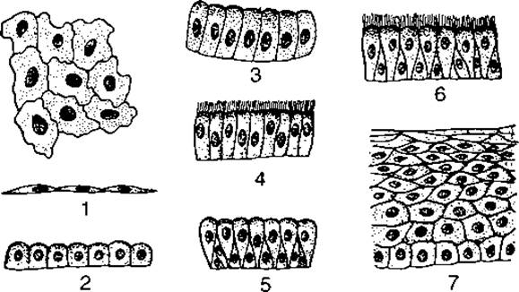
Fig. 4. Types of epithelium:
1 — simple squamous; 2 — low columnar; 3 — high columnar; 4 — ciliated; 5 — pseudostratified columnar; 6 — pseudostratified ciliated; 7 — stratified squamous
The mucous membrane of the Small Intestine is lined with a simple columnar epithelium, which performs a trophic (digestive) function and enables the absorption of nutrients into blood vessels.
The mucous membrane of the urinary tracts is covered with transitional epithelium, while the serous membranes of the Peritoneum and Pleura are lined with simple squamous epithelium (mesothelium).
The alveoli and capillaries are lined from the inside with a simple squamous epithelium.
All epithelial tissues share a common structure. They contain a minimal amount of intercellular substance; cells fit tightly together and rest upon a basement membrane. All epithelial tissues possess high regenerative capacity, containing numerous cells that maintain a high rate of mitosis.
Epithelial tissues originate from all three germ layers: the skin epidermis develops from the ectoderm, the epithelium of the digestive tract mucous membranes from the endoderm, and the mesothelium of the peritoneum, pleura, etc., from the mesoderm.
Glandular Epithelium performs a secretory function and is a constituent part of endocrine and exocrine glands. Glands can be unicellular or multicellular.
Unicellular glands are typically located within the thickness of the epithelium, with goblet cells being their most common form, which secrete mucus in The Stomach and intestines.
Multicellular glands are of three types: exocrine, endocrine, and mixed. Exocrine glands have excretory ducts and release their secretions directly into intercellular spaces, from which they enter the BLOOD AND Lymph.
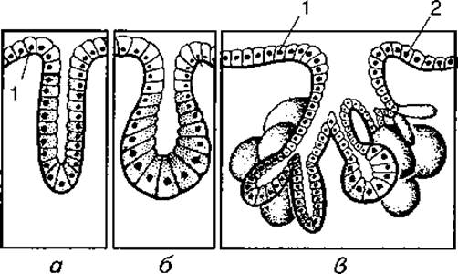
Fig. 5. Diagram of gland structure:
a — simple tubular gland; b — simple alveolar gland; c — complex tubulo-alveolar gland; 1, 2 — glandular epithelium
Endocrine glands lack excretory ducts (hence they are also called ductless glands); their secretion (hormone) enters the blood directly.
Exocrine glands consist of many cells and have a complex structure. Depending on the structure and shape of their terminal portion and excretory ducts, they are classified as tubular, alveolar, and tubulo-alveolar (Fig. 5), as well as simple and complex, branched and unbranched. Simple glands include sweat, intestinal, and Sebaceous Glands; their terminal portion lacks branches. Branched glands consist of several simple units opening into a single excretory duct. The Glands of the stomach, Uterus, and Urethra are built according to this principle. Mixed glands are formed by several branched units that also converge into a single excretory duct. These include the Liver, salivary, lacrimal, Pancreas, mammary, and other glands.
Unlike other tissues, connective tissue is formed by A large number of cells and intercellular substance containing fibers of various structures.
Connective tissue is a component of all human organs and performs supportive, connecting, nutritive, and protective Functions. Depending on the predominance of particular morphological and functional features, connective tissue is subdivided into fibrous, elastic, reticular, cartilaginous, osseous, etc.
Fibrous connective tissue is divided into loose and dense, with dense further categorized into irregular and regular.
The fibers of loose fibrous connective tissue include collagenous and elastic fibers, while its cellular components comprise fibroblasts, fibrocytes, macrophagocytes, plasmocytes, lipocytes, and tissue basophils.
Fibroblasts are large resident cells featuring broad processes, a well-developed rough Endoplasmic reticulum, and a prominent Golgi apparatus. They synthesize the Components of the Extracellular matrix.
Fibrocytes are mature fibroblasts that have completed their developmental cycle and are incapable of further transformation.
Macrophagocytes are large resident cells containing numerous Lysosomes within their protoplasm. Utilizing pseudopodia-like processes for motility, they destroy microorganisms, neutralize toxic substances, and produce immune bodies.
Plasmocytes are immune system cells derived from lymphocytes, which play a key role in antibody production.
Lipocytes are fat cells capable of storing Lipids. Due to significant aggregations of lipocytes, loose fibrous connective tissue can transform into adipose tissue.
Tissue basophils are large cells whose Cytoplasm is packed with granules containing heparin and histamine.
Pigment cells contain melanophores, which impart a specific color (brown or red) to the skin.
Dense Fibrous Connective tissue exists in three forms: dense irregular connective tissue of the skin, dense regular connective tissue of tendons, and elastic connective tissue. These tissues contain fibrocytes and fixed macrophagocytes, as well as collagenous and elastic fibers.
In the dense irregular connective tissue of the skin, collagenous fibers outnumber elastic fibers, forming interwoven bundles that impart strength to the skin.
In contrast to the Cytology/practical/45.html">Dense connective tissue of the skin, the Collagen bundles in the dense regular connective tissue of tendons are thick and arranged in parallel, with fibrocytes interspersed between them. This exceptionally strong tissue forms Muscle tendons and ligaments.
Elastic connective tissue contains a high proportion of elastic fibers, rendering it highly stretchable. It forms the structural framework of large blood vessels, the Trachea, Bronchi, and other structures.
Reticular connective tissue consists of reticular cells and reticular fibers. The cells feature multiple branching processes that connect with neighboring cells, thereby forming a syncytium (network). This tissue constitutes the connective tissue stroma of the Bone Marrow, Spleen, and Lymph Nodes, and is also a component of the mucous membranes of the digestive and respiratory tracts. Under certain conditions, reticular cells can transform into macrophages.
Based on its Development and Structure, blood is classified as a connective tissue—a fluid tissue that integrates biochemical processes occurring within cells and the extracellular matrix, while also performing protective, regulatory, and other functions.
Blood consists of hemocytes, or Blood Cells (45%), and a liquid extracellular matrix known as Blood Plasma (55%). The cellular components include erythrocytes (normally 4.5–5.5 ∙ 1012/L), leukocytes (normally ranging from 4–6 to 8–10 ∙ 109/L), and thrombocytes (180–320 ∙ 109/L) (see color insert).
Erythrocytes perform a respiratory function in the body owing to the presence of Hemoglobin, a complex protein that transports oxygen and carbon dioxide.
Leukocytes are classified into granulocytes and agranulocytes. They serve a protective function in the body, as their cytoplasmic processes (pseudopodia) enable active migration and phagocytosis.
Thrombocytes likewise perform a protective function and play an active role in Blood Coagulation.
Blood plasma consists of 90% Water and 10% dry residue (Proteins, mineral salts, lipids, Enzymes, tissue breakdown products, etc.). Overall, blood contains approximately 80% water and 20% organic substances. The body of an adult human contains roughly 4.5–6 liters of blood, with the total cellular mass accounting for 1/3 to 2/5 of the total blood volume.
Lymph is also a type of connective tissue, consisting of a liquid extracellular matrix (plasma) and cells (predominantly leukocytes). Lymph maintains communication between the internal environment of organs and the blood, transports dietary lipids from the intestines into the Venous system, and participates in the body's immunological reactions. It is estimated that the human body contains about 2.5 liters of lymph. It is a transparent or slightly opalescent fluid with a significantly lower protein concentration than blood plasma.
Depending on The structure of its extracellular matrix, Cartilage tissue is classified into three types: hyaline, fibrous, and elastic cartilage. All three share a common structural Organization, comprising a homogeneous extracellular matrix (cartilage matrix) composed of chondrin, chondromucoproteins, and chondrocytes. Within the cartilage, these cells form isogenous groups located within lacunae. The cartilage matrix contains an Abundance of collagenous and elastic fibers.
Hyaline cartilage is extremely dense and contains numerous fine connective tissue fibers impregnated with chondrin. This cartilage covers the articular surfaces of bones and forms part of the nasal septum, Larynx, trachea, and bronchi.
Fibroblast cartilage (fibrocartilage) is a type of fibrous connective tissue rich in collagen bundles impregnated with chondrin, much like hyaline cartilage. It is found in the intervertebral and interpubic discs, as well as at the insertion sites of tendons into bone.
Elastic cartilage contains a high density of yellowish elastic fibers, making it highly pliable and flexible. It forms the framework of the auricular pinna, the cartilaginous PARTS OF THE auditory tube, the external acoustic meatus, and certain laryngeal cartilages. Unlike fibrocartilage, elastic cartilage does not undergo ossification.
Bone tissue is characterized by high mechanical properties and consists of cells and an extracellular bone matrix containing collagen fibers impregnated with mineral salts. The human Skeleton is built from bone tissue.
There are two types of Muscle tissue: striated (skeletal) and non-striated (smooth).
Striated muscle tissue forms part of skeletal Muscles and has The ability to contract, which drives various essential motor processes. Consequently, skeletal muscles constitute the active component of The Musculoskeletal System. Striated muscle tissue is composed of multinucleated muscle fibers featuring alternating dark and light bands (discs) along their length that exhibit different optical properties. Skeletal muscles contract through innervation by spinal and Cranial Nerves. These are voluntary muscles, meaning they can contract under conscious control.
Unlike striated muscle tissue, Smooth muscle tissue lacks transverse striations. It consists of spindle-shaped cells called myocytes, each containing a single Nucleus. The cells range in length from 15 to 500 µm and have a diameter of 10–20 µm. Smooth muscle cells lack myofibrils, but they do contain contractile myofilaments (protofibrils) measuring 1–2 µm in length and 5–8 nm (thin myofilaments) to 10–30 nm (thick myofilaments) in thickness.
Smooth muscle cells are found in the walls of blood vessels and internal organs. Smooth muscle contractions occur involuntarily, slowly, and rhythmically (for example, peristalsis, changes in vascular lumen, and the ducts of glands).
Nervous Tissue forms the Central Nervous System, peripheral nerves, and their ganglia. Nervous tissue consists of cells known as Neurons (neurocytes) and neuroglia.
Neurons are the primary Structural elements of the central nervous system, forming nerve centers to which sensory neurons transmit nerve impulses from Sensory Organs. These sensory organs inform the centers about their functional state. From the nerve centers, motor neurons deliver commands to effector organs (muscles, glands, blood vessels, and internal organs), which then execute the response. Neurons consist of a cell body and processes, and they vary in size and shape. Based on their function, neurons are classified as sensory, motor, or associative.
Neuroglia performs supportive, trophic, protective, and proliferative functions. Glial cells (gliocytes) outnumber neurons in the central nervous system by 10 to 15 times and possess numerous processes that form a dense network, ensuring extensive interconnections among Nerve Cells.
Last update: 08/08/2026
Editorial and Educational Adaptation: This material has been compiled based on the primary/original source text. The project team performed an editorial review, corrected technical inaccuracies, structured sections, and adapted the content for an educational format.
What was processed:
- elimination of formatting defects (OCR errors, structural breaks, corrupted characters);
- editorial organization of content;
- standardization of terminology in accordance with academic sources;
- verification of factual statements against the original source text.
All mentions of the author, publication year, and origin of the primary text have been preserved in accordance with the source.