Human Anatomy and Physiology - N. I. Fedyukovich 2003
Nervous System
Physiology of the Neuromuscular System
It is well known that under METABOLISM/18.html">The Influence of a stimulus, living Cells and Tissues transition from a state of physiological rest to a state of activity. Among all tissues, nervous and Muscle Tissues exhibit the greatest response to stimulation. The primary properties of nervous and muscle tissues are excitability, excitation, conductivity, refractoriness, and contractility.
Excitability is the ability of living tissue to respond to a stimulus by altering its physiological properties and initiating The process of excitation. Excitation is an active physiological process that arises in tissue under the influence of stimuli and is characterized by changes in metabolic rates, energy release, Muscle tissue contraction, secretion, and Nerve Impulse generation. Conductivity is the ability of living tissue to conduct waves of excitation (biopotentials). Refractoriness is a temporary decrease in tissue excitability resulting from excitation. Lability is the capacity of tissue, dependent on its metabolic characteristics, to be excited a certain number of times per unit of time.
Stimuli capable of eliciting a response from excitable tissues are classified as electrical, chemical, mechanical, and thermal. Based on biological characteristics, stimuli can be adequate or inadequate, and based on intensity, they are classified as subthreshold, threshold, and suprathreshold.
The intensity of the stimulus plays a decisive role in the onset of excitation (the law of stimulation). There is a specific relationship between stimulus intensity and the response. The greater the intensity of the stimulus, the higher the response of the excitable tissue, up to a certain limit. The duration of the stimulus is also of great importance. The relationship between stimulus intensity and the duration required to elicit a minimal response is defined by the strength-duration curve (Fig. 140). The minimum current strength (voltage) capable of causing excitation is called the rheobase (ordinate segment OA). In addition to the rheobase, chronaxie is an important parameter of the strength-duration curve. Chronaxie represents the shortest duration of time required for a current of twice the rheobase intensity (abscissa segment OF) to excite the tissue. Chronaxie value indicates The rate of excitation onset in the tissue: the shorter the chronaxie, the faster excitation occurs. The adaptation of excitable tissue to a gradual increase in stimulus intensity is known as accommodation. This is because, as the stimulus intensity rises slowly, active changes occur in the tissue that raise the threshold of stimulation and prevent The Development of excitation. Thus, the rate of increase in stimulus intensity over time is called the stimulus gradient.
Class="center">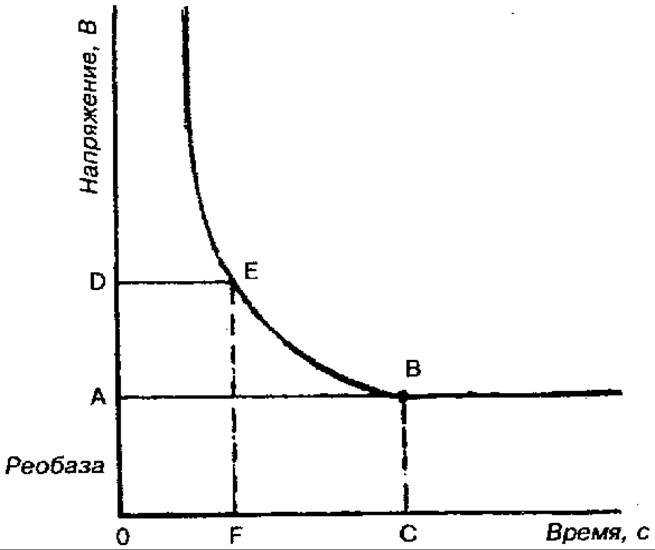
Fig. 140. Strength-duration curve
The law of the stimulus gradient states that the response to a stimulus depends on the rate or steepness of the stimulus increase over a given time: the higher the stimulus gradient, the stronger (up to certain limits) the response of the excitable object.
Resting Potential (Membrane Potential) is the potential difference between the outer surface of The Cell and its internal contents; it is approximately 60—90 мВ depending on the CHARACTERISTICS OF THE specific cell.
An Action Potential (excitation potential) occurs when a stimulus of sufficient strength and duration acts on a section of nerve or muscle fiber. The manifestation of the resulting excitation is a rapid fluctuation in the membrane potential (see Fig. 99). In this state, the excited region has an electronegative charge relative to the unexcited one. An action potential consists of local fluctuations in the membrane potential, the action potential peak (spike), and afterpotentials (negative and positive). The action potential peak is a brief change in the internal potential, featuring a very rapid rising phase and a somewhat slower decay. Following the peak of the action potential, weaker and more prolonged negative, and then positive, afterpotentials are recorded. The duration of the action potential in nerve and muscle fibers is 0.1—5.0 мс. The system of permeating channels for Na+, К+, Сl-, Са2+ ions plays a crucial role in the development of both resting and action potentials. The neural membrane contains specific sodium, potassium, chloride, and calcium channels that allow only the aforementioned ions to pass. These channels possess gating mechanisms and can be open or closed. Determining the state of membrane Ion Channels is essential for resting potential generation, where the leading role belongs to the unequal distribution of potassium ions. Sodium ions play the primary role in action potential generation. Excitation conduction is a specialized function of nerve fibers. The velocity of excitation propagation along them depends mainly on the diameter and histological Structural Features of the nerve fibers. The larger the diameter of the nerve fiber, the higher the velocity of excitation propagation. For example, in a nerve fiber with a diameter of 12— 22 мкм, the propagation velocity of excitation is 70—120 м/с, whereas in a nerve fiber with a diameter of 8—12 мкм, it is only 40—70 м/с.
According to their histological Structure, nerve fibers are divided into myelinated and unmyelinated. A myelinated fiber consists of an axon (axis cylinder) and its covering myelin, or Schwann, sheath. It contains lipid substances that have high electrical resistivity and serve an insulating function. At regular intervals, the myelin sheath is interrupted, leaving exposed areas of the axon about 1 мкм wide (nodes of Ranvier). The surface of the axon is represented by The Plasma Membrane, and its contents by the axoplasm. Unmyelinated fibers lack a myelin sheath and are covered only by Schwann cells. The space between the Schwann cells and the axon is filled with extracellular fluid, allowing the surface membrane of the axon to communicate with the environment surrounding the nerve fiber.
Excitation transmission along myelinated and unmyelinated fibers has its own characteristics. Thus, the transmission of an action potential along a myelinated fiber occurs saltatorily (in leaps) from one node of Ranvier to another, which allows the excitation to propagate without decrement. The velocity of propagation along myelinated fibers is significantly higher than along unmyelinated ones. While the velocity of excitation along motor nerve fibers (covered by a myelin sheath) is 80—120 м/с, along fibers that lack myelin sheaths, it is only 0.5—2.0 м/с. The Propagation of Excitation along a nerve fiber upon stimulation obeys specific laws.
The law of physiological integrity states that the conduction of excitation along a nerve fiber is possible only if not only its anatomical but also its physiological integrity (continuity) is preserved.
The law of bilateral conduction of excitation states that the transmission of excitation occurs in two directions: centripetal and centrifugal.
The law of isolated conduction of excitation states that upon stimulation, excitation is conducted along only one nerve fiber and does not spread to neighboring fibers, which ensures strict coordination of reflex activity. Nerve fibers exhibit low fatigability. This is explained by low Energy Expenditure and rapid recovery processes.
A synapse is a specialized structure that ensures the transmission of a nerve impulse from a nerve fiber to an effector cell, such as a muscle fiber, neuron, or secretory cell.
Synapses are classified according to anatomical and histological principles (neurosecretory, neuromuscular, interneuronal); neurochemical principles (adrenergic, with norepinephrine as the neurotransmitter, and cholinergic, with acetylcholine as the neurotransmitter); and functional principles (excitatory and inhibitory). The Neuromuscular Junction (synapse) consists of three primary structures: the presynaptic membrane, the synaptic cleft, and the postsynaptic membrane. The presynaptic membrane covers the nerve terminal, while the postsynaptic membrane covers the effector cell. The synaptic cleft lies between them. The postsynaptic membrane differs from the presynaptic membrane in that it contains protein chemoreceptors sensitive not only to Neurotransmitters and Hormones but also to drugs and toxic substances. The structure of the neuromuscular junction determines its physiological properties: 1) unidirectional conduction of excitation (from the pre- to the postsynaptic membrane) due to the presence of neurotransmitter-sensitive receptors only on the postsynaptic membrane; 2) synaptic delay in excitation conduction, associated with the low rate of neurotransmitter diffusion compared to the speed of the nerve impulse; 3) low lability and high fatigability of the synapse; 4) high selective sensitivity of the synapse to chemical substances.
Synaptic transmission of excitation is a complex physiological process that occurs in several stages: 1) neurotransmitter synthesis; 2) neurotransmitter secretion; 3) interaction of the neurotransmitter with receptors on the postsynaptic membrane; 4) inactivation (complete loss of activity) of the neurotransmitter. It is known that certain chemical substances, including drugs, can significantly influence synaptic excitation. This phenomenon has found application in clinical practice.
A neuromotor (motor) unit is an anatomical and functional unit of Skeletal Muscle, consisting of an axon (the long process of a Spinal Cord motor neuron) and a specific number of muscle fibers innervated by it. A motor unit can include varying numbers of muscle fibers (from a few to several thousand), depending on the specialization of the muscle. The motor unit Functions as a single entity. Impulses generated by the motor neuron activate all the muscle fibers that comprise it.
The primary function of skeletal Muscles is contraction, which is manifested in various human movements. Skeletal muscles also perform receptor, metabolic, and thermoregulatory functions. They are composed of A large number of multinucleated muscle fibers. The contractile part of a muscle fiber consists of long muscle threads called myofibrils, which run inside the fiber from one end to the other and exhibit transverse striation. This striation is formed by the alternation of dark (anisotropic) A-bands and light (isotropic) I-bands (see Fig. 53). A Z-line runs through the middle of the I-band; two adjacent Z-lines define a sarcomere, the Structural and functional unit. Electron Microscopy of the fibers reveals that the A-band contains a lighter region (H-zone), and in the center, this band is bisected by a dark line, the M-line. The dark band is formed by thick filaments of the protein Myosin, while the light I-band is formed by thin filaments of the protein Actin. The muscle fiber also contains the fibrillar rod-like protein Tropomyosin and the globular protein troponin. The Mechanism of contraction involves the sliding (pulling) of thin filaments along thick ones toward the center of the sarcomere via transverse actin-myosin cross-bridges. The primary source of energy required for Muscle contraction is adenosine triphosphate (АТФ) and the presence of Са2+ and Mg2+ ions. The conversion of chemical energy into mechanical energy occurs in the muscle both without oxygen and with its participation. The anaerobic (oxygen-free) phase is characterized by a series of sequential reactions leading to The breakdown of АТФ and creatine phosphate, and their restoration. The energy released in this process is used for muscle contraction and the recovery (resynthesis) of these substances. The aerobic (oxygen) phase of chemical transformations is associated with The oxidation of lactic acid to carbon dioxide and Water. The resulting energy is used for the further conversion of lactic acid residues into glucose, and subsequently into Glycogen.
The activity of skeletal muscles is regulated by the CNS—specifically the Cerebral Cortex—via sensory, motor, and sympathetic nerve fibers. Skeletal muscles possess the following physiological properties: excitability, conductivity, refractoriness, lability, and contractility. The excitability, propagation velocity of excitation, and lability of muscle tissue are lower than those of Nervous Tissue, while the refractory period is longer than that of nerves. Skeletal muscles can perform work in isotonic, isometric, and auxotonic contraction modes. In the first type of contraction, the muscle fiber primarily shortens while tension remains constant, whereas In the second, the length of the muscle fibers remains unchanged while tension changes. The Nature of skeletal muscle contraction depends on the frequency of stimulation (the frequency of incoming nerve impulses).
Stimulation by a single impulse leads to a single muscle twitch, whereas successive nerve impulses lead to a tetanic contraction, or tetanus.
The physiological properties of smooth muscles are related to their structural characteristics and metabolic rates, and they differ significantly from those of skeletal muscles. Smooth muscles are less excitable than striated muscles. Contraction of smooth muscle occurs more slowly and lasts longer. The refractory period in smooth muscles is more prolonged than in skeletal muscles (up to several seconds). A characteristic feature of smooth muscles is their capacity for automatic activity (autorhythmicity), which is provided by neural elements. Smooth muscles are innervated by sympathetic and parasympathetic autonomic nerves and exhibit high sensitivity to certain BIOLOGICALLY ACTIVE SUBSTANCES (acetylcholine, epinephrine, norepinephrine, serotonin, etc.).
Last update: 08/08/2026
Editorial and Educational Adaptation: This material has been compiled based on the primary/original source text. The project team performed an editorial review, corrected technical inaccuracies, structured sections, and adapted the content for an educational format.
What was processed:
- elimination of formatting defects (OCR errors, structural breaks, corrupted characters);
- editorial organization of content;
- standardization of terminology in accordance with academic sources;
- verification of factual statements against the original source text.
All mentions of the author, publication year, and origin of the primary text have been preserved in accordance with the source.