Human Anatomy and Physiology - N. I. Fedyukovich 2003
Nervous System
Physiology of the Central Nervous System
PHYSIOLOGY OF THE Cerebral Cortex. The cerebrum, or Telencephalon, is one of the most complex human Organs. The Functions of this division of the CNS differ significantly from those of the Brainstem and Spinal Cord. They form The basis of the physiology of Higher Nervous Activity. By higher nervous activity, I. P. Pavlov meant behavior—activity aimed at adapting the Organism to changing environmental conditions and maintaining equilibrium with the environment. Through his research, I. P. Pavlov not only proved the reflex activity of the cerebral cortex but also discovered a qualitatively new, higher type of Reflexes: conditioned reflexes. It was subsequently established that conditioned reflexes are the elementary acts that make up The behavior of humans and animals. At the same time, experimental studies showed that damage to the cerebral cortex leads to the permanent loss of acquired reactions developed during an individual's life—namely, conditioned reflexes. Morphological confirmation of physiological and clinical data came from The Theory of the heterogeneous Cytology/cytology/26.html">Structure of Different areas of the cerebral cortex, known as cortical cyto- and myeloarchitectonics. Detailed studies resulted in the creation of specialized maps of the cerebral cortex, reflecting the distribution of cortical ends of analyzers.
An analyzer is a neural mechanism consisting of a receptive receptor apparatus, Nerve Impulse pathways, and a cerebral center where all stimuli coming from the environment and The Human Body are analyzed. Different analyzers are closely interconnected; consequently, analysis and synthesis occur in the cortex, leading to the generation of response reactions that regulate all types of human activity. It is known that the cerebral cortex contains a Nucleus and scattered elements occupying a specific area. The cerebral cortex represents a collection of nuclei of various analyzers, interspersed with scattered elements of adjacent analyzers.
Thus, in accordance with the cytoarchitectonic maps of the human cerebral hemispheres, the cortical ends of various analyzers (nuclei) can be localized relative to the gyri and lobes of the hemispheres (Fig. 141).
Class="center">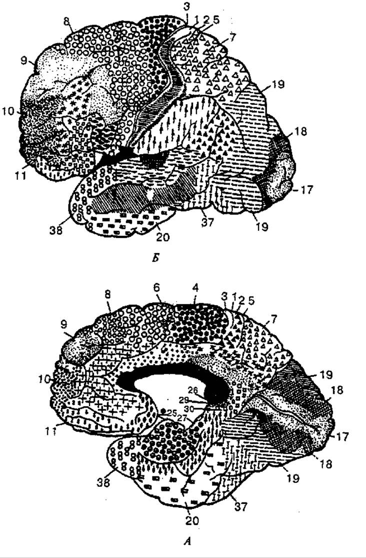
Fig. 141. Cytoarchitectonic areas of the cerebral cortex (according to Brodmann, 1925):
A - lateral surface; B - medial surface; numbers - cortical areas
In the cortex of the postcentral gyrus and the superior parietal lobule, there are Nerve Cells that form the Nucleus of the cortical analyzer of general sensation (thermal, pain, tactile) and Proprioception. Sensory pathways projecting to the cerebral cortex decussate at the level of the spinal cord and Medulla Oblongata. As a result, the postcentral gyrus of each hemisphere is connected to the opposite half of the body. In the postcentral gyrus, all receptive fields of different body parts have their own projection onto the region of the cortical end of the general sensation analyzer. The Nucleus of the motor analyzer is located mainly in the motor cortex and is localized in the precentral gyrus, which lies anterior to the central (Rolandic) sulcus. It is known that the upper PARTS OF THE precentral gyrus and the paracentral lobule contain a nucleus from which impulses travel to the Muscles of the lower trunk and limbs.
The lower part of the precentral gyrus contains the nucleus of the motor analyzer, which regulates The activity of the facial muscles.
The posterior parts of the middle frontal gyrus contain the nucleus of the analyzer that provides the function of conjugate Rotation of the HEAD and eyes to the opposite side.
The inferior parietal lobule contains the nucleus of the motor analyzer, whose function is to execute all purposeful complex movements.
The cortex of the superior parietal lobule contains the nucleus of the cutaneous analyzer, a type of sensation characterized by the function of recognizing objects by Touch. The cortical ends of this analyzer are located in both the right and left hemispheres. Damage to these areas of the cortex leads to the loss of The ability to recognize objects by touch.
The nucleus of the visual analyzer is located on the Medial surface of the occipital lobe. The cortex of the left occipital lobe receives projections from the receptors of the lateral half of the left retina and the medial half of the right retina, respectively. Damage to the visual analyzer leads to complete loss of Vision or visual memory, with an impaired ability to navigate unfamiliar spaces.
The nucleus of the Auditory Analyzer is localized in the superior margin of the temporal lobe. It receives pathways from receptors on both the left and right sides. Consequently, unilateral damage to the nucleus does not cause a complete loss of the ability to perceive sounds. Bilateral damage results in "cortical deafness." The olfactory analyzer nucleus is located on the Inferior surface of the temporal lobe, in the region of the uncus (the terminus of the parahippocampal gyrus).
The Senses of Smell and Taste are closely interrelated, which is explained by the proximity of their analyzers. The analyzer nuclei of the hemispheres are connected to receptors on both the left and right sides of the body.
The posterior part of the middle frontal gyrus contains the nucleus of the motor analyzer of writing. Damage to this area leads to the loss of fine motor movements required for writing letters and numbers.
The nucleus of the motor analyzer of speech articulation is located in the posterior parts of the inferior frontal gyrus (Broca's area). Damage to this area leads to the loss of motor ability in the muscles involved in speech production. The inferior frontal gyrus also contains the nucleus of the language analyzer associated with singing; its damage causes the loss of the ability to remember musical phrases. The superior temporal gyrus contains the nucleus of the cortical analyzer whose damage leads to musical deafness.
The projection areas of the cortex occupy a small portion of The surface of the human cerebral cortex compared to the association areas. The latter have no direct connection to either Sensory Organs or muscles; instead, they establish connections between different cortical regions. They integrate and combine all impulses entering the cortex into complex acts of learning (reading, language, writing), logical thinking, and memory, enabling purposeful behavioral responses.
Disruptions of the association areas lead to agnosia (the inability to recognize objects) and apraxia (the inability to perform learned movements). For example, damage to the lateral surface of the occipital lobe—the visual association area—results in visual agnosia, where the patient is unable to read text or recognize a familiar person. In the case of damage to the speech association areas of the cerebral cortex, aphasia (loss of speech) may occur. Aphasia can be sensory or motor.
Sensory aphasia (Wernicke's aphasia) is characterized by impaired comprehension of spoken language, while the ability to repeat words spoken by another person is preserved; it is observed in lesions of the pathways between the posterior and middle parts of the superior temporal gyrus (Wernicke's area) and other areas of the cerebral cortex. Motor aphasia occurs with damage to the posterior third of the left inferior frontal gyrus (Broca's area): the patient understands spoken speech but is unable to speak.
ELECTRICAL PHENOMENA IN the cerebral cortex. In humans and other vertebrates, spontaneous electrical oscillations characterized by a specific periodicity can be recorded using specialized instruments. These continuous oscillations reflect the elementary activity of the cortex and are referred to as an Electroencephalogram—EEG (Fig. 142).
Two Methods are commonly used to record an EEG: bipolar and monopolar. In bipolar recording, both recording electrodes are placed on the scalp, are active, and register the potential difference between two points on the cortex. In monopolar recording, one electrode is fixed On the surface of the head (active), and the other is placed on the earlobe (indifferent). The electrode placement for EEG recording is standardized and includes mandatory leads from the frontal lobes, motor cortex, parietal, and occipital lobes.
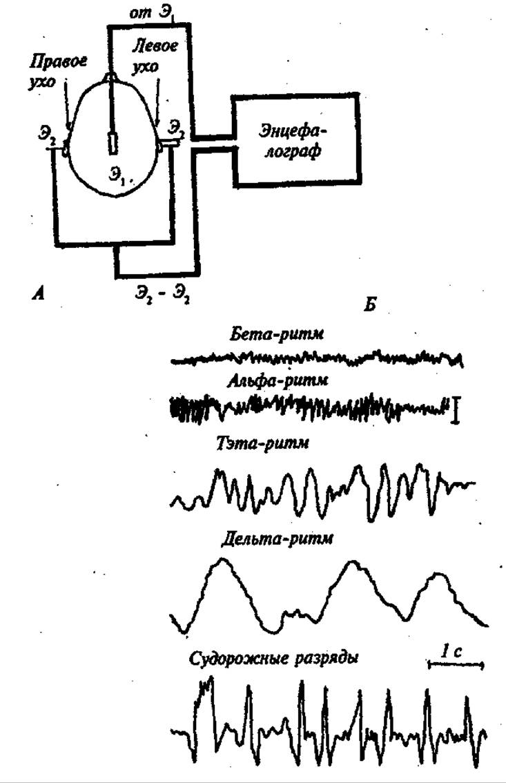
Fig. 142. Electroencephalography.
A — EEG recording setup; B — main EEG rhythms; Э1 — active electrode; Э2 — indifferent electrodes
When analyzing an EEG, the frequency, amplitude, shape, and duration of its electrical oscillations are taken into account. In a relaxed adult in the absence of external stimuli, the EEG shows regular waves with a frequency of 8—13 Hz and an amplitude of about 50 μV. These waves are designated as the alpha rhythm and are most pronounced in the occipital lobes of the cortex. The transition from a state of rest to activity (mental work, light perception, etc.) is accompanied by the disappearance of the alpha rhythm and the appearance of rapid (14—30 Hz), low-amplitude (25 μV) oscillations of the beta rhythm. If a person in a state of rest transitions to Sleep rather than active work, slower and higher-amplitude waves (compared to the alpha rhythm) appear in their EEG, specifically the theta rhythm (4—7 Hz) — 100—150 μV and the delta rhythm (0.5— 3.5 Hz) - 250-300 μV.
Under normal conditions, theta and alpha rhythms are not detected in an awake individual. Cessation of cerebral Blood supply leads to the disappearance of its electrical activity within just 15s.
Thus, EEG and the analysis of its frequency spectrum allow for the Assessment of the functional state of the CEREBRAL CORTEX AND are widely used in clinical practice.
Last update: 08/08/2026
Editorial and Educational Adaptation: This material has been compiled based on the primary/original source text. The project team performed an editorial review, corrected technical inaccuracies, structured sections, and adapted the content for an educational format.
What was processed:
- elimination of formatting defects (OCR errors, structural breaks, corrupted characters);
- editorial organization of content;
- standardization of terminology in accordance with academic sources;
- verification of factual statements against the original source text.
All mentions of the author, publication year, and origin of the primary text have been preserved in accordance with the source.