Human Anatomy Part 1 - K. A. Dyubenko, A. K. Kolomiytsev, Yu. B. Chaykovsky 2002
Special Section
Urinary System, systema urinarium - General Overview
Urinary Organs - Kidneys - Kidney Structure
On a coronal section, the Kidney consists of an outer layer (the renal cortex, cortex renalis, yellowish-red in color) and an inner layer (the renal medulla), medulla renalis, which is lilac-red in color (Fig. 302). The renal cortex, cortex renalis, is located at the periphery of the organ and is 0.5 cm thick. Alternating darker and lighter radially arranged stripes are visible within the renal cortex. The darker areas of the cortex are termed the convoluted or glomerular part, pars convoluta, whereas the lighter, cone-shaped areas are termed the radiate part, pars radiata.
The renal medulla, medulla renalis, is formed by 14-16 renal pyramids, pyramides renales, whose apices are directed toward the renal sinus and whose bases face The surface of the kidney. Between the renal pyramids lie 10-15 renal columns, columnae renales, which represent extensions of the cortical substance.
The apices of the pyramids terminate in renal papillae, papillae renales, which feature 12-25 papillary foramina, foramina papillaria, representing the continuation of the collecting tubules of the pyramids. The renal papillae project into the lumen of the minor renal calyces, calyces renales minores, which number from 8 to 10. The minor renal calyces unite in groups of 2-3 to form the major renal calyces, calyces renales majores, which open into the renal pelvis, pelvis renalis. A renal pyramid, together with the cortical tissue adjoining its base, is referred to as a renal lobe, lobi renales.
Class="center">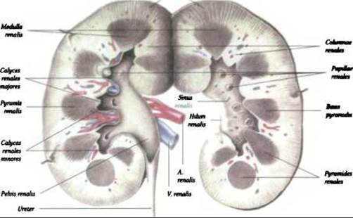
Fig. 302. Kidney, rеn, coronal section (after R. D. Sinelnikov)
The renal pelvis is located within the renal sinus posterior to the renal vein and artery. The walls of the renal calyces and the renal pelvis consist of three layers: an inner mucous membrane (mucosa), a middle muscular layer, and an outer adventitial layer. In the region of the minor renal calyx, where it encompasses the renal papilla, a thickened layer of smooth Muscle Cells is formed. This layer, together with Blood Vessels, Lymphatic vessels, and nerve fibers, constitutes the so-called fornical apparatus of the kidney. It plays a crucial role in regulating the flow of urine from the papillary ducts into the renal calyces.
The renal parenchyma is composed of renal tubules, tubuli renales, the walls of which are formed by epithelium. Together with blood capillaries, they form the Structural and functional unit of the kidney—the nephron, nephron (Fig. 303). The nephron comprises a renal corpuscle and a tubular system. Each kidney contains approximately 1 million nephrons. Nephrons produce primary urine by filtration from Blood Plasma, which then enters the capsular space. Based on their Location, nephrons are classified into cortical and juxtamedullary nephrons, the latter accounting for 20%. (A more detailed Structure OF THE nephron is provided in textbooks of Histology).

Fig. 303. Structure of the nephron (diagram after E. F. Kotovsky)
The nephron comprises: the glomerular capsule, capsula glomerularis, also known as Bowman's capsule1; the proximal convoluted tubule, tubulus contortus proximalis; the proximal straight tubule, tubulus rectus proximalis; the thin segment, tubulus attenuatus, which includes a descending limb, pars descendens, and an ascending limb, pars ascendens; the distal straight tubule, tubulus rectus distalis; and the Distal convoluted tubule, tubulus contortus distalis.
1 Oleksandr Mykhailovych Shumliansky (1748-1795), 60 years before W. Bowman (the English surgeon), described the capsule of the renal corpuscle and introduced the term vascular glomerulus, glomerulus.
The thin segment and the distal straight tubule form the loop of the nephron, ansa nephrica, also known as Henle's loop. The renal corpuscle, corpusculum renale, has a diameter of approximately 200 µm. It comprises the vascular (Malpighian) glomerulus, glomerulus, and the surrounding capsule, capsula glomeruli (Fig. 304). The glomerular capsule resembles a double-walled cup formed by outer and inner layers, between which lies a slit-like space that continues into the proximal opening of the nephron tubule.
The medullary ray, along with its surrounding glomeruli, as well as the proximal and distal convoluted tubules of the nephrons that open into a branched collecting duct, constitute the renal lobule, lobulus renalis (A. Ham, D. Cormack, 1983).
Blood supply to the Kidneys. The kidneys are supplied by the renal Arteries, aa. renales, which branch off the Abdominal Aorta: the left at the level of the first lumbar vertebra, and the right at the level of the second lumbar vertebra. Frequently, the kidneys receive additional renal arteries1, 2 originating from the aorta or the iliac arteries, a clinical factor that must be considered in urology.
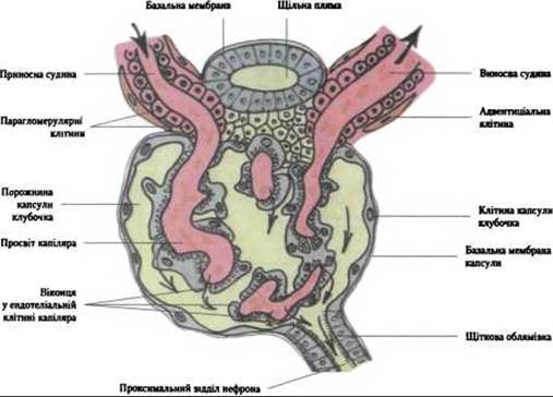
Fig. 304. Ultramicroscopic structure of the renal corpuscle (after E. F. Kotovsky)
1 Friedrich Gustav Jacob Henle (1809-1885) was a German anatomist who taught anatomy in Zurich and served as director of the Anatomical Institute in Göttingen. F. Henle described the loops of the renal tubules (Henle's loops), which reabsorb Water, glucose, and salts from the primary urine.
2 Accessory renal artery, a. renalis accessoria
Within the renal hilum, the renal arteries divide into anterior and posterior branches, which give rise to interlobar arteries, aa. interlobares, running between the pyramids (Fig. 305). In terms of their branching pattern and distribution territory, the interlobar arteries largely correspond to the segmental Arteries of the kidney. The anterior branch gives rise to four segmental arteries: the superior segment artery, segmentum superius; the anterosuperior segment artery, segmentum anterius superius; the anteroinferior segment artery, segmentum anterius inferius; the posterior segment artery, segmentum posterius; and the inferior segment artery, segmentum inferior. The posterior branch continues as the posterior segment artery. The segmental arteries give rise to arcuate arteries, aa. arcuatae, and interlobular arteries, aa. interlobulares. The arcuate and interlobular arteries give rise to the afferent glomerular arterioles, or afferent vessels, vas afferens, which form the capillary glomeruli, glomeruli. These form the renal corpuscle, corpusculum renale. The vascular glomerulus contains up to 50 capillary loops that lack transverse anastomoses. Each terminal capillary loop continues into a venous vessel. The capillary network of the vascular glomerulus is known as a marvelous arterial net, rete mirabile, because the capillaries are interposed between two arterial vessels (the afferent and efferent arterioles).
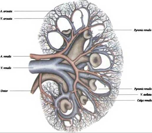
Fig. 305. Blood vessels of the kidney. Corrosion cast (after F. Kadar)
The afferent glomerular arteriole, arteriola glomerularis afferens [vas afferens], possesses a well-developed internal elastic membrane, external to which lie smooth muscle cells. As it approaches the renal corpuscle, it transforms into specialized (juxtaglomerular) cells whose Cytoplasm contains granules, indicating a secretory function. The endothelium of the glomerular capillaries features a perforated cytoplasm.
The efferent glomerular arteriole, arteriola glomerularis efferens, or efferent vessel, vas efferens, conducts blood toward the renal tubules, where it forms a network of peritubular capillaries (the peritubular capillary network).
Juxtaglomerular arteries—namely, arteries with a single capillary network in the renal cortex—are the exception, although they occur where glomeruli are absent or sparse. For instance, in the medulla, straight arterioles form dense capillary networks between the renal tubules and the collecting ducts of the pyramids.
The venous System of the kidney originates from the tubular capillaries of the cortex, from which blood drains into stellate venules, venulae stellatae, which converge to form radially disposed interlobular Veins, venae interlobulares. Straight venules, venulae rectae, are formed from the capillaries of the medulla, while anastomoses between interlobular veins and straight venules form arcuate veins, v. arcuatae, and subsequently interlobar veins, vv. interlobares, which unite to form the renal vein, v. renalis, emptying into the INFERIOR VENA CAVA.
Lymph drainage (according to F. A. Stefanis) occurs via superficial and deep lymphatic vessels. The deep vessels emerge from the renal parenchyma around the papillae, accompanying the corresponding arteries and veins. They are divided into three groups: anterior — located in front of the renal vein; middle — situated between the vein and the artery; and posterior — behind the renal artery. The superficial vessels drain lymph from the adipose and fibrous capsules of the kidney. As a rule, they empty into the deep vessels in the region of the renal hilum.
The lymphatic Vessels of the kidney drain into the para-aortic and lumbar Lymph Nodes. From these nodes, the lymph flowing from the kidney is directed to lymph nodes located in the upper Abdominal cavity along the aorta, the inferior vena cava, and in the cleft between the medial and intermediate crura of the Diaphragm. The renal lymphatic vessels have numerous anastomoses with the lymphatic vessels of the Adrenal gland, Pancreas, Liver, Testis, and Ovary.
The Innervation of the kidneys is provided by the renal plexus (plexus renalis). Its sources of formation include the greater and lesser splanchnic nerves (nn. splanchnici major et minor), Branches of the lumbar Sympathetic trunk (truncus sympathicus), branches of the celiac and superior mesenteric plexuses, and the aortorenal ganglia. Afferent innervation is mediated by sensory ganglia of the Vagus nerve and spinal ganglia containing sensory Neurons. Efferent nerve fibers of the Autonomic Nervous system (sympathetic and parasympathetic) reach the smooth muscle Cells of the walls of the renal blood vessels, calyces, and renal pelvis. At the renal hilum, the renal plexus divides into peri-arterial plexuses that accompany the renal vessels and penetrate the renal parenchyma along with them. In the medulla and cortex, nerve fibers entwine the renal pyramids and lobules, accompany the afferent glomerular arterioles, and reach the glomerular capsules. Cytology/practical/64.html">Unmyelinated nerve fibers approach the walls of the renal tubules and renal calyces.
Last update: 08/08/2026
Editorial and Educational Adaptation: This material has been compiled based on the primary/original source text. The project team performed an editorial review, corrected technical inaccuracies, structured sections, and adapted the content for an educational format.
What was processed:
- elimination of formatting defects (OCR errors, structural breaks, corrupted characters);
- editorial organization of content;
- standardization of terminology in accordance with academic sources;
- verification of factual statements against the original source text.
All mentions of the author, publication year, and origin of the primary text have been preserved in accordance with the source.