Human Anatomy Part 1 - K. A. Dubenko, A. K. Kolomiytsev, Yu. B. Chaykovsky 2002
Special Section
Urinary System, systema urinarium - General Overview
Urinary Organs - Kidneys - Topography of the Kidneys
Skeletotopy. The Kidneys are located in the retroperitoneal space, spatium retroperitoneale, extraperitoneally, on either side of the lumbar Vertebral Column, at the level of the XII thoracic and the upper three lumbar vertebrae. The right Kidney is situated 2–3 cm lower than the left, and its upper pole does not reach the XI rib. The XII rib projects onto the left kidney approximately at its midpoint, and onto the right kidney between its upper and middle thirds. There are three variants of kidney placement: high, intermediate, and low. In high placement, the kidneys lie behind the XI and XII Ribs; in low placement, they project below the lower margin of the XII rib. In an elongated kidney (Fig. 300) (indicated by hatching), it extends significantly beyond the crista iliaca, whereas a shortened kidney reaches the X–XI vertebrae. In intermediate placement, the left kidney is intersected by the XII rib at its midpoint, while in the right kidney, the XII rib "cuts off" its upper third. The varying height of kidney placement is of substantial importance in surgical Procedures (they are easier to perform when the kidneys are positioned low, whereas a high placement requires a transcostal approach).
Class="center">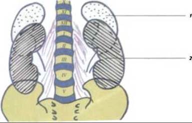
Fig. 300. Variants of kidney placement (according to Helm): 1 - short kidney; 2 - long kidney
The length and direction of the XII rib are of great clinical significance in urological practice. Variants occur where the XII rib ranges from 1.5 cm to 14 cm in length. If the XII rib is long, it runs parallel to the XI rib obliquely downward, whereas a short rib has a more horizontal direction (Fig. 301). With a long XII rib, resection of its lateral third can be performed without damaging the Pleura, whereas with a short rib, resection of the lateral end may result in pleural injury.
Syntopy. Each kidney is situated within a muscular renal bed bounded laterally by the transversus abdominis Muscle, posteriorly by the quadratus lumborum muscle, and medially by the psoas major muscle, which covers THE VERTEBRAL COLUMN. The upper third of the kidneys lies outside this bed and abuts the Diaphragm.
The TOPOGRAPHIC AND ANATOMICAL relations of the right and left kidneys with adjacent Organs differ. A significant portion of the anterior surface of the right kidney is covered by the Liver; closer to the medial margin, the duodenum runs anteriorly, followed inferiorly by the right colic flexure, flexura colica dextra.
The anterior surface of the left kidney contacts The Stomach superiorly, and the Pancreas and loops of the jejunum inferiorly. The lateral margin of the left kidney is covered anteriorly by the Spleen, while the descending colon descends posteriorly along it. By its posterior surface, the kidney is adjacent to the diaphragm, the transversus abdominis muscle, and the quadratus lumborum muscle.
Holotopy. The right kidney projects onto the anterior abdominal wall in the right epigastric, umbilical, and lateral Abdominal Regions (regio epigastrica, umbilicalis et abdominalis lateralis dextra). The left kidney projects into the left epigastric, umbilical, and lateral abdominal regions (regio epigastrica, umbilicalis et abdominalis lateralis sinistra). The kidneys are covered by a Connective Tissue capsule, capsula fibrosa, external to which lies the adipose capsule, capsula adiposa. The anterior and posterior fascial layers enclosing the kidneys form pockets that open downward on both the right and left sides. The renal fascia, fascia renalis, and major Blood Vessels play a crucial role in renal fixation. Fascial bundles pierce the adipose capsule and grow into the fibrous capsule of the kidney and partially into the adventitia of the renal pelvis. Intra-abdominal pressure, the vascular pedicle, and the renal ligaments also play an essential role in stabilizing the kidney.
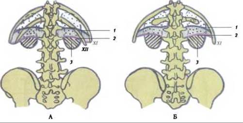
Fig. 301. Relationship of the kidneys to the XII rib at various rib lengths (according to Récamier):
A, B - 1 - inferior border of the pleura; 2 - diaphragm (origin); 3 - projection field of the right kidney
Last update: 08/08/2026
Editorial and Educational Adaptation: This material has been compiled based on the primary/original source text. The project team performed an editorial review, corrected technical inaccuracies, structured sections, and adapted the content for an educational format.
What was processed:
- elimination of formatting defects (OCR errors, structural breaks, corrupted characters);
- editorial organization of content;
- standardization of terminology in accordance with academic sources;
- verification of factual statements against the original source text.
All mentions of the author, publication year, and origin of the primary text have been preserved in accordance with the source.