Human Anatomy, Part 1 - K. A. Dyubenko, A. K. Kolomiytsev, Yu. B. Chaykovsky 2002
Specialized Section
Urinary System, systema urinarium - General Overview
Urinary Organs - Kidneys - X-ray Anatomy of the Kidneys
On plain radiographs, the outer contour of the Kidney, its Blood Vessels, calyces, renal pelvis, and Ureters can sometimes be distinguished by shadow intensity alone, which is achieved only through the artificial administration of contrast agents (Fig. 306).
THE POSITION OF the Kidneys is determined by the distance from their poles to the median horizontal line passing through the midpoint of the body of the second lumbar vertebra.
Class="center">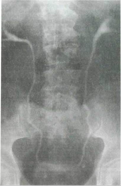
Fig. 306. Excretory urogram at 10 min (68-year-old male) (after V. V. Zhyla). The upper Urinary Tract is clearly outlined along its entire length, along with the Urinary Bladder.
The horizontal distance between the lower poles of the kidneys is typically 11 cm, and between the upper poles, 7 cm. In most cases, the right kidney lies lower than the left, situated at the level of the 12th thoracic to 3rd lumbar vertebrae, whereas the left is located at the level of the 11th thoracic to 2nd lumbar vertebrae. In one-third of individuals, the kidneys are positioned at the same level, and in 5% the left kidney lies lower than the right.
Radiologically, two main kidney shape variants are distinguished: broad and narrow; occasionally, a triangular kidney shape is observed. On radiographs, the right kidney appears narrower than the left. The contours of normal kidneys are even, smooth, and continuous, except for the medial margin where they are not visualized in the region of the renal hilum. According to radiological studies (N. Moell, 1956), normal kidney dimensions average: in males — right kidney 12.9 × 6.2 cm, left kidney 13.2 × 6.3 cm; in females — right kidney 12.3 × 5.7 cm, left kidney 12.6 × 5.9 cm.
There is considerable anatomical variation in the pelvicalyceal system (ampullary, branched, and mixed types) (Fig. 307). The renal pelvis may protrude significantly from the renal sinus or lie deep within its medial margin. Normally, the contours of the pelvicalyceal system are distinct, gently curved, free of protrusions, and transition smoothly into the ureters. Major renal calyces have sharp, funnel-shaped outlines (margins), while minor calyces are arranged in two rows corresponding to the anterior and posterior halves of the kidney. Their detailed examination requires oblique and targeted radiographs.
Computed tomography of the kidneys. The upper pole of the right kidney is located at the level of the lower margin of the 12th thoracic vertebra, and the left is 1–1.5 cm higher than the right.
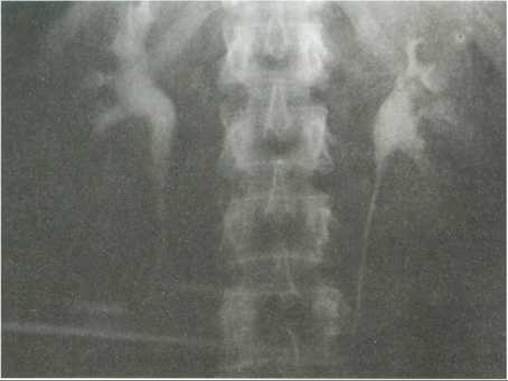
Fig. 307. Anatomical variant of the pelvicalyceal system (after V. V. Zhyla). The papillae of the pyramids are clearly delineated.
The lower poles of the kidneys are located at the level of the III–IV lumbar vertebrae. The renal hilum lies at the level of the lower margin of the first lumbar vertebra. Depending on The Development of adipose tissue, the renal fascia can sometimes be observed in certain individuals, dividing the space surrounding the kidney into pararenal and perirenal spaces. The ureters lie posterior to the vascular pedicle of the kidneys; however, due to their small diameter and low density, they are not visible and can only be detected following contrast enhancement. The medullary substance of the parenchyma is rarely differentiated from the cortical substance, which can be achieved using intravenous contrast enhancement (Figs. 308, 309). The renal sinus is oriented slightly medially relative to the INFERIOR VENA CAVA and aorta. On tomograms, the renal Arteries branch off the aortic wall at a right angle at the level of the I–II lumbar vertebrae (Fig. 310). The left renal artery is slightly higher than the right. The right renal artery originates behind the inferior vena cava. The renal Veins drain into the inferior vena cava at the level of the I–II lumbar vertebrae.
Ultrasonography of the kidneys. Ultrasound imaging of the kidneys is performed in the supine position, both from the back, the anterior abdominal wall, and the lateral body surface.
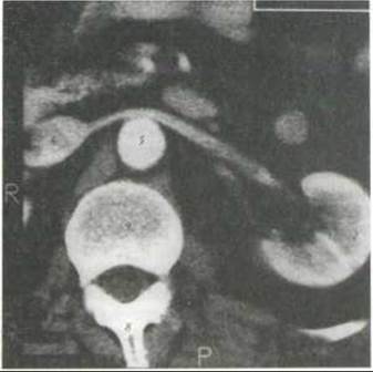
Fig. 308. Computed angiogram of the left kidney at the level of the hilum in the arterial phase, following contrast administration (after O. Ivankov):
1 - renal parenchyma; 2 - cortical layer; 3 - renal artery; 4 - renal vein; 5 - aorta filled with contrast medium; 6 - inferior vena cava; 7 - Spinal Cord; 8 - spinous process; 9 - vertebral body
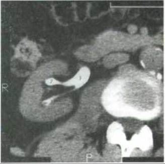
Fig. 309. Computed tomogram of the right kidney at the level of the hilum after intravenous contrast enhancement (after O. Ivankov):
1 - renal parenchyma; 2 - calyces; 3 - renal pelvis; 4 - inferior vena cava; 5 - aorta; 6 - lumbar vertebra; 7 - spinal cord; 8 - spinous process; 9 - psoas Muscle
To determine kidney position, sequential transverse scans are performed at 1–1.5 cm intervals from the lower pole to the upper pole, while longitudinal scans are carried out at similar intervals from the lateral to the medial margin. The right kidney is scanned through the Liver, which provides good acoustic transmission, making its visualization possible from the anterior abdominal wall in all cases. The left kidney is not always visible from the anterior wall and is identified along the anterior axillary line.
Normally, the length of the kidney is 7.5–12 cm, width 4.5–6.5 cm, and thickness 3.5–5 cm. On longitudinal sonograms, the kidney is visualized as an elongated-oval Structure, and on transverse scans, as an ovoid one. A Connective Tissue fascia 1.5 cm thick can be observed On the surface of the kidney.
The renal parenchyma has a delicate structure. Numerous echo-negative structures representing the pyramids are visible between the capsule and the pelvicalyceal system. The diameter of the pyramids ranges from 0.5 to 0.9 cm. On transverse scans, the calyceal complex should be oval or rounded in shape, and on longitudinal scans, elongated, with increased echogenicity in the center of the kidney. The renal pelvis is noticeable in the region of the renal hilum and appears as a spindle-shaped structure on transverse sonograms. The size of the renal pelvis tends to exhibit significant individual variations. Normally, its anteroposterior dimension on sonograms is 1–1.5 cm.
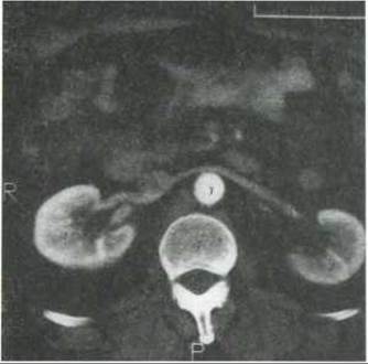
Fig. 310. Computed angiogram of the kidneys at the level of the hilum in the arterial phase (after O. Ivankov):
1 - right kidney; 2 - left kidney; 3 - aorta filled with contrast medium; 4 - inferior vena cava; 5 - renal vein; 6 - renal artery; 7 - lumbar vertebral body
Last update: 08/08/2026
Editorial and Educational Adaptation: This material has been compiled based on the primary/original source text. The project team performed an editorial review, corrected technical inaccuracies, structured sections, and adapted the content for an educational format.
What was processed:
- elimination of formatting defects (OCR errors, structural breaks, corrupted characters);
- editorial organization of content;
- standardization of terminology in accordance with academic sources;
- verification of factual statements against the original source text.
All mentions of the author, publication year, and origin of the primary text have been preserved in accordance with the source.