MODERN BOTANY - P. RAVEN - 1990
SECTION IV. DIVERSITY
CHAPTER 13. FUNGI
Major Groups of Fungi
Phylum Zygomycota
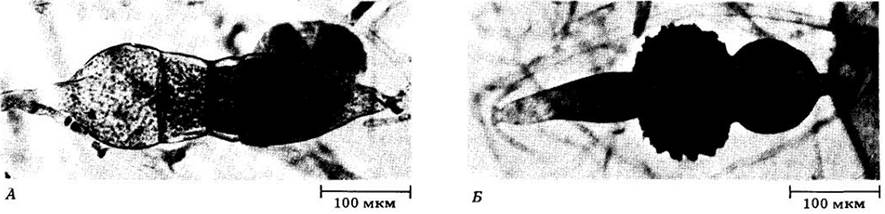
The vast majority of zygomycetes (about 600 described species) live in the soil as saprotrophs on decaying PLANT AND ANIMAL remains. Some are parasites of plants, insects, or small soil-dwelling animals, while several forms cause serious diseases in humans and domestic animals. The term "zygomycetes" reflects a key characteristic of these Fungi: The formation of specialized sexual structures known as zygosporangia through the fusion of two multinucleate gametangia (Fig. 13-9). Within the zygosporangium, the Gametes (in this case, simply nuclei) fuse to form one or more diploid nuclei (zygotes). The zygosporangium is sometimes referred to as a zygospore, though we prefer to avoid this term because it often contains numerous zygote nuclei. Asexual reproduction via spores produced in one or more distinct sporangia on hyphae is common to nearly all zygomycetes. Most members of this phylum possess coenocytic (multinucleate, aseptate) mycelia, within which a rapid cytoplasmic streaming is frequently observable.
Class="center">Fig. 13-9. The mold Rhizopus stolonifer colonizes the surfaces of moist aerial substrates. A. Gametangia: structures where gametes are formed. B. Zygosporangium: the resting stage associated with sexual reproduction (dark central mass). It contains one or more diploid zygotes.
One of the most widespread zygomycetes is Rhizopus stolonifer, which forms a cotton-like black mold On the surface of moist, carbohydrate-rich foods and other similar organic matter left exposed to the air. This species poses a serious threat to stored fruits and vegetables, as it can spoil them quite rapidly. The life cycle of R. stolonifer is illustrated in Fig. 13-10. The mycelium is composed of three distinct types of haploid hyphae. The bulk of the mycelium consists of rapidly growing coenocytic hyphae that ramify primarily within the substrate, absorbing nutrients. Arising from these are arching hyphae called stolons, which produce rhizoids wherever their tips Touch the substrate. Sporangia develop at the tips of sporangiophores, which in turn emerge as vertical branches directly above the rhizoids. Initially, a sporangium appears as a Swelling packed with numerous nuclei; it is then walled off from the sporangiophore by a septum. Its Cytoplasm fragments According to the number of nuclei, and a Cell wall forms around each spore. As they mature, the sporangia turn black, giving the mold its characteristic color. Upon dispersal, each spore is capable of germinating to establish a new mycelium.
Fig. 13-10. In Rhizopus stolonifer, sexual reproduction involves only strains of different mating types, commonly designated as "-" and "+" (although these strains are morphologically indistinguishable, they are represented here in two contrasting colors). When two compatible strains come into close proximity, Hormones are released that induce the hyphal tips to form specialized outgrowths. These make contact, develop into gametangia, and become walled off from the rest of the mycelium by septa (see Fig. 13-9, A). The Cell walls between the contacting gametangia dissolve, and the two multinucleate protoplasts merge. Plus and minus nuclei fuse in pairs, yielding a young zygosporangium containing several diploid nuclei. The zygosporangium subsequently develops a thick, rough, black wall and enters a dormant period that often lasts for several months. Meiosis occurs during germination, at which point the zygosporangium germinates to produce a sporangium similar to that formed during asexual reproduction, and the life cycle begins anew.

One of The most significant groups of zygomycetes, including the genus Glomus and several closely related forms, colonizes plant roots to form mycorrhizae, as described below.
Phylum Ascomycota
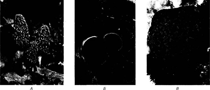
The ascomycetes encompass approximately 30,000 species, including many widespread and economically important organisms such as the blue-green, red, and brown Molds responsible for food spoilage. Among them is the orange-pink fungus Neurospora, which played a pivotal role in the advancement of modern genetics. Ascomycetes also cause A number of severe plant diseases, such as powdery mildew (primarily on leaves), chestnut blight (caused by Endothia parasitica, accidentally introduced to America from northern China), and Dutch elm disease (caused by Ceratocystis ulmi, a fungus of European origin). This phylum additionally includes diverse Yeasts, edible morels, and truffles (Fig. 13-11). Overall, the group remains relatively understudied, with thousands of new species—undoubtedly including economically significant ones—awaiting formal scientific description.
Fig. 13-11. Ascomycetes. A. The common morel, Morchella esculenta, one of the most popular edible fungi. Mushroom hunters traditionally search for it in spring, when oak leaves reach "the size of a mouse's ear." Although morels were first brought into culture in 1983, they are not yet cultivated on a commercial scale. B. Sarcoscypha coccinea,
a striking fungus featuring an open ascocarp (apothecium). C. The highly prized edible ascocarp of the black truffle (Tuber melanosporum). In truffles, spore-bearing structures develop underground and remain enclosed, releasing ascospores only after the ascocarp decays or is broken open by burrowing animals. Truffles form mycorrhizae (predominantly with oaks). They are hunted with the aid of specially trained dogs or pigs, and recently, small-scale commercial cultivation has begun by inoculating the roots of tree seedlings with fungal spores.
Characteristics of Ascomycetes
With the exception of unicellular yeasts, ascomycetes possess a filamentous body. Their hyphae are typically septate—meaning they are divided by cross-walls perforated by pores through which cytoplasm and nuclei can migrate. The Cells of the vegetative mycelium may be uninucleate or multinucleate. Some ascomycete species are homothallic (capable of self-Fertilization, or completing a sexual cycle within a single strain), whereas others are heterothallic and require the presence of both plus and minus strains for sexual reproduction.
The majority of ascomycetes reproduce asexually by producing specialized spores called conidia (derived from a Greek word meaning "fine dust"). Usually multinucleate, these spores originate from cells (Fig. 13-12) that are pinched off at the tips of modified hyphae known as conidiophores.
Sexual reproduction in ascomycetes invariably involves the formation of an ascus (plural, asci) containing haploid ascospores.
Fig. 13-12. Conidia are the typical asexual spores produced by ascomycetes. This electron micrograph shows the stages of conidiogenesis in the fungus Nomuraea rileyi infecting an armyworm caterpillar. A. Scanning electron micrograph illustrating various stages of conidial development. B. Transmission electron micrograph of conidia. In this species, conidia are formed in a basipetal sequence (from tip to base).
These unique reproductive structures distinguish ascomycetes from all other fungi (Fig. 13-13). Asci typically develop within a complex, macroscopic Structure formed by tightly interwoven hyphae called an ascocarp. The ascocarp may be open and cup-shaped (an apothecium, Fig. 13-11, B), closed and spherical (a cleistothecium, Fig. 13-13, B), or flask-shaped with a narrow pore for ascospore release (a perithecium, Fig. 13-13, C). Asci generally line the inner surface of the ascocarp, a fertile layer referred to as the hymenium, or hymenial layer (Fig. 13-14).
Fig. 13-13. A. Electron micrograph of asci of Ascodesmus nigricans containing maturing ascospores. B. Ascocarp of Erysiphe aggregata containing enclosed asci and ascospores. This completely closed type of ascocarp is termed a cleistothecium. C. Ascocarp of Chaetomium erraticum containing enclosed asci and ascospores. A small apical pore is clearly visible. This variant of ascocarp featuring a small opening is called a perithecium.
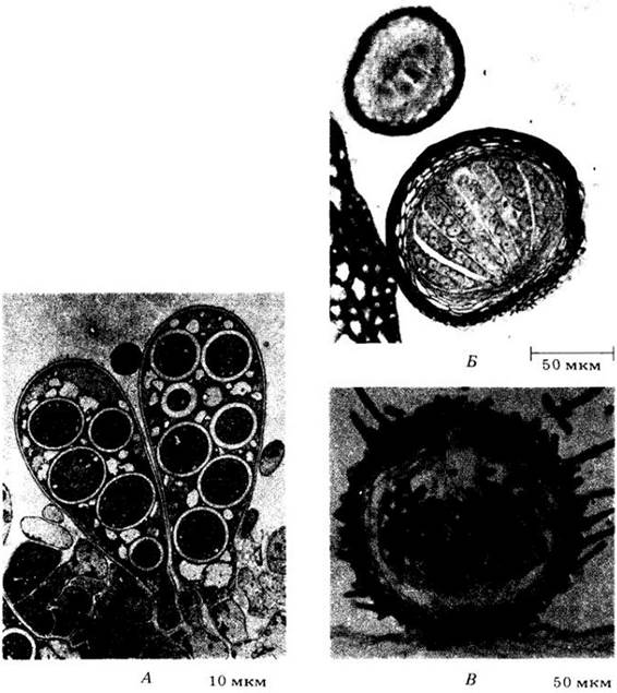
Fig. 13-14. Section through the hymenial layer of a morel (Morchella); asci with ascospores are visible
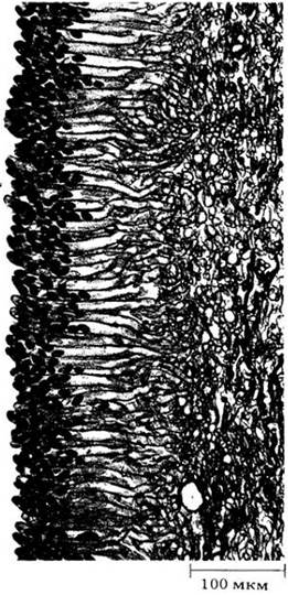
Fig. 13-15. Typical Life Cycle of an ascomycete. Asexual reproduction involves the formation of specialized, usually multinucleate spores called conidia. The sexual process includes the formation of asci and ascospores resulting from meiosis, which occurs in the ascus immediately after karyogamy

Fig. 13-15 illustrates the life cycle characteristic of an ascomycete. Mycelium formation begins with the germination of an ascospore; soon it begins to form conidiophores. During the growing season, a huge number of conidia are produced; these are responsible for the dispersal of the fungus.
The asci arise on the same mycelium as the conidia, preceded by the formation of multinucleate gametangia—the antheridium and ascogonium. Male nuclei from the antheridium pass into the ascogonium through an outgrowth of the latter called the trichogyne. Thus, plasmogamy takes place—the fusion of two protoplasts.
Within the shared cytoplasm, the male nuclei find paired, genetically distinct female nuclei without fusing with them. Next, ascogenous hyphae begin to grow and elongate from the ascogonium, into which pairs of nuclei migrate as the hyphae develop; synchronous mitotic divisions occur within these hyphae and the ascogonium. Cell Division in the developing ascogenous hyphae leads to the formation of dicaryons, i.e., cells with two haploid nuclei.
The ascus is initially initiated at the tip of a developing ascogenous hypha consisting of dicaryons. One of its binucleate cells grows out in the form of a hook, and its two nuclei divide in such a way that their spindles lie roughly along the axis of the hypha, parallel to each other. Two daughter nuclei approach each other at the tip of the hypha, a third ends up at the "tip" of the hook, and the remaining one lies near the septum at its base. Two septa then form, dividing the hook cell into three cells, the middle of which develops into the ascus. It is here that karyogamy takes place: the two nuclei fuse to form a diploid nucleus (the zygote), the only one in the ascomycete life cycle (except for the parasexual process). Shortly after karyogamy, the "young" ascus begins to elongate. The diploid nucleus then divides meiotically and usually once again mitotically. If this mitosis occurs, the mature ascus contains eight spores; if mitosis is absent, it contains four. The resulting haploid nuclei, along with adjacent regions of cytoplasm, separate from one another to give rise to ascospores. In most ascomycetes, the mature ascus swells and eventually bursts, shooting its spores into the air. They typically travel about 2 cm, but in some species the dispersal distance reaches 30 cm.
Unicellular Ascomycetes: Yeasts
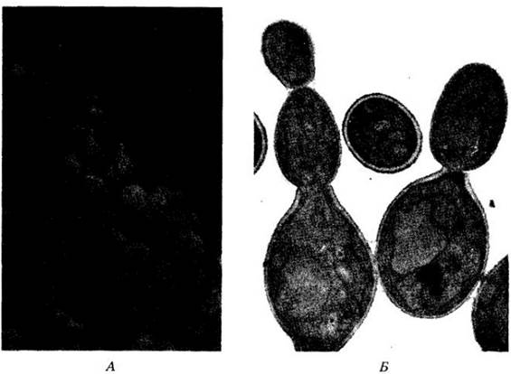
Yeasts are predominantly unicellular organisms that reproduce asexually by fission or budding (Fig. 13-16, A), and more rarely by spores. The sexual process consists of the fusion of two cells or ascospores to form a zygote. The zygote may produce diploid buds or function as an ascus, dividing meiotically to form four haploid nuclei. Mitosis may then follow. Inside the zygote (i.e., now an ascus), walls form around the nuclei, producing four or eight ascospores that are released upon The breakdown of the ascus wall (Fig. 13-16, B). The ascospores either bud asexually or fuse with other cells to repeat the sexual process.
Yeasts play a vital role in human life due to their ability to ferment CARBOHYDRATES, breaking down glucose to produce ethyl alcohol and CO2. As a result, they are used in winemaking (source of ethanol), baking (source of CO2), and brewing, where both products are required. Many economically useful Yeast strains have been obtained through Selection and crossbreeding. Today, Introduction/32.html">Genetic Engineering techniques are used to further improve them by introducing beneficial genes from other organisms (see Ch. 30). In modern winemaking, pure yeast strains are added to relatively sterile grape juice; previously, wild strains residing on the grape clusters were used to make wine (see Fig. 16-17). Sometimes the bouquet of a wine is determined directly by the grapes, but in most cases it depends on the fungus used. In brewing, the wort is first sterilized by heating and then fermented with a pure yeast culture. As a rule, strains of a single species, Saccharomyces cerevisiae, are used in The production of wine, cider, sake, and beer, although others find application. In fact, virtually this single species is currently used in baking (Fig. 13-16, A). Some yeasts are pathogenic to humans, causing diseases such as thrush and cryptococcosis.
Fig. 13-16. Yeasts. A. Budding of cells in baker's yeast (Saccharomyces cerevisiae). B. Asci with ascospores in Schizosaccharomyces octosporus

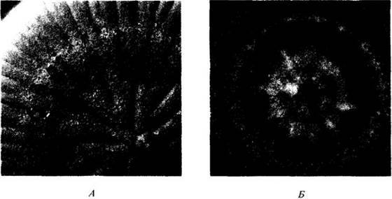
Fig. 13-17. Penicillium and Aspergillus—two widespread genera of imperfect fungi. A. Culture of Penicillium notatum, the fungus that produces penicillin; differences in coloration between the growing and sporulating PARTS OF THE mycelium are visible. B. Culture of Aspergillus fumigatus, a fungus that causes respiratory disease in humans. The concentric pattern of growth resulting from successive "pulses" of sporulation is clearly noticeable
A number of species, particularly Saccharomyces cerevisiae, are valuable objects for genetic research. Like the bacterium Escherichia coli, this species has become a favorite Organism for studying METABOLISM, genetics, and development—in this case, in Eukaryotic cells. Scientists have recently even managed to synthesize a functional yeast microchromosome that is transmitted through successive mitotic cycles; this has not yet been achieved with other eukaryotes. Detailed study of yeasts, combined with the ease of experimenting with their genetic material, will undoubtedly greatly enhance The Importance of these organisms in the industries of the future (see Fig. 13-3).
Most yeasts are ascomycetes, though a few genera belong to the Basidiomycetes, such as Cryptococcus, which causes cryptococcosis, as well as some non-pathogenic Candida species. The simplified and typically unicellular structure of yeasts is evidently the result of the evolution of more complex mycelial fungi. The reduction of the original structure in these organisms has gone so far that it is difficult to determine the evolutionary affinities of yeasts with other groups. However, to the best of our knowledge, they all evolved from multicellular ancestors, with unicellular forms having arisen multiple times among fungi, even within the ascomycetes. Yeasts comprise 39 genera and about 350 species. They are distributed in A wide variety of terrestrial and aquatic habitats where they find a suitable carbon source.
Fungi imperfecti (Deuteromycetes)
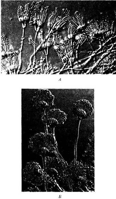
Imperfect fungi, or deuteromycetes, include about 25,000 described species for which no sexual process is known (Fig. 13-17, 13-18), which is explained either by insufficient study or by the loss of sexual reproduction during evolution. Most such "imperfect" fungi are essentially ascomycetes that produce only conidia, though some can be assigned to basidiomycetes based on their characteristic septa and clamp connections (see Fig. 13-29). Parasexual cycles, widespread among imperfect fungi, maintain genetic Variability and probably partially compensate for the absence of a sexual process. Overall, Classification here is based on the mode of conidium formation (Fig. 13-12, 13-18). Imperfect fungi are an artificial group uniting diverse organisms and are maintained largely for the convenience of form identification. It can be viewed as a class not equivalent in nature to other fungal classes.
Fig. 13-18. Conidiophores of imperfect fungi used in their classification: branched in Penicillium (A) and gathered in dense bundles in Aspergillus (B)
Many imperfect fungi are of great economic importance. For example, certain Representatives of the genus Penicillium impart an appearance, odor, and flavor highly prized by gourmets to specific cheese varieties. One such mold—P. roquefortii—was first discovered in caves near the French village of Roquefort. According to legend, a peasant boy left his lunch—a piece of ordinary fresh cheese—in one of these caves, and upon returning a few weeks later, found in its place a sharp, aromatic cheese with colored Veins. The name "Roquefort" can only be applied to cheeses produced in this region of France. Another species of this genus, P. camembertii, imparts specific properties to Camembert cheese. In the East, fermenting soybeans with Aspergillus oryzae yields soybean paste (miso), while fermenting them with a mixture of A. oryzae and A. soyae produces soy sauce (shoyu). Lactic acid Bacteria and yeasts are also used to impart the final properties to the product (soybean curd, tofu, and tempeh, a similar protein-rich food in tropical Asian countries, are prepared by fermenting soybeans with zygomycetes of the genera Mucor and Rhizopus, respectively). A. oryzae is also important for the Initial Stages of sake production—a traditional alcoholic beverage in Japan; the yeast S. cerevisiae is used in subsequent stages of this process. In industry, citric acid is produced in large quantities from Aspergillus colonies grown in a strongly acidic medium. The problem of protein enrichment of animal feed by fermenting it with A. oryzae is currently being investigated in Europe and the USA.
Antibiotics—special substances synthesized by imperfect fungi—inhibit the growth of other organisms (such as bacteria) and are used in the Treatment of many human diseases. The first antibiotic was discovered in 1928 by Alexander Fleming, who noticed that a Penicillium spore that had contaminated a growing culture of Staphylococcus on nutrient Agar completely inhibited the growth of this bacterium. Ten years later, Howard Florey and his colleagues at Oxford University isolated pure penicillin, and later large-scale production of this medicinal drug was established in the USA. During World War II, the demand for it was so great that production increased from a few million units in 1942 to 700 billion units in 1945. Penicillin is effective in treating a wide range of diseases caused by Gram-positive bacteria, including Pneumonia, scarlet fever, Syphilis, Gonorrhea, diphtheria, rheumatism, and many others. Many antibiotics used as medications undoubtedly also play an important ecological role in nature, enabling the organisms that synthesize them to win out in competition with other creatures.
Not all compounds produced by imperfect fungi are beneficial to humans. For example, aflatoxins—a group of closely related secondary metabolites from Aspergillus flavus and A. parasiticus—are highly toxic and carcinogenic. Sometimes these fungi colonize stored food, which is why they remain a constant focus of public health attention. Another group of fungal toxins is trichothecenes, produced by certain genera of imperfect fungi. They inhibit Protein Synthesis in eukaryotes.
One group of imperfect fungi, the dermatophytes (from the Greek dermatos meaning Skin and phyton meaning plant), includes the pathogens responsible for ringworm, athlete's FOOT, and other cutaneous infections. These conditions are particularly prevalent in tropical regions. The pathogenic stages here correspond to asexual reproduction, but most of these fungi are now classified as ascomycetes, even though classification continues to be based on their disease-causing forms. The feet are affected in warm, humid conditions, but the infection typically resolves quickly if a person switches from enclosed shoes or boots to sandals and keeps their feet dry. Although about 2 million bacteria normally inhabit every 1 cm2 of the SOLE OF THE foot, competing with dermatophytes for nutrients, the fungi win this competition by growing directly into epidermal cells and poisoning the bacteria with secreted antibiotics. Fungi make much more active use of keratin, the tough fibrous protein that fills epidermal cells before they slough off. However, resistant bacterial strains can sometimes emerge and, through rapid proliferation, cause severe infections.
During World War II, more soldiers were evacuated from the South Pacific due to skin infections than from battle wounds. Many fungi act as agents of specific infections; for example, Candida albicans, a yeast-like species under certain conditions, causes thrush and other mucosal lesions. Fungal spores are constantly inhaled by humans, and some cause internal diseases that can occasionally be very severe or even fatal, particularly when the Lungs are involved.
In recent years, mycoses have become significantly more widespread in humans than before. Certain compounds, notably cyclosporin (see p. 184)—first isolated from the imperfect fungus Tolypocladium inflatum—are now routinely administered to transplant patients to suppress normal immune responses that threaten to reject the transplanted Organs or Tissues. However, this also increases susceptibility to fungal and other infections. Certain chemical agents, such as those used in the treatment of acute leukemia, similarly reduce the body's resistance to fungal infections. As a result, attention to the Prevention and treatment of these infections is steadily growing.
Lichens are symbiotic associations between ascomycetes and certain genera of green Algae or cyanobacteria1. Their autotrophic components, which supply nutrients, are protected from extreme environmental stresses by their fungal symbionts. As a result of such symbiotic relationships, lichens are able to colonize some of the most hostile habitats on Earth (Fig. 13-19). They comprise approximately 20,000 species of morphologically diverse fungi—distinctly different from the roughly 30,000 species of other ascomycetes—and about 26 genera of photosynthetic organisms. The most common photobionts are the green algae Trebouxia, Pseudotrebouxia, and Trentepohlia, and the cyanobacterium Nostoc, which together serve as the autotrophic partners in roughly 90% of all lichen species.
1 About a dozen basidiomycete species also associate with algae, but they are closely related to free-living basidiomycetes and differ markedly from other lichen-forming fungi.
Lichens are exceptionally widespread in nature, inhabiting environments worldwide from arid deserts to the Arctic: on bare soil, tree trunks, sun-baked rocks, fences, and wind-swept alpine peaks (Figs. 13-20, 13-21, and 13-22). Some are so small they are barely visible to the naked eye, while others, such as reindeer "moss," form ankle-deep carpets covering vast areas. The species Verrucaria serpuloides is permanently submerged in seawater. Lichens are frequently pioneer organisms on newly exposed rocky substrates. More than 350 species inhabit Antarctica (Fig. 13-19)—along with only two species of vascular plants—and seven lichen species have been found at 86° S, near the South Pole itself! Their soil-forming activity on bare rock represents the initial stage of biological succession (see Chapter 31) in these regions. Lic lichen species with cyanobacterial autotrophs are of particular importance because they contribute to N2 fixation in the soil, serving as a primary source of available nitrogen in many areas.
Fig. 13-19. In this dry, seemingly lifeless region of Antarctica (A), lichens (B) live just beneath the sandstone surface. A fractured rock face reveals differently colored bands corresponding to distinct biological zones. The black and white zones are formed by lichens, whereas the lower green zone consists of
unicellular green algae. The Temperature in this part of Antarctica rises to roughly 0°C in summer, and can drop to —60°C in winter.

Lichens display a color palette ranging from white to black through various shades of red, orange, brown, yellow, and green. They contain numerous unusual chemical compounds, and many species are used worldwide as sources of Dyes. For instance, the characteristic color of Harris tweed was historically produced by dyeing wool with substances of lichen origin. Furthermore, many lichens have Applications in medicine and perfumery, and some are used as food by certain human populations.
Fig. 13-20. Crustose lichen on a bare rock surface in central California.

Fig. 13-21. A. Parmelia perforata, a foliose lichen covering a hummingbird nest on a dead tree branch in Mississippi. B. Usnea, a fruticose lichen that often hangs in masses from tree branches. Remarkably similar in appearance and occupying the same ecological niche is "Spanish moss" (Tillandsia usneoides), common in the southern United States, which is actually a true flowering plant belonging to the bromeliad family.
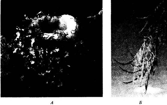
Fig. 13-22. Several fruticose lichens. A. Teloschistes chrysophthalmus. B. Cladonia cristatella, 1 — 2 cm in height. C. Cladonia subtenuis, the so-called "reindeer moss," which is actually a lichen. Species of this group, abundantly represented in the Arctic, accumulated radioactive fallout from atmospheric nuclear weapons testing. Reindeer feeding on these lichens concentrated the radioisotopes even further, passing them on to humans and other animals that consume reindeer meat or derived products, particularly milk and cheese.

Lichens have long been studied by biologists due to the fascinating Nature of the relationship between their fungal and algal symbionts. The fungus apparently determines the overall shape of the organism, but recent findings indicate that the same fungus combined with different algae can produce morphologically distinct structures that were traditionally assigned to different genera. While the algae or cyanobacteria found in lichens also occur as free-living organisms, lichen-forming fungi generally exist only in Symbiosis with them.
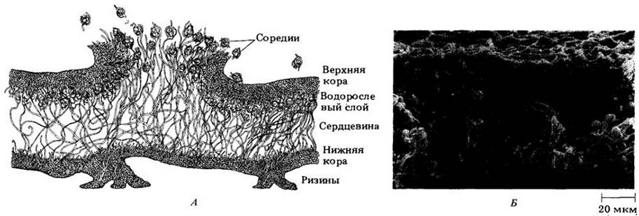
Some lichens produce specialized structures called soredia, which consist of fungal hyphae enclosing algal cells or cyanobacteria (Fig. 13-23). The dispersal of these soredia AIDS in the colonization of new habitats by lichens. Lichen fungi frequently form ascocarps similar to those observed in free-living species, differing mainly in their prolonged longevity and capacity to produce spores over several years.
Fig. 13-23. A. Cross section through the lichen Lobaria verrucosa. In the simplest case, such a section reveals a cortex composed of fungal hyphae tightly enveloping algal cells. In more complex forms, hyphae and algae organize into thalli with a definite growth form and characteristic internal structure. Four distinct layers are clearly visible in the lichen shown here: an upper protective cortex of heavily gelatinized hyphae; an algal layer composed of algal cells and loosely interwoven thin-walled hyphae; a medulla—a thick layer of loosely arranged, colorless, slightly gelatinized hyphae (accounting for about two-thirds of the thallus thickness, this layer likely Functions for storage and is formed of large-celled hyphae); and a lower cortex, which is thinner than the upper one and bears specialized attachment structures (rhizines) for anchoring to the substrate. Soredia—lichen fragments containing algal cells and fungal hyphae—serve for propagation into new habitats. B. Scanning electron micrograph of a Cytology/practical/72.html">Cross section of Cladonia cristatella (see Figs. 13-22, B and 13-25), showing the upper cortex, algal layer, and medulla. This species lacks a lower cortex.
The Biology of Lichens
How are lichens able to survive environmental conditions so hostile to virtually any other life form? It was once believed that the secret to their success lay in the fungal symbiont protecting the alga or cyanobacterium from desiccation. However, one of the most critical factors in their survival appears to be, paradoxically, their ability to dry out extremely rapidly. Lichens frequently exist in a nearly dehydrated state, with Water content dropping to 2–10% of their dry mass. Upon drying, Photosynthesis ceases; entering this state of suspended animation enables some species to withstand intense solar radiation, high temperatures, and severe cold. The cessation of photosynthesis is largely due to the upper cortex of the lichen thickening and becoming more opaque upon drying, thereby blocking The entry of solar radiation. A hydrated lichen is much more susceptible to damage from intense light and extreme temperatures than a dry one.
Upon wetting by rain, lichens rapidly absorb water (3 to 35 times their dry weight). If a dry, brittle lichen is immersed in water, it becomes soft and pliable within minutes through simple physical imbibition, much like blotting paper.
Judging by photosynthetic activity, a lichen is most viable when it begins to dry out after being fully saturated. Photosynthesis proceeds most intensely at water contents between 65% and 90% of maximum water-holding capacity; below this level, as water loss continues, photosynthetic rates decline. In many habitats, lichen Hydration fluctuates wildly over the course of a day, and for many species, photosynthesis is possible for only a few hours—typically early in the morning following hydration by fog or dew. This results in an exceptionally slow growth rate, with the radius increasing by only 0.1 to 10 mm per year. Based on size measurements, the age of certain mature lichens is estimated at 4,500 years or more. These organisms achieve their most vigorous GROWTH AND DEVELOPMENT in coastal environments or mountainous areas subject to heavy fogs.
Apparently, lichens absorb certain mineral elements from their substrate (as evidenced by the fact that a number of species inhabit only specific rock types, soils, or tree trunks), but most elements are captured from the atmosphere and rainwater. The uptake of elements from rainwater occurs very rapidly and is accompanied by their concentration. Lichens play a vital role in ecosystem functioning, yet they are particularly sensitive to toxic substances because they cannot excrete absorbed elements into the environment. Toxins cause the destruction of chlorophyll in the cells of algae or cyanobacteria. Lichen growth serves as a highly sensitive indicator of airborne pollutants, and they are increasingly utilized in monitoring atmospheric pollution, particularly around major cities. Lichens react most sharply to sulfur dioxide, which likely rapidly destroys the already small amount of chlorophyll they possess.
Both the "health" of a lichen and its chemical composition are used to indicate habitat "quality." On this basis, it is possible to track the presence of heavy metals or other pollutants around industrial centers. Many lichens are capable of binding heavy metals on the exterior of their cells, thereby preventing damage.
When nuclear tests were conducted in the atmosphere, lichens were used to monitor radioactive fallout. Today, it is believed that their application is useful in monitoring radioactive contamination potentially resulting from satellite re-entry, especially when this occurs in remote areas that are difficult to survey by other means.
Nature of the Relationship Between Fungi and Photosynthetic Organisms
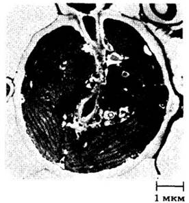
What is The Nature of the relationship between the two components of a lichen? It is clear that the fungus obtains organic carbon from the alga or cyanobacterium, given that the lichen behaves as a typical autotrophic organism dependent only on light, air, and mineral nutrients. Indeed, The transport of organic carbon from the alga or cyanobacterium to the fungus has been demonstrated using 14C-labeled carbon dioxide. In lichens containing cyanobacteria of the genus Nostoc, Nitrogen Fixation by the autotroph and its transfer to the heterotrophic component are also important. Within the lichen, fungal hyphae form a dense network around the internal cells. Widely distributed haustoria (specialized hyphae of parasitic fungi) penetrate the photosynthetic cells (Fig. 13-24); other specialized organs, appressoria, press tightly against The surface of these cells and penetrate them with projections. The interaction between the fungus and the autotrophic organism strongly influences the metabolism of the latter. For instance, green algae secrete large quantities of the sugar alcohols D-sorbitol and D-ribitol only under the Influence of the fungal partner within the lichen.
Fig. 13-24. A haustorium of the fungal lichen component Strigula elegans penetrates its partner, the green alga Cephaleuros virescens. Closely packed Chloroplasts are clearly visible within it.
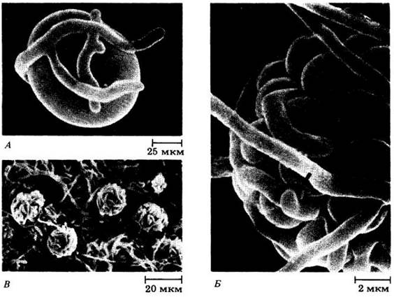
In some cases, it is possible to separate the photosynthetic and fungal components of a lichen and grow them in pure culture. Under these conditions, the fungus forms compact colonies that bear no resemblance to the symbiotic organism. Its growth requires a wide range of complex carbohydrates, and spore-forming structures generally do not develop. Conversely, algae or cyanobacteria isolated from the lichen grow faster in a free-living state. Thus, it can be suggested that the partnership in a lichen represents a controlled parasitism of the fungus on the autotroph rather than a true symbiosis. Nevertheless, a lichen is not simply the sum of its components, but a fully independent organism. When grown together in culture, the fungus apparently takes control of the photosynthetic partner first, leading to The Emergence of the characteristic morphological traits of a mature lichen (Fig. 13-25).
Fig. 13-25. Scanning electron micrograph of Early stages of interaction between the fungal and algal Components of the lichen Cladonia cristella in laboratory culture. The photosynthetic partner in this lichen (see Fig. 13-23, B) is the green alga Trebouxia. A. An algal cell surrounded by fungal hyphae. B. Penetration of fungal haustoria into algal cells. C. A mixed group of hyphae and algal cells developing into a mature lichen.
Phylum Basidiomycota
The most familiar fungi belong to this large phylum, which includes about 25,000 described species, including edible and poisonous mushrooms, stinkhorns, puffballs, bracket fungi, and two Major Groups of plant pathogens: rusts and smuts (Fig. 13-26). The defining feature of basidiomycetes is the formation of basidiospores on the outer surface of a club-shaped spore-bearing structure, the basidium (Fig. 13-27). In nature, they reproduce primarily via such spores. Although large basidiomycetes are the most thoroughly studied group of fungi, undescribed species vastly outnumber known ones here as well.
Fig. 13-26. Corn smut is a common plant disease in which the fungus Ustilago maydis forms black, powdery masses of spores on corn ears. Smuts belong to the class Teliomycetes within the phylum Basidiomycota.
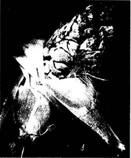
Fig. 13-27. Scanning electron micrograph of the apex of a basidium of the species Aleurodiscus amorphus, showing elongated, warted basidiospores attached to four sterigmata.

The mycelium of basidiomycetes is always septate, and during their life cycle, most species pass through two distinct phases: Primary and secondary. Upon germination, basidiospores produce a primary mycelium. Initially, it may be multinucleate, but septa soon form, dividing it into monokaryotic (uninucleate) cells. Typically, secondary mycelium arises through the fusion of primary hyphae belonging to different mating types (in which case it is heterokaryotic), though occasionally septa fail to form following nuclear division, resulting in a homokaryotic state. In either case, the outcome is a dikaryotic mycelium (with binucleate cells), since karyogamy does not immediately follow plasmogamy.
The septa of secondary basidiomycete mycelium are perforated, but the pores are flanked by thick, barrel-shaped structures (with the exception of rusts and smuts) (Fig. 13-28). Nuclei cannot pass through such pores.
Fig. 13-28. Transmission electron micrograph of a characteristic septum in the secondary mycelium of the wood-rotting basidiomycete Laetisaria arvalis. As is typical for basidiomycetes, the septum is perforated by a pore.

In heterokaryotic secondary mycelia where nuclear migration apparently still occurs, the septal pores are simple and broad, similar to those observed in ascomycetes (see Fig. 13-7).
The apical cells of the secondary mycelium typically divide with the formation of a clamp connection (Fig. 13-29). These clamp connections, which ensure the distribution of each nuclear type between daughter cells, are a hallmark feature of basidiomycetes.
Fig. 13-29. A. In basidiomycetes, dikaryotic hyphae are distinguished by the presence of clamp connections bypassing the septa; these clamps form during cell division and presumably ensure the correct distribution of two genetically distinct types of nuclei within the basidiocarp. B. Clamps and a septum of characteristic structure in a basidiomycete hypha.

It is the secondary mycelium that forms basidiocarps—fleshy spore-bearing structures such as those of mushrooms or puffballs. Their formation sometimes requires light; as light exposure occurs, the secondary mycelium differentiates into specialized hyphae that perform various functions within the basidiocarp. Such mycelium is sometimes referred to as tertiary.
Basidiomycetes are divided into three classes: Hymenomycetes, Gasteromycetes, and Teliomycetes.
Class Hymenomycetes
This class includes edible and poisonous agarics, coral fungi, and bracket fungi (Figs. 13-4, 13-30). It encompasses all forms that produce basidiospores on a basidiocarp (in gasteromycetes, they form inside the basidiocarp). The basidia of Hymenomycetes always develop on a hymenium (hence the group's name) rather than from isolated cells, as seen in Teliomycetes. The basidiocarps of hymeno- and gasteromycetes are analogous to the ascocarps of ascomycetes.
Fig. 13-30. Basidiomycetes of the class Hymenomycetes. A. The fly agaric *Amanita muscaria* at various Stages of development, showing spore-bearing gills. A typical feature of this genus, which contains many poisonous species, is the ring-like collar on the stipe and a cup-like structure at its base. B. The bay bolete *Suillus bovinus*. The spore-bearing part of the basidiocarp features a spongy, porous structure. C. The artist's conch *Ganoderma tsugae*. Fungi of this type cause the majority of wood decay cases. D. The coral fungus *Hericium coralloides*. The hymenium, the external spore-bearing layer containing basidia, covers the entire surface of the basidiocarp.

Some hymenomycetes possess club-shaped, aseptate (i.e., non-partitioned) basidia, each typically bearing four basidiospores on tiny projections called sterigmata (Figs. 13-27, 13-31). Others, the gelatinous-looking jelly fungi (Fig. 13-32), feature septate basidia similar to those of teliomycetes.
Fig. 13-31. The ink cap *Coprinus*, a common agaric. A. A cross-section through the spore-bearing gills. Their relatively dark edges correspond to the hymenium. B. A cross-section through the hymenial layer showing developing basidia and basidiospores. C. Nearly mature basidiospores attached to a basidium by sterigmata.
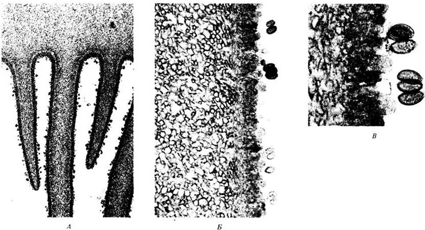
Fig. 13-32. A jelly fungus on a dead log in an Amazonian rainforest (Brazil). Jelly fungi are the only group of hymenomycetes with septate basidia.

In everyday terms, a "mushroom" (agaric) refers to a basidiocarp typically consisting of a cap atop a stipe (stalk). The mass of hyphae within the basidiocarp generally forms distinct tissues and layers. In early stages, it may be enveloped by a membrane that ruptures as the mushroom grows. In some genera, remnants of this membrane remain visible on the upper surface of the cap or at the Base of the stipe, where a cup-like structure (volva) is formed. In many hymenomycetes, the underside of the cap consists of radially radiating gills that bear the hymenium (Fig. 13-31); in others, the arrangement differs. For instance, in coral fungi, it covers spines or finger-like outgrowths of the basidiocarp (Figs. 13-30, D). In bracket fungi, boletes, and similar species, the hymenium lines numerous pores on the underside of the cap (Fig. 13-30, B).
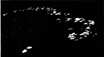
In relatively uniform habitats, such as clearings or meadows, the mycelium spreads underground outward and laterally, forming a ring of basidiocarps on the surface that can reach up to 30 m in diameter. This occurs because in an open space, the mycelium grows uniformly in all directions while dying off in the center; spore-bearing structures form only along its outer edge, where development is most active because the soil in this zone is richest in nutrients. As a result, rings of basidiocarps emerge, expanding in diameter as the mycelium grows. Popularly known as "fairy rings" (see Fig. 13-36).
Gilled mushrooms are the best-known basidiomycetes. *Agaricus campestris*, the field mushroom, is particularly abundant. Its relative, *Agaricus bisporus* (the button mushroom), is one of the few agarics that can be commercially cultivated. Today, it is grown in more than 70 countries, with total crop revenues reaching 14 billion dollars annually. Along with the East Asian shiitake mushroom (*Lentinus edodes*), it accounts for about 86% of global mushroom production. Other species are also cultivated, but the majority of edible hymenomycetes are gathered in large quantities from the wild and regularly marketed. Not all agarics are edible; for example, many poisonous forms belong to the gilled group. Thus, the genus *Amanita* includes both the most dangerous and several edible species. Even a tiny ingested piece of the death cap (*Amanita phalloides*) can be fatal to humans. Some basidiomycetes contain substances that induce hallucinations in humans (Fig. 13-34).
The typical life cycle of an agaric is illustrated in Fig. 13-33. Basidia form on the hymenium lining the gills as the terminal cells of dikaryotic hyphae. Soon, the young basidium enlarges, and karyogamy occurs. This is almost immediately followed by meiosis of each diploid nucleus, yielding four haploid nuclei. These four nuclei migrate individually into the sterigmata, which expand at the tip to form uninucleate basidiospores. The reproductive capacity of a single mushroom is immense—a single basidiocarp disperses billions of spores.
Fig. 13-33. Life cycle of an agaric (class Hymenomycetes). Primary mycelia develop from basidiospores. Secondary dikaryotic mycelia arise from primary ones. In some cases, they form through the fusion of hyphae of different mating types and are heterokaryotic. The secondary mycelium divides and differentiates to produce tertiary mycelium, which constructs the basidiocarp.

Fig. 13-34. Mushrooms play a special role in the religious ceremonies of certain indigenous tribes in southern Mexico and Central America. During these rituals, the natives ingest specific hallucinogenic basidiomycetes. One of the best-known such mushrooms, *Psilocybe mexicana*, is shown here growing in a pasture near Huautla de Jiménez (Oaxaca, Mexico). The shaman María Sabina consumes this mushroom during a nocturnal religious ceremony. Psilocybin, the chemical substance that induces colorful visions in those who eat these "sacred" mushrooms, is a structural analog of the hallucinogens LSD and mescaline (see Fig. 29-50).

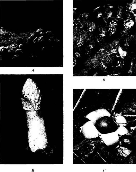
Fig. 13-35. Basidiomycetes of the class Gasteromycetes. A. The puffball *Lycoperdon ericetorum*. Spores are released through a pore at the apex of each spherical basidiocarp and dispersed by the wind. B. The stinkhorn *Phallus impudicus*. Basidiospores are embedded in a foul-smelling, sticky mass at the apex of the basidiocarp. Flies (such as *Mydaea urbana*, shown here) visit these fungi in search of food and disseminate spores that adhere in large numbers to their legs and bodies. C. The fluted bird's nest fungus *Cyathus striatus*. Their cup-shaped basidiocarps contain basidiospores that are splashed out and dispersed by raindrops. D. The collared earthstar *Geastrum triplex*. The outer layer of this "puffball" unfolds into star-like lobes.
Class Gasteromycetes
Gasteromycetes produce basidiospores within basidiocarps that remain entirely closed, at least during early developmental stages. This class likely evolved from hymenomycetes. Common representatives include stinkhorns, earthstars, false truffles, bird's nest fungi, and puffballs (Fig. 13-35). Stinkhorns (Fig. 13-35, B) feature a unique Morphology. They develop underground as tight "witches' eggs" and, upon maturation, produce above-ground receptacle structures consisting of a stalk bearing a gleba, which represents the outer wrinkled hymenium. The gleba is covered in a foul-smelling, sticky spore mass that attracts flies and beetles, which in turn disperse the spores via their feces.
Puffballs are familiar gasteromycetes. When mature, their internal tissue dries out, and touching the fungi causes them to release a cloud of spores (Fig. 13-35, A). A single puffball can produce several trillion basidiospores. Bird's nest fungi (Fig. 13-35, C) begin their development like puffballs, but after the breakdown of most of their internal structure, they come to resemble tiny bird nests.
Class Teliomycetes
This class comprises two major orders: rusts (Uredinales) and smuts (Ustilaginales). Its representatives differ from other basidiomycetes in that they do not form basidiocarps. However, they do produce dikaryotic hyphae and basidia, which are septate, much like those of jelly fungi in the class Hymenomycetes. Rusts and smuts (see Fig. 13-26) form their spores in clusters known as sori, which are smaller in rusts. As plant pathogens, these fungi hold immense economic significance, causing billions of dollars in crop damage worldwide each year.
Fig. 13-36. An incomplete "fairy ring" formed by the mushroom Agaricus arvensis. Some fairy rings have been found to be about 500 years old. Due to the depletion of essential soil nutrients, the grass inside the ring is often shorter and lighter in color than the grass outside it
The life cycle of many rust fungi is extremely complex and remains a subject of ongoing research by phytopathologists seeking ways to control them. Until recently, these fungi were considered obligate parasites of vascular plants, but researchers have successfully cultured several species in artificial media. Some smut fungi are also capable of completing their entire developmental cycle in laboratory conditions. The life cycle of the cereal stem rust, Puccinia graminis—one of the 7,000 species of the order Uredinales—is well understood. Numerous strains of this species parasitize wheat and other cereals, including barley, oats, rye, and various wild grasses. P. graminis is a persistent cause of wheat crop losses. In a single year, losses in Minnesota, North and South Dakota, and the Canadian prairie provinces totaled nearly 8 million metric tons. As early as the first century AD, Pliny wrote of this rust as "the greatest scourge of cereal crops." Today, phytopathologists have significantly mitigated the damage it causes by breeding resistant wheat varieties; however, parasite Mutations and recombination make any success short-lived. Parasexual processes leading to somatic recombination are among the key factors driving the emergence of new pathogenic strains of cereal stem rust.
P. graminis is a heteroecious parasite, meaning it requires two different hosts to complete its life cycle (Fig. 13-37). Autoecious parasites, by contrast, develop on a single host species. P. graminis can inhabit a cereal host indefinitely, reproducing exclusively through asexual means. For sexual reproduction, however, part of its life cycle must be spent on barberry (Berberis) and the other part on a cereal. Consequently, past efforts were made to eradicate the rust by destroying barberry plants. For example, the British colony of Massachusetts passed a law stating that "every person on whose land barberry bushes shall grow shall cause the same to be destroyed and grubbed up on or before the 13th day of June, 1760."
Fig. 13-37. Life cycle of the cereal stem rust fungus Puccinia graminis. This heteroecious parasitic fungus spends part of its life cycle on barberry and part on cereals (in this case, wheat). In early spring, barberry is infected by mononuclear basidiospores. Plus and minus hyphae originating from the basidiospores give rise to plus and minus spermogonia. When spermatia of one strain contact receptive hyphae of the opposite mating type, plasmogamy occurs, leading to the formation of aecia. Dikaryotic (n + n) aeciospores produced in the aecia subsequently infect wheat, where they soon give rise to red streaks—uredinia containing unicellular dikaryotic urediniospores. These spores are produced throughout the summer, repeatedly infecting wheat crops. As autumn approaches, the red streaks transform into dark teliopustules containing bicellular dikaryotic teliospores, which are non-infectious. In early spring, the nuclei in each cell of the teliospore fuse in pairs (karyogamy). When the two teliospore cells germinate, meiosis occurs, and each gives rise to a basidium bearing four haploid basidiospores

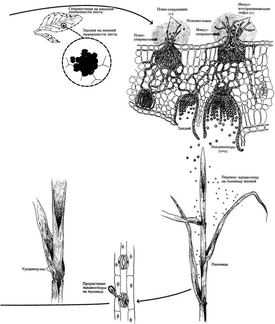
Infection of the barberry plant occurs in spring, when haploid basidiospores infect the plant, forming flask-shaped spermogonia primarily on the upper surface of its leaves. This parasitic form includes plus and minus strains; similarly, the basidiospores and the spermogonia that arise from them are also designated as "+" or "-". Each spermogonium contains Two Types of hyphae: one produces chains of small cells called spermatia, while the other forms so-called receptive hyphae. Spermatia are expelled through the ostiole of the spermogonium. When a plus-spermatium from one spermogonium contacts a minus-receptive hypha of another, or vice versa, plasmogamy takes place, producing dikaryotic hyphae that grow downward from the spermogonium and form aecia predominantly on the lower leaf surface. Chains of dikaryotic aeciospores develop within the aecia and subsequently infect wheat.
The first external manifestation of infection on wheat is the appearance of rust-colored streaks on the leaves and stems (the "red stage"), known as uredinia, which contain unicellular dikaryotic urediniospores. These spores are produced all summer long, infecting neighboring wheat plants and serving as the primary mechanism for the spread of rust in the world's cereal-growing regions. In late summer and early autumn, the reddish sorus structures gradually darken into teliopustules containing bicellular dikaryotic teliospores (the "black stage"). These spores overwinter without infecting either wheat or barberry. In early spring, prior to germination, the two haploid nuclei within each cell of the teliospore fuse into a single diploid nucleus. Upon germination, meiosis takes place—presumably in the short cylindrical basidia emerging from the two teliospore cells. The resulting nuclei are separated by septa, migrate into the sterigmata, and develop into basidiospores. This marks the completion of the annual developmental cycle.
In certain regions, the life cycle of the cereal stem rust is shortened due to the persistence of the uredinial stage whenever actively growing plant tissues are continuously available within the dispersal range of the urediniospores. On the North American plains, these spores migrate northward from winter wheat crops in the southwestern states and Mexico until they reach southern Manitoba. Later generations are dispersed westward as far as Alberta and, by late summer, drift southward to "overwinter," presumably along the eastern slope of the Rocky Mountains. Under such conditions, the survival of the rust no longer depends on the alternate host. Conversely, in Eurasia, where the northward transport of urediniospores is blocked by extensive latitudinally oriented mountain ranges, barberry remains essential for the parasite's survival.
Last update: 07/08/2026
Editorial and Educational Adaptation: This material has been compiled based on the primary/original source text. The project team performed an editorial review, corrected technical inaccuracies, structured sections, and adapted the content for an educational format.
What was processed:
- elimination of formatting defects (OCR errors, structural breaks, corrupted characters);
- editorial organization of content;
- standardization of terminology in accordance with academic sources;
- verification of factual statements against the original source text.
All mentions of the author, publication year, and origin of the primary text have been preserved in accordance with the source.