Human Biochemistry, Volume 1 - Murray R. 1993
Metabolism of Proteins and Amino Acids
Catabolism of the Carbon Skeletons of Amino Acids
Amino Acids Forming Acetyl-Coenzyme A
All Amino Acids that yield Pyruvate (Alanine, Cysteine, cystine, Glycine, hydroxyproline, Serine, and Threonine) can be converted into acetyl-CoA. In addition, 5 Amino acids are converted into acetyl-CoA without The intermediate formation of pyruvate. These include the aromatic amino acids phenylalanine, Tyrosine, and Tryptophan, the basic amino acid Lysine, and the neutral branched-chain amino acid leucine.
Tyrosine
A. General reaction sequence. Five consecutive enzymatic reactions convert tyrosine into fumarate and acetoacetate (Fig. 31.13): 1) Transamination yielding p-hydroxyphenylpyruvate; 2) oxidation accompanied by migration of the three-carbon side chain and decarboxylation, leading to The formation of homogentisate; 3) oxidation of homogentisate to maleylacetoacetate; 4) isomerization of maleylacetoacetate to fumarylacetoacetate; and 5) Hydrolysis of fumarylacetoacetate yielding fumarate and acetoacetate. Acetoacetate can subsequently undergo thiolytic Cleavage to yield acetate and acetyl-CoA.
Several intermediates of tyrosine METABOLISM were identified through The Study of the human genetic disorder alkaptonuria. Patients with alkaptonuria excrete homogentisate in their urine, and a wealth of useful information has been gained by administering putative precursors of homogentisate to these patients.
Class="center">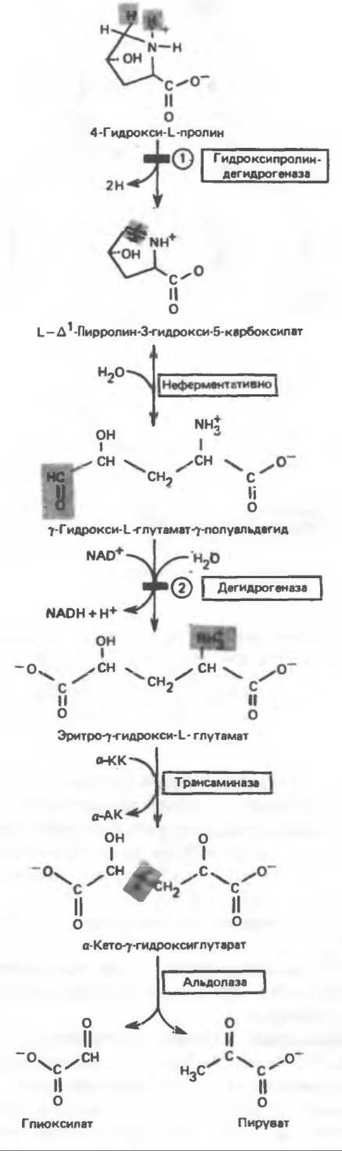
Fig. 31.12. Intermediates of α-hydroxyproline Catabolism in mammalian Tissues. α-KA — α-keto acid, α-AA — α-amino acid. Circled numbers indicate probable sites of metabolic blocks in: 1 — hydroxyprolinemia, 2 — type II hyperprolinemia.

Fig. 31.13. Intermediates of tyrosine catabolism. Except for the reaction catalyzed by β-ketothiolase, all other reactions are discussed in the text. To help the reader trace The Fate of each carbon atom, they are numbered (see also Fig. 31.15). α-KG — α-ketoglutarate, Glu — glutamate, PLP — Pyridoxal phosphate. Circled numbers indicate probable sites of metabolic blocks in 1 — type II tyrosinemia, 2 — neonatal tyrosinemia, 3 — alkaptonuria, and 4 — type I tyrosinemia or tyrosinosis.
B. Transamination of tyrosine. The transamination of tyrosine to form p-hydroxyphenylpyruvate is catalyzed by tyrosine-α-ketoglutarate transaminase, an inducible enzyme found in mammalian Liver.
C. Oxidation of p-hydroxyphenylpyruvate to homogentisate. Although the reaction (Fig. 31.13) appears to be a simple hydroxylation of p-hydroxyphenylpyruvate accompanied by the oxidative removal of the carboxyl carbon, it actually involves the coupled migration of the side chain and ring hydroxylation. p-Hydroxyphenylpyruvate hydroxylase is a copper-containing metalloprotein similar to tyrosinase. Although various reducing agents besides ascorbate can serve as Cofactors for the reaction in vitro, patients with scurvy excrete partially oxidized products of tyrosine metabolism in their urine.
D. Conversion of homogentisate to fumarate and acetoacetate. The oxidative reaction leading to the Cleavage of the benzene ring of homogentisate and the formation of maleylacetoacetate is catalyzed by homogentisate oxidase, an iron-containing metalloprotein from mammalian liver.
The conversion of maleylacetoacetate to fumarylacetoacetate is a cis-trans isomerization about the double bond, catalyzed by maleylacetoacetate cis,trans-isomerase, a (—SH)-dependent enzyme of mammalian liver. The hydrolysis of fumarylacetoacetate, catalyzed by fumarylacetoacetate hydrolase, yields fumarate and acetoacetate. The latter can then be converted into acetyl-CoA and acetate in a reaction catalyzed by β-ketothiolase (see Chapter 23).
Disorders of tyrosine catabolism. Severe defects in catabolism lead to tyrosinemia, tyrosinuria, and phenolaciduria.
A. Type I tyrosinemia (tyrosinosis). Tyrosinosis is characterized by the accumulation of metabolites that depress The activity of several Enzymes and transport systems. The Pathophysiology of this disorder is therefore quite complex. The defective enzymes are presumably fumarylacetoacetate hydrolase (Fig. 31.13) and maleylacetoacetate hydrolase.
Both acute and chronic forms of tyrosinosis are known. The acute form typically presents in infancy, manifesting as diarrhea, vomiting, a "cabbage-like" odor, and failure to thrive. If untreated, death occurs at 6 to 8 months of age due to Liver failure. Chronic tyrosinemia features similar but more moderate symptoms, with death ensuing around 10 years of age. Plasma tyrosine levels are elevated to 6–12 mg/100 mL, along with increased levels of certain Other Amino Acids, notably Methionine. Treatment involves a diet low in tyrosine and phenylalanine, and in some cases, methionine as well.
B. Type II tyrosinemia (Richner–Hanhart syndrome). The putative cause of the metabolic defect in type II tyrosinemia is a deficiency of hepatic tyrosine transaminase (Fig. 31.13). Clinical manifestations include elevated plasma tyrosine levels (4–5 mg/100 mL), characteristic ocular and Skin lesions, and mild mental retardation. Cases of self-mutilation and impaired fine motor coordination have also been reported. Tyrosine is the only amino acid whose concentration is elevated in the urine. Nevertheless, Glomerular Filtration and tubular reabsorption of tyrosine remain within normal limits. Urinary metabolites include p-hydroxyphenylpyruvate, p-hydroxyphenyllactate, p-hydroxyphenylacetate, N-acetyltyrosine, and tyramine (Fig. 31.14).
C. Neonatal tyrosinemia. This condition is attributed to a deficiency of p-hydroxyphenylpyruvate hydroxylase (Fig. 31.13). Blood levels of tyrosine and phenylalanine are elevated, as are urinary levels of tyrosine, p-hydroxyphenylacetate, N-acetyltyrosine, and tyramine. Treatment involves a low-protein diet.
D. Alkaptonuria. This inherited metabolic disorder was described in medical literature as early as the 16th century and was fully characterized in 1859. The disease is of considerable historical interest because it was through its study that Garrod formulated THE CONCEPT OF inborn errors of metabolism. The most striking clinical manifestation of this condition is the darkening of urine upon exposure to air. In later Stages of the disease, generalized pigmentation of Connective Tissue (ochronosis) develops, along with Arthritis. The underlying cause is a deficiency of homogentisate oxidase (Fig. 31.13). The enzyme substrate, homogentisate, is excreted in the urine and forms a dark brown pigment upon air oxidation. Over 600 cases have been reported, with an estimated incidence of 2 to 5 cases per million newborns.

Fig. 31.14. Alternative catabolites of tyrosine. p-Hydroxyphenylacetaldehyde is an intermediate in The oxidation of tyramine to p-hydroxyphenylacetate.
Alkaptonuria is inherited in an autosomal recessive manner. At present, no diagnostic Methods are available to identify heterozygotes. Although The Mechanism of ochronosis is not fully established, it is believed to result from the oxidation of homogentisate by polyphenol oxidase, yielding benzoquinonearray acetate, which subsequently polymerizes and binds to connective tissue macromolecules.

Phenylalanine
Phenylalanine is first converted into tyrosine through the action of phenylalanine hydroxylase (see Fig. 29.11). The isotope labeling pattern in the amphibolic products fumarate and acetoacetate (Fig. 31.15) is identical to that observed in the corresponding catabolic products of tyrosine (Fig. 31.13).
Metabolic Disorders of phenylalanine catabolism. The primary metabolic defects involve the blockade of the conversion of phenylalanine to tyrosine (see Fig. 29.12). Three main causes can be distinguished: phenylalanine hydroxylase deficiency (type I hyperphenylalaninemia, or classical phenylketonuria), dihydropteridine reductase deficiency (types II and III hyperphenylalaninemia), and defects in dihydrobiopterin Biosynthesis (types IV and V hyperphenylalaninemia). Disorders of Other types have also been documented (Table 31.3).
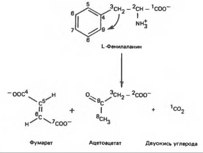
Fig. 31.15. Catabolic fate of phenylalanine carbon atoms. Isotope label distribution in the final catabolites of phenylalanine (and tyrosine).
The primary consequence of untreated type 1 hyperphenylalaninemia (classical phenylketonuria, PKU) is intellectual disability (scoring below 70 on standard tests for older children). Additional clinical symptoms include seizures, psychoses, eczema, and a "mousy" odor. With early Diagnosis and prompt treatment, these symptoms can be prevented. Due to the availability of experimental disease models and the feasibility of preventing otherwise inevitable intellectual disability through dietary management, PKU serves as a paradigm for studying this condition in other metabolic disorders. In classical PKU, an inherited disorder with an incidence of 1 in 10,000 newborns, the level of hepatic phenylalanine hydroxylase component 1 (see Fig. 29.12) is about 25% of normal, and the enzyme is unresponsive to the regulatory action of phenylalanine.
The patient's Organism is unable to convert phenylalanine into tyrosine, resulting in the formation of alternative phenylalanine catabolites (Fig. 31.16). These include phenylpyruvic acid (a product of phenylalanine deamination), phenyllactic acid (a product of phenylpyruvic acid reduction), and phenylacetic acid, which is formed via decarboxylation and oxidation of phenylpyruvic acid. Most of the phenylacetate in the liver is conjugated with glutamine and excreted in the urine as the conjugate phenylacetylglutamine. Table 31.4 lists the concentrations of phenylalanine metabolites in the blood and urine of patients with phenylketonuria. The presence of the keto acid phenylpyruvate in urine gave the disease its name—phenylketonuria.
Table 31.3. Types of hyperphenylalaninemia 1)
|
Type |
Disease name |
Cause of disease |
Treatment method |
|
I |
Phenylketonuria |
Absence of Phe hydroxylase |
Low-Phe diet |
|
11 |
Persistent hyperphenylalaninemia |
Reduced level of Phe hydroxylase |
Observation or periodic diet therapy |
|
111 |
Transient mild hyperphenylalaninemia |
Hydroxylase deficiency during development |
Same as above |
|
IV |
Dihydropteridine reductase deficiency |
Absence or deficiency of dihydropteridine reductase |
DOPA, 5-hydroxytryptophan, carbi-DOPA |
|
V |
Abnormal dihydrobiopterin function |
Impairments in the dihydrobiopterin synthesis system |
DOPA, 5-hydroxytryptophan, carbi-DOPA |
|
VI |
Persistent hyperphenylalaninemia and tyrosinemia |
Tyrosine catabolism |
Low-Phe diet |
|
VII |
Transient neonatal tyrosinemia |
Inhibition of p-hydroxyphenylpyruvate oxidase |
|
|
VIII |
Hereditary tyrosinemia |
Deficient activity of 1. p-hydroxyphenylpyruvate dioxygenase 2. cytoplasmic tyrosine aminotransferase |
Low-Tyr diet |
|
Fumarylacetoacetate deficiency |
Low-Tyr diet plus Glutathione injections |
1) Adapted with modifications from Tourian A., Sidbury J. B.: Phenylketonuria and hyperphenylalaninemia (p. 273) in: Stanbury J. B. et al. (eds): The Metabolic Basis of Inherited Disease, 5th ed. McGraw-Hill, 1983.
When normal phenylalanine Catabolic pathways are blocked, several alternative catabolic reactions come to the forefront; these normally occur in the healthy liver but usually play a minor role. In patients with phenylketonuria, phenylpyruvate, phenyllactate, phenylacetate, and phenylacetylglutamine appear in the blood and urine (Fig. 31.16). Although the presence of phenylpyruvate in the urine of PKU patients can be detected by a simple biochemical assay, a definitive diagnosis requires demonstrating elevated plasma phenylalanine levels.
The Progressive development of intellectual impairment in children with phenylketonuria can be prevented by administering a very low-phenylalanine diet. This diet can be discontinued upon reaching six years of age, as elevated concentrations of phenylalanine and its derivatives no longer exert adverse effects on the Brain by this time.
Plasma phenylalanine levels can be measured using an automated micromethod requiring no more than 20 µL of blood samples. It should be noted that abnormally high phenylalanine levels in children with phenylketonuria are detectable only by the third or fourth day of life due to their minimal Dietary Protein Intake. Furthermore, false-positive results may occur in premature infants owing to the delayed development of certain phenylalanine-metabolizing enzymes. Detection of elevated urinary phenylpyruvate using ferric chloride as a reagent serves as a useful, though not entirely reliable, mass screening test.
Table 31.4. Phenylalanine metabolites accumulating in plasma and urine of patients with phenylketonuria
|
Metabolite |
Plasma level (mg/100 mL) |
Urine level (mg/100 mL) |
||
|
normal |
in phenylketonuria |
normal |
in phenylketonuria |
|
|
Phenylalanine |
1–2 |
15–63 |
30 |
300–1000 |
|
Phenylpyruvate |
0.3–1.8 |
300–2000 |
||
|
Phenyllactate |
290–550 |
|||
|
Phenylacetate |
Elevated levels |
|||
|
Phenylacetylglutamine |
200–300 |
2400 |
||
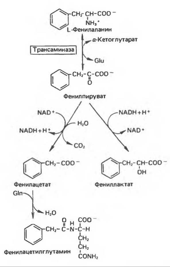
Fig. 31.16. Alternative pathways of phenylalanine catabolism of particular importance in phenylketonuria. These reactions also occur in the liver of healthy individuals, but they are of little significance when phenylalanine hydroxylase Functions normally. Glu — glutamate, Gln — glutamine.
Following the administration of phenylalanine to patients with phenylketonuria, blood levels of this amino acid remain elevated for a prolonged period, indicating a reduced tolerance to phenylalanine. Furthermore, an abnormally low tolerance to administered phenylalanine and elevated levels of the latter between meals are also characteristic of the heterozygous parents of phenylketonuric children. Thus, the defective Gene responsible for phenylketonuria can also be detected in phenotypically normal heterozygous parents.
Lysine
Lysine is an exception to the general rule that the initial step in Amino Acid Catabolism is the removal of the a-amino group by transamination. In mammalian tissues, neither the a- nor the ε-amino group of L-lysine participates in transamination. In the mammalian body, the carbon Skeleton of L-lysine is converted into a-aminoadipate and a-ketoadipate (Fig. 31.17). It was initially believed that L-lysine degradation proceeded via the formation of pipecolic acid, a cyclic imino acid. In the liver, D-lysine is indeed converted to pipecolate; however, The breakdown of L-lysine occurs via the formation of saccharopine (Fig. 31.18), an intermediate in fungal lysine biosynthesis.
L-Lysine first condenses with a-ketoglutarate, releasing a molecule of Water to form a Schiff base. This compound is subsequently reduced to saccharopine in the presence of the corresponding dehydrogenase (Fig. 31.18), followed by the oxidation of saccharopine by another dehydrogenase. Cleavage of the product by water yields L-glutamate and L-a-aminoadipate-δ-semialdehyde. The net effect of this series of reactions is equivalent to the removal of the ε-amino group of lysine by transamination; L-lysine and a-ketoglutarate are converted into a-aminoadipate-δ-semialdehyde and glutamate. NAD+ and NADH serve as cofactors in this process, with no net oxidation or reduction ultimately taking place.
During further catabolism, a-aminoadipate is converted into a-ketoadipate via transamination, which is then likely followed by The oxidative decarboxylation of a-ketoadipate to yield glutaryl-CoA. Lysine is both a glucogenic and a ketogenic amino acid; however, the exact Nature of the glutaryl-CoA catabolites formed in the mammalian organism has not yet been established.
Metabolic disorders of lysine catabolism. Two rare metabolic disorders of lysine catabolism have been described. Both result from enzymatic defects in the pathways leading to the degradation of lysine to acetoacetyl-CoA, and in both cases, the primary defect likely blocks the conversion of L-lysine and a-ketoglutarate into saccharopine (Fig. 31.18).
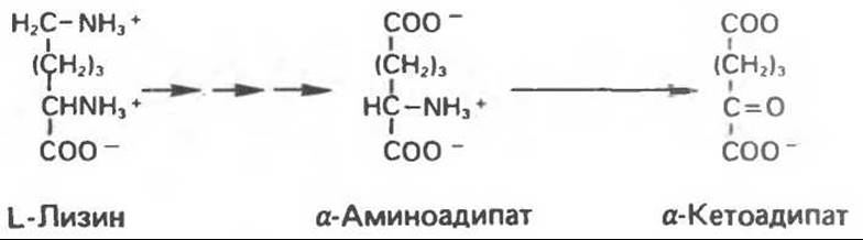
Fig. 31.17. Conversion of L-lysine to a-aminoadipate and a-ketoadipate. Multiple consecutive arrows indicate intermediate reactions.
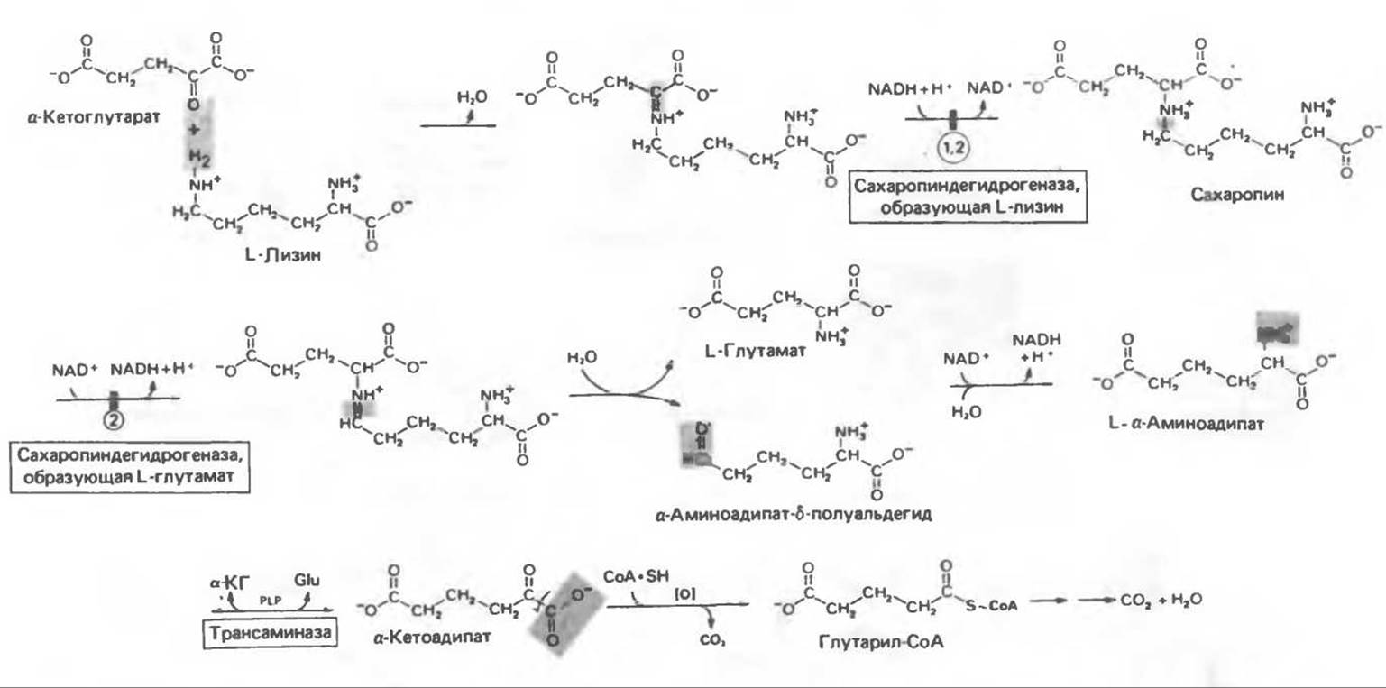
Fig. 31.18. Catabolism of L-lysine (a-KG — a-ketoglutarate, Glu — glutamate, PLP — pyridoxal phosphate). Numbers in circles indicate probable sites of metabolic blocks in 1 — periodic hyperlysinemia with accompanying hyperammonemia, 2 — persistent hyperlysinemia without accompanying hyperammonemia.
A. Periodic hyperlysinemia and accompanying hyperammonemia. In periodic hyperlysinemia, even normal dietary protein intake leads to elevated tissue concentrations of lysine. Secondary to hyperlysinemia is hyperammonemia, which develops As a result of competitive inhibition of hepatic arginase activity by lysine (see Fig. 30.13).
Increased fluid intake and a reduced dietary lysine content help lower hyperammonemia and mitigate its clinical manifestations. Conversely, a lysine load triggers a severe crisis and a comatose state. Data on the genetic nature of the disease are currently lacking.
B. Persistent hyperlysinemia without concomitant hyperammonemia. Clinical and biochemical findings varied considerably among 12 patients with persistent hyperlysinemia. Mental retardation was observed in some, but not all, patients. Concomitant hyperammonemia was absent even after a lysine load. In several cases, an accumulation of lysine catabolites was detected in biological fluids. Persistent hyperlysinemia is considered an autosomal recessive trait. Along with a blocked conversion of lysine and a-ketoglutarate to saccharopine in certain patients, there appears to be a defect in the conversion of saccharopine to L-glutamate and a-aminoadipate-8-semialdehyde (Fig. 31.18).
Tryptophan
Owing to The Diversity of its associated metabolic reactions and products, tryptophan was one of the first amino acids to be classified as essential. Studies on Neurospora mutants and Pseudomonas Bacteria, as well as the isolation of tryptophan metabolites from urine, have provided invaluable insights into the details of Tryptophan Metabolism.
Upon dietary administration of [14C]-tryptophan, the bulk of the isotope is incorporated into Proteins, yet a substantial portion is excreted in the urine as various catabolites. The carbon atoms of the side chain and aromatic ring can be completely converted into amphibolic intermediates via the kynurenine-anthranilate pathway (Fig. 31.19), which plays a pivotal role in tryptophan degradation and its conversion into nicotinamide.
Tryptophan oxygenase (tryptophan pyrrolase) catalyzes the cleavage of the indole ring with the incorporation of two atoms of molecular oxygen into the resulting N-formylkynurenine. This enzyme is an iron-porphyrin-containing metalloprotein; its synthesis in the liver is induced by adrenocorticosteroids and tryptophan. A significant portion of the synthesized enzyme exists in a latent form and requires activation. Tryptophan stabilizes the oxygenase against Proteolytic Enzymes. It is subject to feedback inhibition by nicotinic acid derivatives, including NADPH.
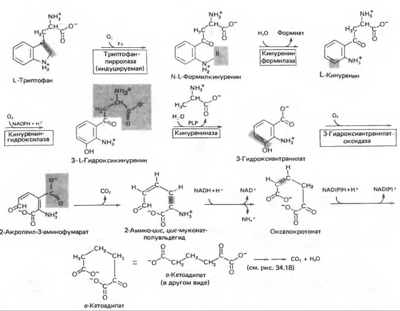
Fig. 31.19. L-tryptophan catabolism. PLP, pyridoxal phosphate.
The hydrolytic removal of the formyl group from N-formylkynurenine is catalyzed in mammalian liver by kynurenine formylase. Hydrolysis in H218O results in the incorporation of an 18O atom into the resulting formate. The enzyme also catalyzes analogous reactions with various arylformylamines.
The product of the reaction catalyzed by kynurenine formylase is kynurenine (Fig. 31.19). It can be deaminated via a Transamination reaction involving The transfer of the side-chain amino group to a-ketoglutarate. The resulting keto derivative undergoes spontaneous cyclization, transforming into kynurenic acid. This compound is a byproduct of kynurenine catabolism and is not part of the primary degradative pathway (Fig. 31.19).
Further metabolism of kynurenine involves its conversion into 3-hydroxykynurenine and subsequently into 3-hydroxyanthranilate. Hydroxylation proceeds with the participation of molecular oxygen through an NADPH-dependent hydroxylation reaction analogous to the phenylalanine hydroxylation pathway (see Chapter 29).
Kynurenine and hydroxykynurenine are converted into hydroxyanthranilate by the pyridoxal-phosphate-containing enzyme kynureninase. Vitamin B6 deficiency leads to a partial loss of the capacity to catabolize these kynurenine derivatives; in extrahepatic tissues, they are converted into xanthurenate (Fig. 31.20). This catabolite, normally absent, appears in the urine of humans, monkeys, and rats under conditions of dietary vitamin B6 deficiency. Under these circumstances, the administration of excess tryptophan leads to The excretion of xanthurenate in the urine.
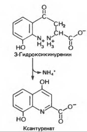
Fig. 31.20. Formation of xanthurenate under conditions of vitamin B6 deficiency. The Conversion of the tryptophan catabolite 3-hydroxykynurenine to 3-hydroxyanthranilate is impaired (Fig. 31.19), leading to the diversion of the bulk of the catabolite into xanthurenate.
In many animals, the Conversion of Tryptophan to nicotinic acid obviates the dietary requirement for this vitamin. In rats, rabbits, dogs, and pigs, dietary tryptophan can completely replace the vitamin; in humans, as well as in several other animal species, excessive dietary tryptophan intake increases the urinary excretion of nicotinic acid derivatives (e.g., N-methylnicotinamide). In vitamin B6 deficiency, impaired biosynthesis of nicotinic acid from tryptophan can lead to defective Synthesis of the pyridine NUCLEOTIDES NAD+ and NADP+. If a sufficient amount of nicotinic acid is administered, normal pyridine nucleotide synthesis resumes even in the absence of vitamin B6.
Metabolic disorders of tryptophan catabolism. Hartnup disease, an inherited metabolic disorder of tryptophan, is characterized by a pellagra-like skin rash, intermittent cerebellar ataxia, and mental retardation. The urine of affected patients contains markedly elevated levels of indolylacetate (a-N-[indole-3-acetyl]glutamine) and tryptophan.
Last update: 06/08/2026
Editorial and Educational Adaptation: This material has been compiled based on the primary/original source text. The project team performed an editorial review, corrected technical inaccuracies, structured sections, and adapted the content for an educational format.
What was processed:
- elimination of formatting defects (OCR errors, structural breaks, corrupted characters);
- editorial organization of content;
- standardization of terminology in accordance with academic sources;
- verification of factual statements against the original source text.
All mentions of the author, publication year, and origin of the primary text have been preserved in accordance with the source.