Basics of Medical Genetics - Buzhiyevska T.I. 2001
Hereditary Diseases
Disorders of Other Types of Metabolism
The hereditary pathology of mucopolysaccharide METABOLISM is known as mucopolysropolysaccharidosis, which occurs in at least 8 distinct forms. This group of inherited Connective Tissue disorders is characterized by involvement of The Musculoskeletal System, Internal Organs, eyes, and Nervous system. Just a few months after birth, a child may exhibit the following changes: hypertelorism, exophthalmos, macrocephaly, a prominent forehead, a depressed nasal bridge, a broad Nose, and thick Lips (gargoylism). Inguinal and umbilical hernias also develop. Notable features include a short neck and limbs, as well as spinal and Chest deformities, and joint contractures. Hepatosplenomegaly and Congenital Heart defects are also detected. The Various Forms of Mucopolysaccharidoses are caused by impaired glycosaminoglycan metabolism at the level of different Enzymes.
Laboratory analysis reveals increased urinary excretion of glycosaminoglycans (dermatan sulfate, keratan sulfate, heparan sulfate). Histochemical examination shows metachromasia in fibroblasts and lymphocytes.
Hurler syndrome (type I) manifests as dwarfism, a lumbar gibbus, profound intellectual disability, and heart defects. The affected child typically does not survive past the age of 10. The primary biochemical defect is an L-iduronidase deficiency. This condition is inherited in an autosomal recessive manner.
Hunter syndrome (type II) is characterized by a milder clinical course. Intellect is preserved, and life expectancy is quite significant (up to 60 years). The syndrome is driven by a deficiency of the enzyme iduronate-2-sulfatase. It is inherited in an X-linked recessive manner.
Sanfilippo syndrome (type III) has a relatively mild clinical course, presenting with the characteristic symptoms of mucopolysaccharidosis and mental retardation. Behavioral changes are prominent: the child becomes agitated, aggressive, and unable to concentrate; Sleep patterns are disrupted, and Hearing deteriorates. The syndrome exhibits biochemical heterogeneity (types A, B, C, and D) due to deficiencies in different enzymes. Affected individuals generally live to 20–30 years of age. Sanfilippo syndrome is inherited in an autosomal recessive manner.
Morquio–Brailsford syndrome (type IV) is marked by multiple skeletal deformities and hearing loss. Affected individuals have a very short neck and short stature. Corneal clouding, Osteoporosis, along with cardiac and neurological abnormalities, are also observed. Life expectancy is up to 20 years. The primary biochemical defect is a deficiency of galactosamine-6-sulfatase or beta-D-galactosidase. This disorder is inherited in an autosomal recessive manner.
Scheie syndrome (type V) manifests at birth with hernias and corneal clouding. Other, moderately severe symptoms typical of mucopolysaccharidosis develop during school age. These include hand stiffness resulting in claw-hand deformity, aortic valve disease, and impaired hearing and Vision. The biochemical defect is an alpha-L-iduronidase deficiency (possibly an allelic variant of Hurler syndrome). Scheie syndrome is inherited in an autosomal recessive manner.
Maroteaux–Lamy syndrome (type VI) manifests at 2–3 years of age. Growth retardation is observed, and all symptoms characteristic of mucopolysaccharidosis progressively worsen. Tibial bowing occurs. Affected individuals typically do not survive past the age of 20. The biochemical defect is an arylsulfatase B deficiency. It is inherited in an autosomal recessive manner.
Sly disease (type VII) is characterized by clinical symptoms very similar to those of Maroteaux–Lamy syndrome, though some variants present without hepatosplenomegaly. The biochemical defect is a beta-D-glucuronidase deficiency. It is also inherited in an autosomal recessive manner.
Winchester syndrome (pseudorheumatoid mucopolysaccharidosis) manifests with dwarfism, joint contractures, corneal clouding, osteoporosis, and rheumatoid-like joint destruction. It is inherited in an autosomal recessive manner.
Treatment for all mucopolysaccharidosis disorders is purely symptomatic: hormonal agents, high doses of retinol, and a low-ascorbic-acid diet. Enzyme replacement therapy Methods are currently under development.
Inherited Disorders of purine and pyrimidine metabolism, depending on the level at which the metabolic pathway is disrupted, can either present with extreme severity from birth—resulting in severely compromised viability and intellect, along with marked neurological damage (Lesch–Nyhan syndrome)—or mimic diseases of old age (such as Gout).
Lesch–Nyhan syndrome is a congenital pathology characterized by spastic cerebral palsy, psychomotor retardation, self-injurious behavior (autoaggression), and features typical of gout: elevated Blood uric acid levels, Hematuria, hyperuricosuria, nephropathy, and acute Arthritis. The primary biochemical defect is a deficiency of hypoxanthine-guanine phosphoribosyltransferase (HGPRT), which disrupts DNA Synthesis. Lesch–Nyhan syndrome is inherited in an X-linked recessive manner. There is currently no pathogenetic treatment available, though direct Gene Therapy for this condition is under active development.
Xanthinuria manifests as Nephrolithiasis, and some children experience nocturnal enuresis complicated by Pyelonephritis. Serum uric acid levels and urinary uric acid excretion are decreased. The primary biochemical defect is reduced xanthine oxidase activity. It is inherited in an autosomal recessive manner. Affected patients are advised to maintain a high fluid intake and limit dietary meat consumption.
Gout is characterized by joint pain, restricted mobility, and joint deformation, though without acute inflammatory joint changes. It is often accompanied by Kidney stones and ankylosis. Abnormal uric acid metabolism is observed, along with reduced activity of hypoxanthine-guanine phosphoribosyltransferase. The Selection/32.html">Genetic heterogeneity of the disease accounts for variants with autosomal dominant and X-linked recessive inheritance (allelic to Lesch–Nyhan syndrome), as well as forms with polygenic predisposition.
Adrenogenital syndrome (AGS) is a group of heterogeneous disorders caused by Genetic Defects in androgen hormone metabolism, belonging to the broader category of inherited steroid metabolism disorders. Five Clinical forms of this pathology have been described depending on the level of the biochemical pathway disruption: deficiencies of 21-alpha-hydroxylase, 11-beta-hydroxylase, 17-alpha-hydroxylase, 20,22-desmolase, or 3-beta-hydroxysteroid dehydrogenase. Consequently, this endocrine disorder, like several other hereditary endocrinopathies, is an enzymopathy. The first two defects are the most common. The disease originates during the Prenatal period of development.
Adrenogenital syndrome may present with or without salt wasting. Newborn females exhibit masculinization, hyperpigmentation, and low sex Chromatin levels with a 46,XX karyotype. Without specific hormone replacement therapy, secondary sexual development does not occur. In boys, the initial clinical symptom is precocious Puberty accompanied by short stature. Partial 21-alpha-hydroxylase deficiency does not disrupt electrolyte balance, nor does it alter cortisol and aldosterone levels. A complete enzymatic block (absence of 21-alpha-hydroxylase) leads to the salt-wasting form of adrenogenital syndrome, which manifests as vomiting, dehydration, tachycardia, hyponatremia, and hyperkalemia. Urinary levels of 17-ketosteroids and pregnanetriol are elevated.
Adrenogenital syndrome with arterial Hypertension. In girls, much like the syndrome variant described above, masculinization of the external genitalia is observed, whereas boys present with scrotal hyperpigmentation. Arterial hypertension develops during childhood, while internal reproductive organs develop normally. The primary biochemical defect is an 11-beta-hydroxylase deficiency, leading to elevated urinary levels of 17-oxosteroids and 17-hydroxycorticosteroids, alongside decreased blood levels of cortisol and aldosterone.
Both described forms share an autosomal recessive pattern of inheritance. Appropriate defect-specific hormone replacement therapy is prescribed, and the patient must remain under the continuous supervision of an endocrinologist. A more comprehensive Overview of all 5 types of AGS and their various clinical presentations can be found in the works of O.A. Benikova, T.I. Buzhievska, and O.M. Sylvanska (1993).
Pituitary dwarfism (panhypopituitary dwarfism) is associated with a deficiency of all tropic Pituitary Hormones and manifests as proportional growth retardation, along with reduced thyrotropic and adrenocorticotropic Functions. Complete growth arrest, absence of puberty, and varying degrees of oligophrenia may occur. Both sporadic autosomal recessive and X-linked recessive inheritance patterns have been described. Comprehensive hormone replacement therapy can be quite successful (Figs. 7 and 11).
Among inherited metabolic defects of the enzymopathy type, Porphyrias are widely known; these are a group of disorders caused by impaired Synthesis of Porphyrins and heme. As a result, various porphyrins accumulate in body Tissues and are partially excreted in urine and feces. Porphyrias are classified into erythropoietic (impaired porphyrin synthesis in Bone Marrow erythroblasts) and hepatic (impaired synthesis in the Liver) forms.
Class="center">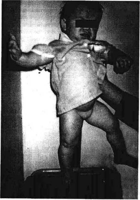

Fig. 11. a — Pituitary dwarfism (proband 0.3 in the pedigree shown in Fig. 7). An 11-year-old girl who cannot walk or speak; her psychophysical development corresponds to that of an 11-month-old child; b — Proband 0.3 (13 years old) and 0.8 (2 years old) from the pedigree presented in Fig. 7, following appropriate treatment for two years
In erythropoietic protoporphyria, the primary symptom is photodermatosis, which is inherited in an autosomal dominant manner. Treatment consists primarily of protecting the Skin from sunlight exposure.
Hepatic porphyrias present with various clinical forms caused by deficiencies in corresponding enzymes. Acute intermittent porphyria (Swedish hereditary porphyria) has been described; it may manifest from childhood throughout life, remain latent, or mimic acute appendicitis attacks, yet feature pronounced Central Nervous System symptoms up to a comatose state. Hemolytic attacks can be triggered by: certain medications (barbiturates, sulfonamides, diphenyl, griseofulvin, etc.), pesticide exposure, fasting, infections, estrogens (2–3 days prior to menstruation), progesterone, or oral contraceptives. Autosomal dominant inheritance is characteristic.
Treatment involves prescribing a diet rich in CARBOHYDRATES and Proteins, as well as the discontinuation of medications that trigger attacks.
Variegate porphyria (South African porphyria) is characterized by photosensitivity (blisters, vesicles, scarring, skin pigmentation, hypertrichosis, scleroderma-like features) and neurological symptoms. It is marked by an increased excretion of delta-aminolevulinic acid in the urine. An autosomal dominant (AD) inheritance pattern is characteristic. Treatment is the same as for acute intermittent porphyria.
Last update: 08/08/2026
Editorial and Educational Adaptation: This material has been compiled based on the primary/original source text. The project team performed an editorial review, corrected technical inaccuracies, structured sections, and adapted the content for an educational format.
What was processed:
- elimination of formatting defects (OCR errors, structural breaks, corrupted characters);
- editorial organization of content;
- standardization of terminology in accordance with academic sources;
- verification of factual statements against the original source text.
All mentions of the author, publication year, and origin of the primary text have been preserved in accordance with the source.