Harper's Illustrated Biochemistry, Volume 1 - Murray R. 1993
Metabolism of Proteins and Amino Acids
Catabolism of the Carbon Skeleton of Amino Acids
Pyruvate-Forming Amino Acids
Below is the general pathway for The conversion of the carbon skeletons of Alanine, Cysteine, Glycine, Threonine, and Serine into Pyruvate. All carbon atoms of glycine, alanine, cysteine, and serine, but only two carbon atoms of threonine, are incorporated into pyruvate. Subsequently, Pyruvate can be converted into acetyl-CoA.
Class="center">
Glycine
The amphibolic intermediates generated from glycine include pyruvate, СО2, and N5-, N10-methylenetetrahydrofolate. In The formation of pyruvate from glycine, glycine is first converted to serine via a reaction catalyzed by serine hydroxymethyltransferase (Fig. 31.5), and then pyruvate is formed from serine (a reaction catalyzed by serine dehydratase) (Fig. 31.7; see also the "Serine" section below).
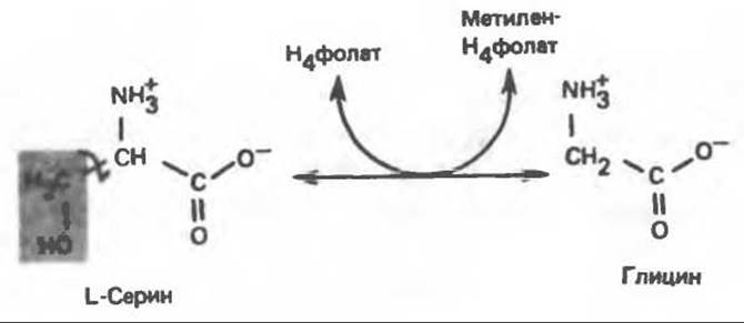
Fig. 31.5. Readily reversible reaction catalyzed by serine hydroxymethyltransferase. H4folate — tetrahydrofolate.
The major catabolic pathway for glycine in vertebrates is the conversion catalyzed by the glycine synthase complex, which yields СО2 and NH+4, while transferring the methylene group to tetrahydrofolate to form N5-, N10-methylenetetrahydrofolate. This reversible reaction (Fig. 31.6) is analogous to the conversion of pyruvate into acetyl-CoA by the Enzymes of the pyruvate dehydrogenase complex. Both complexes are located in Liver Cell/35.html">Mitochondria and exist as macromolecular aggregates. The glycine Cleavage reaction occurs in the liver of most vertebrates, including humans and other mammals, as well as birds and reptiles.
Apparently, in humans and many other vertebrates, this reaction serves as the primary catabolic pathway not only for glycine, but also for serine (see the "Serine" section below).
Metabolic Disorders of Glycine Catabolism.
Two Types of glycine metabolic disorders are discussed below.
A. Glycinuria. Glycinuria has been observed in only a single family. It is characterized by elevated urinary excretion of glycine and is associated with a tendency to form renal oxalate stones, while urinary oxalate levels remain within the normal range. Glycinuria appears to be inherited as a dominant trait, likely linked to the X chromosome. Plasma glycine levels remain normal, whereas The amount of glycine excreted in the urine reaches 600–1000 mg∙day-1. This leads to the Conclusion that glycinuria is associated with impaired renal tubular reabsorption of glycine.
B. Primary hyperoxaluria. Primary hyperoxaluria is characterized by a persistently high rate of urinary oxalate excretion, independent of dietary oxalate intake. As the disease progresses, bilateral oxalate urolithiasis develops, followed by nephrocalcinosis and recurrent Urinary Tract infections. Death occurs in childhood or early adulthood due to renal failure or Hypertension. The excess oxalate is evidently of endogenous origin, presumably formed from glycine (via deamination of glycine to glyoxylate, an oxalate precursor). The metabolic defect is thought to involve impaired glyoxylate METABOLISM, specifically its conversion to formate or (via Transamination) to glycine. As a result, excess glyoxylate is oxidized to oxalate. This inherited metabolic disorder—primary hyperoxaluria—can likely be explained by a combination of glycine transaminase deficiency and impaired oxidation of glyoxylate to formate.
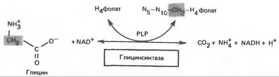
Fig. 31.6. Reversible cleavage of glycine by the mitochondrial glycine synthase complex. PLP — Pyridoxal phosphate.
Alanine
Transamination of L-alanine (Fig. 31.7) yields pyruvate, which can subsequently undergo decarboxylation to form acetyl-CoA.
Presumably for the same reasons discussed in relation to glutamate and aspartate catabolism, no metabolic disorders of L-alanine catabolism have been identified.
Serine
The conversion of serine into pyruvate, catalyzed by serine dehydratase (a pyridoxal phosphate-dependent protein), involves the elimination of Water and the hydrolytic removal of the ammonium group from the resulting intermediate (Fig. 31.7). Rat and guinea pig livers are rich in serine dehydratase; in these species, the enzyme-mediated conversion of serine to pyruvate is of major physiological significance, whereas in humans and many other vertebrates, serine is degraded primarily to glycine and N5, N10-methylenetetrahydrofolate. This reaction is catalyzed by serine hydroxymethyltransferase (Fig. 31.5). Further catabolism of serine proceeds via the glycine catabolic pathway (Fig. 31.6).
Cystine
Like nitrogen and carbon, sulfur undergoes a continuous cycle in the biosphere, driven by the metabolic activities of prokaryotic and eukaryotic organisms. Mammals lack systems for fixing sulfur into organic forms; they participate in the cycle by catabolically converting organic sulfur compounds into inorganic ones. For example, humans excrete approximately 20–30 mmol of sulfur per day, at least 80% of which is in the form of inorganic sulfate.
The principal metabolic pathway for cystine in mammals is its conversion to cysteine via a reaction catalyzed by cystine reductase (Fig. 31.8). Subsequent cystine catabolism is identical to that of cysteine (see below).

Fig. 31.7. Conversion of alanine and serine into pyruvate. Alanine transaminase and serine dehydratase utilize Pyridoxal phosphate as a cofactor. The reaction catalyzed by serine dehydratase involves the elimination of H2O from serine, leading to the formation of an unsaturated amino acid. The latter rearranges into an α-imino acid, which undergoes spontaneous Hydrolysis to yield pyruvate and ammonia. Consequently, water does not appear in the overall equation for the reaction catalyzed by serine dehydratase. Glu — glutamate, α-KG — α-ketoglutarate.

Fig. 31.8. Reaction catalyzed by cystine reductase.
Cysteine
In mammals, cysteine is catabolized via two primary pathways: the direct oxidative (cysteine sulfinate) pathway and the transamination (3-mercaptopyruvate) pathway (Fig. 31.9). It was initially hypothesized that a third pathway involving cysteine desulfhydrase also existed, which has been shown to function in Bacteria. However, this pathway is unlikely to operate in mammals, as cysteine desulfhydrase activity has not been detected in their Tissues.
A. Direct oxidative pathway of cysteine catabolism. The conversion of cysteine to cysteine sulfinate (Fig. 31.9) is catalyzed by cysteine dioxygenase, an enzyme that requires Fe2+ and NAD(P)H to function. Further catabolism of cysteine sulfinate likely involves transamination to yield β-sulfinylpyruvate. It is possible that the mammalian tissue transaminase utilizing cysteine sulfinate as an amino group donor is identical to the classical glutamate:aspartate transaminase. It should be noted, however, that the putative transamination product, β-sulfinylpyruvate, has not yet been identified in the cysteine sulfinate catabolic system. The Conversion of the postulated β-sulfinylpyruvate intermediate to pyruvate presumably proceeds non-enzymatically.

Fig. 31.9. Catabolism of L-cysteine via the direct oxidation pathway (cysteine sulfinate pathway, left) and the transamination pathway (3-mercaptopyruvate pathway, right). β-Sulfinylpyruvate is enclosed in parentheses because the formation of this intermediate has not been experimentally proven. The oxidation of sulfite in the final reaction of the direct oxidative pathway is catalyzed by sulfite oxidase.
B. Transamination (3-mercaptopyruvate) pathway of cysteine catabolism. The reversible transamination of cysteine to 3-mercaptopyruvate (thiolpyruvate) is catalyzed by specific cysteine transaminases or by aspartate and glutamate transaminases from mammalian liver and Kidney (Fig. 31.9). 3-Mercaptopyruvate can be further reduced in a reaction catalyzed by L-Lactate dehydrogenase. The resulting product, 3-mercaptopolactate, is a normal component of human urine in the form of a mixed disulfide with cysteine; the levels of the latter are elevated in the urine of patients with mercaptolactate-cysteine disulfiduria. An alternative conversion of 3-mercaptopyruvate proceeds via the abstraction of H2S to yield pyruvate (Fig. 31.9).
Disorders of sulfur-containing Amino acid metabolism. Table 31.2 summarizes the data on disorders of sulfur amino acid metabolism. Some of these are discussed below.
A. Cystinuria (cystine-lysinuria). In this inherited metabolic disorder, urinary excretion of cystine is 20 to 30 times higher than normal. The excretion of Lysine, Arginine, and Ornithine is also significantly increased. Cystinuria is considered a consequence of impaired transport processes in the Kidneys. The marked increase in the urinary excretion of lysine, arginine, and ornithine alongside cystine in cystinuric patients suggests that the reabsorption of all four of these Amino Acids is impaired, possibly occurring via a shared reabsorption "site"; therefore, the term cystine-lysinuria is currently preferred over the term cystinuria.
Because cystine is sparingly soluble, patients with cystinuria may develop cystine stones in the renal tubules. In the absence of such complications, cystinuria follows a relatively benign course and often remains undiagnosed.
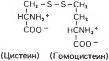
Fig. 31.10. Mixed disulfide of cysteine and homocysteine.
The mixed disulfide of L-cysteine and L-homocysteine has also been detected in the urine of cystinuric patients (Fig. 31.10). This compound is somewhat more soluble than cystine; to the extent that it forms instead of cystine, the tendency toward cystine stone formation is reduced.
B. Cystinosis (cystine storage disease). Cystinosis is also an inherited disorder characterized by the accumulation of cystine crystals in numerous tissues and Organs (particularly the reticuloendothelial system). This condition is typically accompanied by generalized Aminoaciduria, meaning an increased urinary excretion of all amino acids. A range of other renal Functions are also severely impaired, and death usually occurs at an early age due to ACUTE RENAL FAILURE. Recent findings indicate that the underlying cause of the disease is a lysosomal dysfunction.
C. Homocystinuria. The incidence of Inherited Disorders of Methionine catabolism is estimated at 1 in 160,000 newborns. Homocystine is excreted in the urine (up to 300 mg per day), occasionally accompanied by S-adenosylmethionine.
Plasma methionine levels are elevated. At least four metabolic defects can cause homocystinuria (Table 31.2). The Clinical symptoms of type 1 homocystinuria include thrombosis, Osteoporosis, dislocation of the lens of the eye, and frequently, mental retardation. Two forms of the disorder are known: vitamin B6-responsive and vitamin B6-unresponsive. A low-methionine, high-cystine diet can prevent pathological changes if initiated at an early age. Other types of homocystinuria are associated with defects in the remethylation cycle (Table 31.2).
Threonine
Threonine is cleaved by threonine aldolase into acetaldehyde and glycine, and acetaldehyde is subsequently converted into acetyl-CoA (Fig. 31.11). Glycine catabolism was discussed above.
Table 31.2. Inborn errors of sulfur-containing amino acid metabolism
|
Disorder |
Defective enzyme |
Comments |
|
Homocystinuria I |
Cystathionine β-synthase |
Fig. 29.10, reaction 1 |
|
Homocystinuria II |
N5, N10-methylenetetrahydrofolate reductase |
|
|
Homocystinuria III |
Low N5-methylenetetrahydrofolate:homocysteine methyltransferase activity due to impaired methylcobalamin synthesis |
|
|
Homocystinuria IV |
Low N5-methylenetetrahydrofolate:homocysteine methyltransferase activity due to impaired intestinal cobalamin absorption |
- |
|
Hypermethioninemia |
Hepatic methionine adenosyltransferase 1) |
Fig. 31.22 |
|
Cystathioninuria |
Cystathionase |
Fig. 29.10, reaction 2 |
|
Sulfituria (sulfocystinuria) |
Sulfite oxidase |
Legend to Fig. 29.8 |
|
Cystinosis |
Lysosomal dysfunction |
|
|
3-Mercaptopyruvate-cysteine disulfiduria |
3-Mercaptopyruvate sulfurtransferase |
Fig. 31.9 |
|
Methionine malabsorption syndrome |
Impaired intestinal absorption of methionine |
1) Cystathioninuria, tyrosinemia, and fructose intolerance may also be observed.
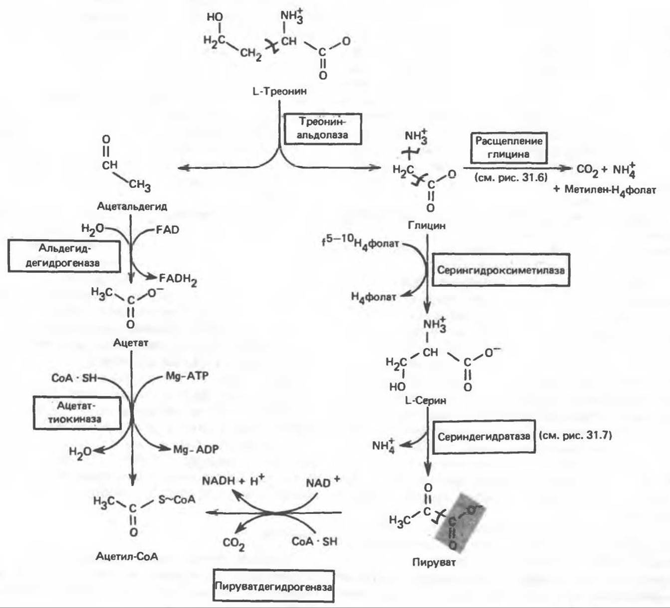
Fig. 31.11. Conversion of threonine and glycine into serine, pyruvate, and acetyl-CoA. f5-10-H4folate — formyl [5–10] tetrahydrofolic acid.
Hydroxyproline
4-Hydroxy-L-Proline is converted into pyruvate and glyoxylate (Fig. 31.12). A mitochondrial dehydrogenase catalyzes the conversion of hydroxyproline to L-Δ1-pyrroline-3-hydroxy-5-carboxylate. The latter exists in non-enzymatic equilibrium with γ-hydroxy-L-glutamate γ-semialdehyde, which is formed via The addition of water. The semialdehyde is oxidized to the corresponding carboxylic acid, erythro-γ-hydroxy-L-glutamate, followed by transamination to yield α-keto-γ-hydroxyglutarate. Subsequent aldol cleavage yields glyoxylate and pyruvate.
Metabolic disorders of hydroxyproline catabolism. Hyperhydroxyprolinemia is a metabolic disorder characterized by high plasma levels of hydroxyproline. It is inherited in an autosomal recessive manner and is caused by a deficiency of 4-hydroxyproline dehydrogenase (Fig. 31.12). Unlike type II hyperprolinemia, proline catabolism is not impaired here, since the defective enzyme is involved only in the catabolism of hydroxyproline. This metabolic disorder does not affect Collagen metabolism and, much like hyperprolinemia, appears to be benign.
Last update: 06/08/2026
Editorial and Educational Adaptation: This material has been compiled based on the primary/original source text. The project team performed an editorial review, corrected technical inaccuracies, structured sections, and adapted the content for an educational format.
What was processed:
- elimination of formatting defects (OCR errors, structural breaks, corrupted characters);
- editorial organization of content;
- standardization of terminology in accordance with academic sources;
- verification of factual statements against the original source text.
All mentions of the author, publication year, and origin of the primary text have been preserved in accordance with the source.