Human Biochemistry, Vol. 1 - Murray R. 1993
Bioenergetics and Metabolism of Carbohydrates and Lipids
Lipid Transport and Storage
Metabolism of Plasma Lipoproteins — Synthesis of Triacylglycerols and Formation of VLDL (Fig. 26.7)
Comparative studies carried out on hepatectomized and control animals have shown that the Liver is the primary source of Blood Plasma Lipoproteins derived from endogenous sources. Triacylglycerols formed in the liver serve as the direct precursors of those incorporated into VLDL. The Fatty acids required for The Biosynthesis of these triacylglycerols are either synthesized within the liver from acetyl-CoA (derived mainly from CARBOHYDRATES) or supplied as free fatty acids from the bloodstream. Under conditions of adequate Nutrition, the former pathway predominates; FATTY ACID BIOSYNTHESIS proceeds actively, and the blood level of free fatty acids remains low. Since under normal conditions triacylglycerols do not accumulate in the liver, it can be concluded that they are transported out of the liver as part of VLDL almost immediately upon formation. Conversely, during starvation, high-fat diets, or Diabetes Mellitus, blood free fatty acid levels rise, leading to an increased uptake of these acids by the liver. Under these conditions, Lipogenesis is inhibited, and free fatty acids become the primary source for the triacylglycerols of The Liver and VLDL. The biosynthetic reactions of triacylglycerols and Phospholipids are described above (see Chapter 25). The hepatic synthesis of triacylglycerols and secretion of VLDL are enhanced under the following conditions: 1) consumption of a high-carbohydrate diet (particularly one rich in sucrose or fructose), 2) elevated levels of free fatty acids in the blood, 3) ethanol consumption, and 4) high Insulin and low Glucagon concentrations.
Class="center">
Fig. 26.7. Synthesis of very low-density lipoproteins (VLDL) and potential sites of action of factors causing hepatic triacylglycerol accumulation and fatty liver. EFA — Essential Fatty Acids; FFA — free fatty acids; HDL — high-density lipoproteins; Apo-A — apolipoprotein A; Apo-B — apolipoprotein B; Apo-C — apolipoprotein C; Apo-E — apolipoprotein E. The indicated pathways underlie the processes schematically shown in Fig. 26.3. B
Fatty liver (Fig. 26.7)
For various reasons, Lipids, primarily triacylglycerols, can accumulate in the liver. Excessive fat accumulation is considered a pathological condition. When hepatic fat accumulation becomes chronic, fibrotic changes occur in the liver Cells, leading to cirrhosis and impaired liver function.
Two MAIN TYPES OF fatty liver are observed. 1. Type I fatty liver results from an increased plasma free fatty acid concentration, caused either by fat mobilization from adipose tissue or by the Hydrolysis of triacylglycerols in lipoproteins or chylomicrons by extrahepatic lipoprotein lipase. The uptake and Esterification of free fatty acids by liver cells increase. The amount of lipoproteins produced by the liver proves insufficient to handle the incoming fatty acids, which consequently accumulate within the liver as triacylglycerols, causing fatty degeneration. Liver triacylglycerol levels increase significantly during starvation and prolonged high-fat diets. In many instances (e.g., during starvation), the secretory capacity of the liver for VLDL is also impaired. In uncontrolled diabetes mellitus, sheep Pregnancy toxemia, and bovine Ketosis, hepatic lipid infiltration can be so extensive that the liver enlarges and appears pale.
2. Type II fatty liver is typically caused by a metabolic block in plasma lipoprotein formation, resulting in the accumulation of triacylglycerols. This disorder may be triggered by: a) a block in the Synthesis of the protein moiety of lipoproteins, b) a block in the assembly of lipoproteins from lipids and apoproteins, c) an insufficient supply of phospholipids required for lipoprotein Structure, or d) Impairment of the secretory mechanism itself.
Fatty liver degeneration in rats, which develops under conditions of Choline deficiency, has been studied in detail; consequently, choline has been termed a lipotropic factor. Because choline is synthesized via The transfer of mobile methyl groups from Methionine during Transmethylation (see Chapters 31 and 32), a shortage of mobile methyl groups supplied by methionine can lead to a choline deficiency. Several mechanisms have been proposed to explain the action of choline as a lipotropic factor, including a reduction in the Synthesis of phospholipids required for lipoprotein formation.
The antibiotic puromycin inhibits Protein Synthesis AND induces fatty liver degeneration along with a significant decrease in VLDL concentration in rats. Substances such as ethionine (α-amino-β-mercaptobutyric acid), carbon tetrachloride, chloroform, phosphorus, lead, and arsenic act in a similar manner. Although choline does not protect the Organism against the effects of these substances, it apparently promotes recovery. It is highly probable that carbon tetrachloride causes either a direct impairment of the lipoprotein secretion mechanism itself or a disruption in the interaction between lipids and apoproteins during lipoprotein assembly. The action of carbon tetrachloride is direct and associated with the transformation of its molecule. This process evidently generates free radicals that trigger Lipid Peroxidation, thereby destroying the lipid membranes of The Endoplasmic reticulum. Administration of vitamin E provides some protective effect against carbon tetrachloride-induced peroxidation processes. The action of ethionine is believed to result from a reduced availability of ATP. Specifically, when methionine in S-adenosylmethionine is replaced by ethionine, a portion of the adenine becomes bound, which limits ATP synthesis. Orotic acid also induces fatty liver degeneration. Because VLDL accumulates in the Golgi apparatus under these conditions, it is thought that orotic acid disrupts the glycosylation of lipoproteins and thereby inhibits their release, leading to a substantial decrease in the plasma concentration of lipoproteins containing apolipoprotein B.
Vitamin E deficiency increases the extent of necrotic areas in the liver during fatty liver degeneration caused by choline deficiency. A protective effect is observed upon the administration of Vitamin E or selenium-containing compounds. Fatty liver degeneration can also arise from a deficiency of Proteins, as well as essential Fatty Acids and Vitamins (e.g., pyridoxine and pantothenic acid). When essential Fatty acids are deficient, phospholipid synthesis is impaired; consequently, substances such as Cholesterol, which compete for available free fatty acids involved in their esterification, can also induce fatty liver degeneration.
Ethanol METABOLISM pathway
Alcoholism is likewise accompanied by hepatic fat accumulation and hyperlipidemia, ultimately leading to liver cirrhosis. The precise mechanism of long-term alcohol action remains incompletely understood. Although it is not yet clear whether the additional mobilization of free fatty acids plays a significant role in fat accumulation, A number of studies have shown that administering a single toxic dose of alcohol to rats results in elevated levels of free fatty acids. However, during chronic alcohol consumption, the fat accumulating in the liver originates not from adipose tissue, but from endogenous biosynthesis. Protein synthesis in the liver is not impaired following alcohol consumption. There is compelling evidence indicating an enhanced hepatic synthesis of triacylglycerols, a decrease in Fatty acid oxidation, and a reduced activity of The Citric Acid Cycle. These effects are believed to be driven by The oxidation of ethanol in the hepatic Cytosol, mediated by Alcohol dehydrogenase and resulting in an excess production of NADH.
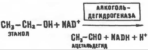
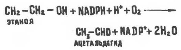
The resulting NADH competes with the reducing equivalents of Other Compounds for the Respiratory Chain, thereby inhibiting their oxidation. An increase in the [NADH]/[NAD+] ratio shifts the malate ⇄ oxaloacetate equilibrium to the left, which can suppress The activity of The Citric Acid cycle. The overall consequence of impaired fatty acid oxidation is an enhanced esterification of fatty acids to form triacylglycerols, which appears to be the primary cause of fatty liver degeneration. The oxidation of ethanol yields acetaldehyde, which is subsequently oxidized in the Cell/35.html">Mitochondria by aldehyde dehydrogenase, with acetate as the end product. Other Aspects of alcohol action include enhanced lipogenesis and cholesterol synthesis from acetyl-CoA. The elevated [NADH]/[NAD+] ratio also leads to an increased [lactate]/[Pyruvate] ratio, precipitating hyperlactatemia, which in turn diminishes the renal capacity to excrete uric acid. This mechanism likely accounts for the exacerbation of Gout associated with alcohol consumption. Although the primary metabolic pathway for ethanol is its alcohol dehydrogenase-catalyzed oxidation, a fraction of ethanol is metabolized by a cytochrome P-450-dependent microsomal system functioning with NADPH and O2. In chronic alcoholism, this system becomes more active, which probably explains the accelerated metabolic conversion of ethanol, as evidenced by elevated blood levels of both acetaldehyde and acetate.
Last update: 06/08/2026
Editorial and Educational Adaptation: This material has been compiled based on the primary/original source text. The project team performed an editorial review, corrected technical inaccuracies, structured sections, and adapted the content for an educational format.
What was processed:
- elimination of formatting defects (OCR errors, structural breaks, corrupted characters);
- editorial organization of content;
- standardization of terminology in accordance with academic sources;
- verification of factual statements against the original source text.
All mentions of the author, publication year, and origin of the primary text have been preserved in accordance with the source.