Molecular Biology of the Cell - Volume 3 - Alberts B., Bray D., Lewis J., Raff M., Roberts K., Watson J. 1994
From Cells to Multicellular Organisms
The Nervous System
Birth, Growth, and Death of Neurons
Structure/149.html">The problem of Nervous system development is unique. How do axons and dendrites extending from billions of Neurons find the right synaptic partners to build an efficiently functioning network? Different types of neurons and sensory Cells, as well as the Muscles they innervate, are typically located at considerable distances from one another in the embryo and are initially unconnected. Therefore, in The first phase of nervous system development, its various parts evolve according to their own "local" programs in accordance with the principles of Cell Differentiation common to all body tissues, as discussed in Chapter 16. The next phase involves a type of morphogenesis unique to The Nervous System. At this stage, a preliminary yet ordered pattern of connections between PARTS OF THE nervous system is established by means of axons and dendrites growing in the appropriate directions. Originally isolated parts can now interact with each other. In the third and final phase, which continues into adult life, the previously established connections are refined and perfected through the interaction of distant components, guided by the electrical signals transmitted and received by these components.
19.7.1. Neurons are formed according to specific programs of Cell Division [48, 49]
In almost all animals, from nematodes to vertebrates, The formation of neurons obeys three fundamental rules, with rare exceptions: 1) mature neurons do not divide; 2) after the complete set of neurons characteristic of the adult is formed, no stem cells capable of producing new neurons remain; 3) neurons in each small region of the developing nervous system form according to their own division program, independent of The Cell groups with which they will later establish neural connections.
In vertebrates, the nervous system develops from two cell complexes of ectodermal origin: the neural tube (Section 16.1.9) and the neural crest (Section 16.6.5). The Central Nervous System (Brain AND SPINAL cord) develops from the neural tube, whereas the neural crest serves as the source for most neurons and supporting Cells of the Peripheral Nervous System. In addition, certain sensory neurons develop from thickenings, or placodes, of the HEAD ectoderm, including those that innervate the ear and Nose (Fig. 19-55).
The neural tube, which is the primary focus here, initially consists of a single-layered epithelium—subsequently the source of both neurons and glial cells of the central nervous system (Fig. 19-56). Later, this epithelium thickens and becomes a more complex structure comprising multiple layers of various cell types. The proliferation process here has been studied using 3H-thymidine, which is incorporated into cells currently in the S phase of the Cell Cycle. The tissue was fixed either immediately after label administration to determine which cells were dividing, or at a later stage to find out which mature cells develop from the precursor cells that were dividing at the time of 3H-thymidine injection. Such studies have been particularly successful in determining the "birthdates" of various neurons: since differentiated neurons do not divide, each neuronal precursor must undergo its final division on a specific day before beginning its maturation as a nerve cell. It has been shown that in both vertebrates and invertebrates, the "birthdates" of neurons of a given type typically fall within that strictly limited developmental window after which no more neurons of this type are produced. Each region of the developing neural tube has its own cell division program, and typically neurons with different "birthdates" are destined for different functions. Since stem cells cease to exist once neuron formation is complete and connections between THEM begin to form, the number of neurons can thereafter only decrease As a result of cell death (see Section 19.7.11 below).
Class="center">
Fig. 19-55. Schematic drawing of an early (2.5-day) chicken embryo, showing the structures from which the nervous system develops. The neural tube (highlighted in pink) has already closed, except for the caudal region, and lies beneath the ectoderm, of which it was initially a part (see Fig. 16-13). The neural crest (shown in gray) is located dorsally between the roof of the neural tube and the ectoderm. Certain groups of sensory cells and neurons develop from surface thickenings of the ectoderm known as placodes. At this stage, invagination of the statoacoustic placode is virtually complete, forming the otic vesicle—the primordium of the Inner ear and the source of neurons for its associated ganglion; invagination of the olfactory placode forms the nasal lining, including the olfactory neurons responsible for the SENSE OF SMELL. Other cranial placodes give rise to cells of the cranial sensory ganglia, which provide most of the sensory innervation to the Head and Neck. Unlike other sensory cells, the sensory cells of the eye develop from the neural tube.

Fig. 19-56. Scanning electron micrograph of a cross-section through a two-day chicken embryo. The neural tube has almost closed and separated from the ectoderm. At this stage, the neural tube epithelium consists of a single cell layer. (Courtesy of Jean-Paul Revel.)
19.7.2. Radial glial cells form a temporary "scaffold" that guides the migration of immature neurons [50]
An immature neuron, having not yet formed an axon and dendrites, typically migrates from its site of "birth" to another Location. Such neuronal Migrations can be tracked using 3H-thymidine: the label is incorporated into precursor cells undergoing their final division, after which their labeled progeny can be detected elsewhere. For example, motor neurons that will innervate the limbs undergo their final mitosis near the lumen of the neural tube and then migrate toward its periphery, entering the ventral horns of the future Spinal Cord (Fig. 19-57).
Neuronal migration is guided by specialized elements of the neural tube—radial glial cells (Fig. 19-58, A). These are surviving cells of the initial columnar epithelium of the neural tube that elongated further as the tube wall thickened: each of these cells extends from the inner surface of the tube to the outer surface. In some Regions of the developing primate brain, this distance can reach 2 cm. Three-dimensional reconstruction from serial-section electron micrographs reveals that immature migrating neurons closely appose radial glial cells and appear to crawl along them (Fig. 19-58, B and C).

Fig. 19-57. Regions from which spinal cord motor neurons develop, based on autoradiography following the administration of a small dose of tritiated thymidine at an early stage of development. The diagrams show cross-sections of the neural tube of an early embryo (left) and the relatively mature spinal cord developing from it (right). At the later stage, the highest concentration of the label is found in cells that were undergoing their final phase of DNA Synthesis at the time of 3H-thymidine injection. For simplicity, the diagram of the mature spinal cord shows only motor neurons, although many other Nerve Cells are also present in the Gray matter. The White matter (unshaded) mainly contains tracts of axons running along the spinal cord and connecting different regions of gray matter. (In adults, these regions appear white because they contain a high myelin content.) For information on how glial cells form during Organism development, see Chapter 16.

Fig. 19-58. A. Cross-section of a portion of the developing Cerebral Cortex of a monkey (simplified diagram). A Golgi-stained preparation reveals radial glial cells extending from the lumen of the neural tube to its outer surface. B. A more detailed diagram of the region highlighted in pink in diagram A, showing immature nerve cells migrating along the processes of radial glial cells. C. Rendering of one such migrating neuron based on reconstructions from serial electron-microscopic sections. (After P. Rakic, J. Comp. Neurol., 145, 61-84, 1972.)
Radial glial cells persist for many days (in some species, up to several months) as a population of non-dividing cells clearly distinct from neurons and their precursors. Only toward the end of the developmental period do they disappear in most regions of the brain and spinal cord; it has been suggested that many of them transform into astrocytes, though this still requires direct proof. Thus, radial glial cells can be viewed as an auxiliary developmental apparatus—they are essential as "scaffolding" for building complex neural structures, but persist almost nowhere in the mature nervous system.
19.7.3. The type of a neuron and its future connections are determined by the time of its "birth" [50, 51]
There is a definite relationship between the "birthdate" of a neuron in the mammalian central nervous System and Its final destination (this is perhaps an evolutionary "echo" of the strict link between cell genealogy in developing organisms and their final localization in invertebrates such as nematodes—see Section 16.3). For instance, neurons in the cerebral cortex are arranged in layers according to their chronological "birth order," owing to a migration pattern in which later-born cells migrate past earlier-born ones. As they mature, cells located in successive cortical layers begin to differ in shape, size, and patterns of connectivity with other cells. Thus, small pyramidal cells, which appear late, reside in one of the outer layers and send their axons to other cortical areas, whereas large pyramidal neurons and irregular cells, which appear earlier, are located in the inner layers and send their axons beyond the cerebral cortex.

Fig. 19-59. Comparison of neuronal layers in the cerebral cortex of normal mice and reeler mutant mice. In the mutants, a defect in cell migration leads to an inversion of the normal relationship between a neuron's "birthdate" and its final position. Nevertheless, these mispositioned neurons differentiate and establish connections in accordance with their "birthdate."
What determines these differences—the "birthdate" or the final localization site? Mouse lines carrying the reeler mutation help answer this question. In these mutants, named for their unsteady gait, The Mechanism of nerve cell migration is impaired: later-born neurons remain in the inner layer, whereas early-born cells migrate to the outer layer. Despite this positional inversion, the differentiation of cortical cells corresponds to their "birth" time; that is, later-born cells become small pyramidal neurons, whereas early-born cells become large pyramidal or irregular neurons. Consequently, in this case, cell type is determined precisely by the time of its formation rather than its final location (Fig. 19-59). Evidently, the Characteristic Features of neurons indeed depend primarily on their origin, as well as the place and time of their formation.
In turn, the inherent properties of a cell dictate the pattern of its future connections—an important general rule that will be examined in more detail later (Section 19.7.9). Therefore, in reeler mice, neurons that have altered their position establish connections (with very few errors) according to their "birthdate" rather than their cortical position: the axons of small pyramidal cells project to other cortical regions, while the axons of large pyramidal and irregular cells project to areas outside the cortex. To understand how such selective connections are established, we must first examine the mechanism by which axons and dendrites form.
19.7.4. Axons and dendrites elongate thanks to a growth cone at their tips [52]
Typically, an axon and subsequently dendrites begin to grow from the cell body of a nerve cell shortly after the neurons reach their final destinations. This entire sequence of events was first observed in intact embryonic tissue using Golgi staining (Fig. 19-60). This technique, along with other Methods developed later, revealed a distinctive, irregularly shaped Swelling at the tip of the growing nerve process. This structure, termed the growth cone, apparently paves the way through the surrounding tissue, serving simultaneously as both an "engine" and a steering device that guides the process along the proper pathway.

Fig. 19-60. Growth cones in the developing spinal cord of a three-day chick embryo, seen in a transverse section stained by the Golgi method. Most neurons appear to have as yet only a single long process—the future axon. Interneuron growth cones remain within the spinal cord, motor neuron growth cones exit it (to head toward muscles), and sensory neuron growth cones grow into the spinal cord from the outside (from the location of their cell bodies). Many cells in the central regions of the embryonic spinal cord are still dividing and have not yet differentiated into neurons and glial cells. (S. Ramón y Cajal, Histologie du Système Nerveux de l'Homme et des Vertébrés, Paris: Maloine, 1909–1911; reprinted, Madrid: C.S.I.C., 1972.)
Most of our current knowledge about The properties of growth cones has been gained from studying neural tissue in culture. Embryonic nerve cells in vitro extend processes that are difficult to identify definitively as axons or dendrites and have therefore been given the neutral designation of neurites. The growth cone at the tip of each neurite advances at a rate of about 1 mm per day. It is a broad, flattened region of the neurite, resembling a palm with numerous long, thin microspikes, or filopodia, which look like fingers (Fig. 19-61). The filopodia are in constant motion: while some are withdrawn back into the growth cone, others elongate, sway in various directions, Touch the substratum, and may adhere to it. The "webs" between the filopodia are covered by a folded, "corrugated" membrane (Section 11.2.11). Electron Microscopy shows that the microtubules and microfilaments present in the neurite terminate within the growth cone, whereas the broad "palm" of the cone is filled with flattened membrane vesicles and also contains Mitochondria (Fig. 19-62). A dense network of Actin filaments is located in the ruffled margins of the growth cone and within the filopodia. All these microscopic findings suggest that the growth cone advances in a manner reminiscent of the movement of the leading edge of cells such as neutrophils and fibroblasts (Section 11.6.4).

Fig. 19-61. Growth cones: scanning electron micrographs. A. Growth cones at the tip of a neurite from a cultured chick sympathetic neuron. Having once been single, the growth cone has recently divided into two. Note the numerous filopodia and the taut, string-like appearance of the neurite resulting from the advance of the growth cones, which are often the sole points of firm adhesion to the substratum. B. The growth cone of a sensory neuron "crawling" along the inner surface of the epidermis of a Xenopus tadpole in vivo. (A, adapted from D. Bray, Cell Behaviour [R. Bellairs, A. Curtis, G. Dunn, eds.], Cambridge, U.K.: Cambridge University Press, 1982; B, adapted from A. Roberts, Brain Res., 118, 526–530, 1976.)

Fig. 19-62. Electron micrograph of a growth cone section. Numerous irregularly shaped, membrane-bounded vesicles are visible, presumably reflecting a high rate of exocytosis and endocytosis. (Courtesy of Gerald Shaw.)
19.7.5. Materials Required for Growth Accumulate and Are Utilized in the Growth Cone [53]
The growth cone serves not only as the "locomotive" for neurite elongation but also as the site where new Components of the growing cell are incorporated (Fig. 19-63). Because Ribosomes in a neuron are concentrated largely in the cell body, the cell body must be the primary site of synthesis for the Proteins used in neurite growth. New membranes are also synthesized in the cell body and are subsequently transported in the form of small vesicles to the growth cone via fast axonal transport (Section 19.1.4). As these vesicles arrive at the growth cone, they are incorporated into The Plasma Membrane by exocytosis. Although some membrane material is reabsorbed via endocytosis and recycled, the overall amount of membrane increases during neurite growth. Evidence for this mode of growth was obtained by observing the movement of small dust particles attached to the outer surface of a growing neuron: particles On the surface of the growth cone itself move rapidly forward, whereas particles closer to the Base of the neurite remain stationary relative to the cell body even as the process elongates. Microtubules act as the "tracks" for fast axonal transport (Section 11.4.8); presumably, membrane vesicles moving along the microtubules are delivered to the regions where these tracks terminate. Various experiments suggest that microtubules determine where a growth cone can form, as they are capable of regulating membrane delivery.
At the same time, the microtubules themselves must grow, as must the rest of the Cytoskeleton. Tubulin is transported from the cell body via slow axonal transport (Section 19.1.4), but it remains unclear precisely where assembly of subunits into microtubules takes place. It is known, however, that microtubules are generally oriented with their plus ends (the rapidly growing ends, see Section 11.4.2) directed toward the growth cone. Furthermore, growth cones are known to be extraordinarily sensitive to local Treatment with drugs that disrupt microtubule assembly. This suggests that microtubules lengthen by The addition of subunits at the growth cone.
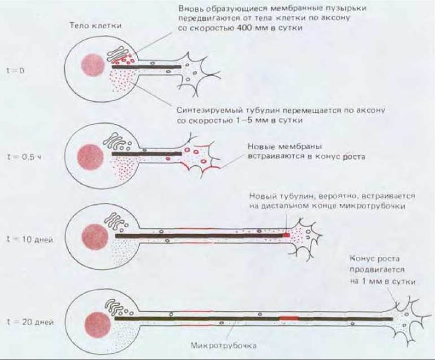
Fig. 19-63. These diagrams illustrate how material required for neurite growth is transported from the cell body to the growth cone, where it is incorporated. For simplicity, only a single microtubule is shown. Microtubules serve as tracks for the fast axonal transport of membrane material. Tubulin is transported from the cell body via slow axonal transport. The precise site where subunits are added to elongate microtubules has not yet been definitively established.
19.7.6. Growth Cone Movement In Vitro Can Be Guided by Selective Adhesion, Chemotaxis, and Electric Fields [52, 54]
"Simplified" cell culture conditions provide an opportunity to investigate mechanisms that might guide growth cone movement in the intact animal. Growth cones, much like neutrophils and fibroblasts, exhibit a preference for surfaces to which they adhere most strongly when selecting a substratum (Fig. 19-64). As they advance, growth cones continuously extend microspikes toward regions ahead and to the sides. Some of these projections may contact a less adhesive substratum and are consequently withdrawn relatively quickly; others encounter a more highly adhesive surface and persist longer. It appears that the microspikes act like tentacles, probing nearby surfaces and steering the growth cone along the path with the strongest adhesive properties.
However, substratum adhesiveness is not the only factor influencing growth cone advance. Surface geometry is also important—for instance, growth cones attached to fibers tend to move along them (a phenomenon known as contact guidance). Substances dissolved in the extracellular fluid also appear to exert a definite influence.
If an embryonic sensory ganglion destined to innervate the jaw, for example, is placed in culture about a millimeter away from a jaw primordium, the neurites will grow preferentially toward that primordium; this suggests that the target tissue secretes molecules that exert a chemotactic effect. An electric field also exerts a strong guiding influence, causing growth cones in Nerve Tissue culture to advance toward the cathode, with a field strength of 7 mV/mm already being effective.
Although such in vitro experiments demonstrate which factors are capable of directing growth cone movement, they do not reveal which specific type of influence plays the primary role in the developing organism.
Is growth cone advance restricted to specific pathways, or are these pathways chosen by trial and error? Some Answers have been obtained by studying growth cone behavior in natural environments.
19.7.7. In Vivo, the Growth Cone Directs Neurite Movement Along a Strictly Defined Pathway ("Pathway Guidance") [55]
Normally, in the living organism, growth cones advance toward their targets along strictly defined pathways. Investigating the mechanisms underlying this process is difficult in most vertebrates; it is somewhat easier to conduct studies in certain invertebrates, such as orthopteran insects, in which the innervation of developing limbs can be traced in detail (Fig. 19-65).
In these insects, the sensory neurons that innervate the limbs arise during embryonic development from specialized cells within the epithelium of the limb buds; the nerve cell bodies remain in the periphery, whereas their axons grow into the central nervous system along precisely defined zigzag pathways. These pathways are pioneered in each limb by one or two pioneer axons, which can be selectively stained using Antibodies or by injecting the fluorescent dye Lucifer Yellow into the cell body. As a result, one can observe that at each turn of the pathway, the growth cones of the pioneers come into contact with specific cells that act as sort of "road signs" (Fig. 19-66). The growth cones form transient Gap Junctions with these cells: if a dye is injected into the axon, these accessory cells also become brightly stained. The microspikes extended by the leading growth cones reach lengths of 50 or even 100 µm, which is sufficient to reach the next signpost cell along the axon's route. Microspikes that make contact with such a cell are stabilized, while the rest are withdrawn. In this manner, the growth cone advances step by step toward the central nervous system. If a signpost cell is destroyed with a laser beam before the growth cone reaches it, the growth cone "gets lost" at that point (Fig. 19-66). Along sections of the pathway where signpost cells are normally absent, the growth cone advances in accordance with the adhesiveness of the basal lamina underlying the limb epithelium (Section 14.2.15). Throughout its journey, the growth cone advances thanks to specific molecules on its surface that enable it to adhere to the appropriate substratum. Some of these adhesion molecules have already been identified.

Fig. 19-64. Substratum Selection by a growth cone can be demonstrated by culturing cells on a dish whose surface is first coated with polyornithine, after which palladium squares are deposited on top. Because cell surfaces are typically negatively charged, cells adhere strongly to polyornithine, which carries a positive charge. Growth cones advance along the polyornithine "tracks" and avoid the palladium; however, if given a choice between palladium and an even less adhesive substratum, they will migrate on the palladium. A. High-magnification phase-contrast micrograph showing growth cones at the boundary between two substrata. B. Lower-magnification view of the same; the pathways chosen by the growth cones are marked by the neurites that trail behind them and remain attached to the polyornithine. (P. Letourneau, Dev. Biol., 44, 92–101, 1975.)
19.7.8. Growth Cones Use Specific Adhesion Molecules to Attach to Cell Surfaces and the Extracellular matrix [56]
Once the initial neurites have paved the way, others follow them by contact: growth cones adhere to preexisting neurites and advance along them. This is a universal tendency observed in both vertebrates and invertebrates. Because strong adhesion exists between neurites and between the neurite and the growth cone, the nerve fibers in an adult animal become bundled into tight parallel fascicles. Large peripheral vertebrate nerves visible to the naked eye are formed in this manner (although individual axons are subsequently insulated from one another by Schwann cell sheaths). Specific integral membrane Glycoproteins that mediate adhesion between neurites have been discovered in vertebrates. Examples include two well-characterized glycoproteins: the so-called neural Cell Adhesion molecule, or N-CAM (Section 14.3.6), and the L1 glycoprotein, also known as the neuron-Glia cell adhesion molecule, or Ng-CAM. Antibodies directed against these proteins—which belong to the immunoglobulin superfamily (Section 14.3.6)—have been shown to suppress the tendency of developing neurites to form bundles and to disrupt normal axon growth (to varying degrees in different regions of the nervous system). N-CAM is present not only on neurons but also on glial cells, as well as on many other cell types, including Muscle cells, during development and regeneration. In the latter case, these molecules may help "attract" motor neuron growth cones to regions where synapses are to be formed.
The movement of growth cones is guided by their adhesion not only to The surface of other cells, but also to various components of the extracellular matrix. An important example is nerve regeneration.
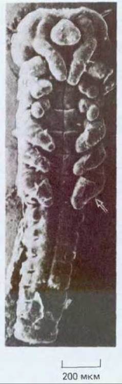
Fig. 19-65. Scanning electron micrograph of an orthopteran embryo (ventral view). Limb buds are visible (indicated by the arrow). (D. Bentley, H. Keshishian, Science, 218, 1082-1088, 1982. Copyright 1982 by the AAAS.)

Fig. 19-66. In the limb of an orthopteran embryo, the initial axons select their growth path with the help of specialized "guidepost" cells. If these cells are destroyed, the axons fail to choose the correct pathway. Later, neurons develop from these guidepost cells, and their axons are directed toward the CNS along the pathways pioneered by the early axons. (After D. Bentley, M. Caudy, Nature, 304, 62-65, 1983.)
When a peripheral nerve is severed, the axons typically regenerate with the help of growth cones formed at the cut ends. These cones advance through tunnels formed by the basal lamina that was previously laid down by Schwann cells enveloping the axons of the distal, degenerated nerve segment. Evidence suggests that a key role in this process is played by Laminin (Section 14.2.15) or a complex of laminin and a heparan sulfate-containing proteoglycan: they bind to receptors located in the growth cone membrane that belong to the integrin family (Section 14.2.17). Laminin and the aforementioned complex accelerate neurite outgrowth in vitro, whereas antibodies against the complex suppress nerve regeneration in vivo.
Although proteins such as N-CAM, Ng-CAM, and laminin appear to play an important role in cell adhesion and in guiding growth cone migration, the properties of these proteins do not provide a clear answer to the central question: why do some growth cones choose one pathway while others prefer another?
19.7.9. The Organization of Neural Connections Is Determined by Differences in Neuronal Properties: The Theory of Neuronal Specificity [57]
In the developing limb of the chick embryo, as in other similar instances, axon growth cones advance along strictly defined pathways (Fig. 19-67). These pathways branch along their course, with different branches leading to different targets. Consequently, at each branch point, the growth cone is faced with a choice among several possible directions. This series of sequential choices is made according to precise rules, resulting in a highly ordered system of connections between neurons and their target cells. Such connections can be traced using the horseradish peroxidase method (Section 19.1.5). It has been shown that the cell bodies of motoneurons innervating the same muscle form a compact group located in the same region of the spinal cord in all individuals, whereas cell bodies innervating different muscles are found in different regions (Fig. 19-68).

Fig. 19-67. Wing buds of an 8-day chick embryo (light micrograph, silver-stained preparation to reveal nerve trajectories). Compare the right wing with the left: the nerves are arranged almost perfectly symmetrically on both sides of the body, indicating the existence of an efficient system guiding nerve growth. Individual growth cones must make a choice at each branch point of the pathway, and this choice is made according to strict rules.

Fig. 19-68. This diagram illustrates how retrograde transport of horseradish peroxidase (HRP) can be used to identify spinal motoneurons innervating a specific muscle. For clarity, the sizes of the neurons shown in cross-section are exaggerated and only three neurons are depicted. In reality, each muscle is supplied by a nerve containing processes from numerous nerve cells (typically several hundred) whose cell bodies are clustered tightly together in the spinal cord.
How is this path selection achieved? Do growth cones arrive at different destinations simply based on their initial position, much like cars on a highway where lane changing is prohibited? To test this hypothesis, a piece of the neural tube was excised from an early embryo and rotated by 180° even before axon outgrowth began. As a result, motoneurons originally destined to innervate muscle A ended up in THE POSITION OF neurons for muscle B, and vice versa. It was found that if the displacement is not too large, the growth cones of the displaced neurons still reach the muscles corresponding to the original position of the neuron in the neural tube, although they are forced to travel along altered pathways to do so (Fig. 19-69). This indicates that neurons destined for different muscles are not equivalent (Section 16.4.6): much like the brain neurons in reeler mice, they differ not only in their location but also in some intrinsic chemical properties. This non-equivalence of neurons is generally referred to as neuronal specificity. As discussed in Chapter 16, Connective Tissue cells in various regions of the limb bud are also non-equivalent, so differences between them could potentially determine the choice of a particular pathway by a given growth cone.
In the central nervous system of both vertebrates and invertebrates, there is also evidence that specific groups of neurons or glial cells bear specific labels that are recognized by other neurons, thereby helping to establish selective neural connections. However, we still know almost nothing about the molecules involved in such processes in either the central or the peripheral nervous system.

Fig. 19-69. The experiment on a chick embryo schematized here demonstrates that motoneurons, even after being displaced, send their axons to the muscles that correspond to the original position of these neurons in the embryonic spinal cord. Note that the axons of motoneurons located at different levels of the spinal cord converge to form a plexus at the base of the limb and then diverge to innervate various muscles. A growth cone passing through the plexus must choose one of many pathways.
19.7.10. Target Tissues Secrete Neurotropic Factors That Regulate the Growth and Survival of Nerve Cells [58]
In the initial part of their journey, the movement of a growth cone is guided by the tissues through which it migrates; as it approaches its destination, the growth cone comes under the Influence of the target—often even before direct contact with it—thanks to the action of neurotropic factors secreted by the target cells. As we have seen in the example of the ganglion innervating the jaw primordium, such factors can act as chemotactic attractants for growth cones. Even more importantly, however, the survival of growth cones, axon branches, and entire neurons depends on them.
Nerve growth factor (NGF) was the first neurotropic factor to be identified and is currently the best understood. It was discovered serendipitously during experiments involving the transplantation of tissues and tumors into chick embryos. Transplants of a certain tumor type were exceptionally densely innervated and induced significant overgrowth of specific groups of peripheral neurons in surrounding areas. Only two categories of neurons were subject to this influence: sensory and sympathetic (a subclass of peripheral autonomic neurons that regulate smooth Muscle contraction and exocrine gland function). Extracts from the tumor also stimulated neurite outgrowth in cultures of these neurons. Further research revealed that a similar stimulating factor is produced in huge quantities by another tissue culture—the Cytology/practical/98.html">Submandibular salivary gland of the adult male mouse. This "trick of nature" remains unexplained, as NGF production by salivary gland cells has no apparent connection to the main function of this factor; nevertheless, it made it possible to obtain pure NGF in quantities sufficient to determine its chemical nature and study its functions. The activity was found to reside in a dimeric protein containing two identical polypeptide chains of 118 amino acid residues each. Once NGF was isolated in pure form, it became possible to produce antibodies that block its action. If anti-NGF antibodies are administered to a mouse whose nervous system development is not yet complete, the majority of sympathetic neurons and some sensory neurons will die.
Similarly, in tissue culture in the absence of NGF, sympathetic neurons and a fraction of sensory neurons die; when NGF is present, they survive and extend neurites (Fig. 19-70). The effects on neuronal survival and neurite outgrowth are two distinct actions of NGF. This can be clearly demonstrated by placing cells in the central compartment of a culture dish separated from two side compartments by a barrier that prevents media mixing but permits neurite outgrowth (Fig. 19-71). When NGF is present in all three compartments, neurites grow into all three. If NGF is absent from one of the side compartments, neurites fail to grow into it. Finally, if NGF is removed from a side compartment where neurites have already grown, they shrink and retract back to the barrier between the compartments. Cells in the central compartment will fail to survive and extend processes unless NGF is present there from the very beginning; however, if NGF is initially added to all compartments and then, after neurites have grown into the side compartments, NGF is removed from the central one, the cells survive and neurite growth in the side compartments continues.
Thus, NGF not only exerts a local effect on distant parts of the cell by maintaining and stimulating the growth of neurites and growth cones, but also serves as a survival factor for The Cell as a whole. Local application to a growth cone produces a direct, rapid effect that does not depend on connection with the cell body; if the NGF-containing medium is replaced with NGF-free medium, growth cones cease movement within 1-2 minutes. In addition to an immediate response to NGF, growth cones of NGF-sensitive cells take it up via endocytosis, and the NGF is subsequently transported in vesicles via retrograde axonal transport to the neuronal cell body, where this factor (or an intracellular second messenger) apparently prevents cell death.

Fig. 19-70. A. Sympathetic ganglion cultured for 48 h with NGF (top) and without it (bottom) (dark-field micrograph). Neurites sprout from sympathetic neurons only in the presence of NGF. Each culture also contains Schwann cells that have migrated out of the ganglion, but NGF has no effect on these cells. B. Behavior of sensory neurons grown for 24 h in medium with NGF (left), in control medium without NGF (middle), and in medium containing Skeletal Muscle extract (right) (phase-contrast micrographs). The sensory neurons in the top row innervate primarily the body surface (though they also send a small number of processes to skeletal muscles and other targets). Most of these cells respond to NGF in the same way as the sympathetic neurons shown in photo A. The sensory neurons in the bottom row normally innervate skeletal muscles (providing sensory feedback). These neurons are insensitive to NGF but respond strongly to the Addition of an extract prepared from skeletal muscle. (A - courtesy of Naomi Kleitman; B - from A.M. Davies. Dev. Biol., 115; 56-67, 1986.)
19.7.11. Cell Death Regulates the Number of Surviving Neurons to Match The amount of Target Tissue [59]
In vertebrates, spinal sensory ganglia are formed in a segmental pattern that corresponds to the series of vertebrae along the spine. Each ganglion consists of a cluster of sensory neurons derived from the neural crest, which send one neurite outward toward the periphery of the body and another inward toward the spinal cord. Initially, the primordia of all ganglia are similar in size, but in the adult animal, the ganglia of body segments associated with limbs contain significantly more neurons than those of other segments (Fig. 19-72). This difference is primarily due to cell death: a substantial proportion of neurons in many ganglia undergo apoptosis. If a limb bud is removed at an early stage of development, the adjacent ganglia decrease in size to match the others; conversely, if an extra, additional limb bud is transplanted into the thoracic region of an embryo, it becomes innervated, and an unusually large number of neurons is preserved in the ganglion at that level. It is believed that the survival of ganglion neurons, proportional to the amount of target tissue, is largely mediated by NGF secreted by this tissue. If an additional amount of NGF is introduced into the embryo at a specific developmental stage, many of the neurons that would normally have died are saved, just as neurons are preserved in the region of an amputated limb bud.
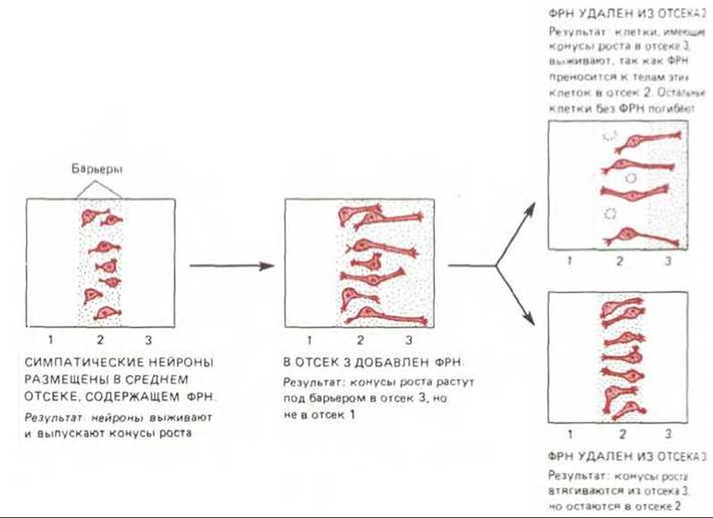
Fig. 19-71. Scheme of a tissue culture experiment demonstrating that sympathetic neurons not only require NGF for their survival, but also direct their growth cones exclusively toward and maintain neurites only in regions where NGF is present. Note that as long as a cell has neurites reaching regions containing NGF, the presence of NGF in the region of the cell body is not required for cell survival.
It might seem excessively wasteful to generate an excess of neurons and then partially destroy them to regulate their numbers. Yet this strategy is widely utilized in vertebrate development for both sensory and motor neurons, in both the central and peripheral nervous systems. For example, about 50% of all motor neurons innervating skeletal muscles die during embryonic development within a few days after establishing contact with their target muscles. Evidently, neuron survival in these systems is regulated by various trophic factors, such as NGF, released by target cells. This strategy offers several important advantages. First, this mechanism automatically corrects for variations in the relative sizes of different body parts. Second, these processes facilitate evolution: if a mutation alters the size of a body part, the number of neurons innervating it will automatically adapt without requiring additional Mutations to rewrite the neurogenic program. Finally, a small number of neurotropic factors, such as NGF, can regulate the quantitative match between A large number of targets and their sources of innervation, even when the wiring diagram is highly complex. Through axonal transport, a factor secreted by a given target is selectively delivered to the cell bodies of the neurons innervating that target, rather than to other neurons that may be located nearby and share similar receptors, but project their axons elsewhere. Thus, cell death regulated by neurotropic growth factors helps establish a precise numerical match between cells in different regions of the nervous system.

Fig. 19-72. Regulation of nerve cell survival in sensory spinal ganglia of a chick embryo. Ganglia and nerves of an 8- to 9-day-old embryo are shown. The size of each ganglion corresponds to the number of surviving neurons, which in turn depends on the amount of tissue innervated by that ganglion. (After V. Hamburger, J. Exp. Zool. 68: 449-494, 1934; J. Exp. Zool. 80: 347-389, 1939.)
19.7.12. Neural connections are continuously formed and dismantled throughout life [60]
Evidence indicates that even in normal, undamaged Nervous Tissue, dendrites and axon terminals are continuously retracted and regrown. For instance, certain neurons in a mature sympathetic ganglion of a mouse can be observed retracting some dendritic branches and sprouting new ones over the course of a month (Fig. 19-73). Under normal conditions, such changes occur slowly and to a limited extent, but if a subset of target cells is deprived of innervation, this mechanism shifts into high gear. This can be demonstrated in skeletal muscle by cutting some of the axons that innervate it; the denervated muscle fibers apparently release a diffusible factor that stimulates the formation of new growth cones from nerve terminals preserved on neighboring muscle cells (Fig. 19-74). This skeletal muscle factor has not yet been identified, but in smooth muscle, NGF has been shown to play a similar role. Denervation leads to an increase in the amount of NGF in smooth muscle (at least partly because fewer nerve terminals are now present to take it up and clear it); this excess NGF stimulates axon growth toward the muscle, thereby restoring innervation.
Clearly, in the intact organism, NGF acts in much the same way as it does in vitro—that is, as a survival factor determining whether cells live or die, and as a local stimulant of growth cone activity that regulates the branching of axonal terminals. The first function is particularly crucial during development, while the second remains important throughout life; however, both lead to the same outcome: adapting innervation to the demands of the target. Accumulating evidence points to the existence of other neurotropic growth factors that perform similar functions for other classes of nerve cells (see Fig. 19-70). In the next section, we will see that such factors likely play a vital role in mediating METABOLISM/18.html">The Influence of electrical activity on The Development of neural circuits.

Fig. 19-73. Dendritic remodeling in neurons of the superior cervical (sympathetic) ganglion of a mouse. In the anesthetized animal, the ganglion was carefully exposed, and a fluorescent dye was microinjected into the cell body of a single nerve cell to reveal its dendrites. The wound was sutured, and after a few days or weeks, the surgery was repeated and the dye was injected into the exact same neuron. The top and bottom panels show two neurons re-stained at different time intervals. The longer the interval between the First and Second injections, the greater the Changes in the pattern of dendritic branching. (Reprinted by permission from D. Purves, R. D. Hadley, Nature, 315: 404-406, 1985. Copyright 1985 Macmillan Journals Limited.)
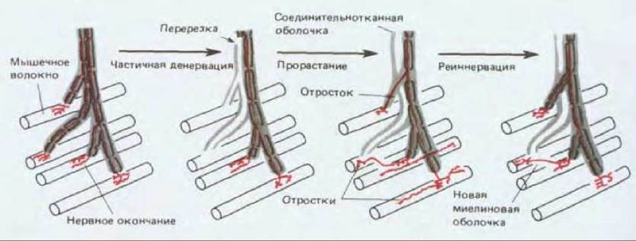
Fig. 19-74. When a fraction of the axons innervating a skeletal muscle is severed, some muscle fibers lose their innervation while neighboring fibers retain theirs. The cut axons degenerate, whereas the intact axons begin to sprout vigorously near the denervated fibers. Within one to two months, those new branches that find their way to vacant synaptic sites on the denervated muscle fiber form stable synapses and restore innervation, while all other branches atrophy. This phenomenon suggests that denervated muscle fibers secrete a "sprouting-promoting factor." (Reproduced by permission from M. C. Brown, R. L. Holland, W. G. Hopkins, Annu. Rev. Neurosci., 4: 17-42, 1981. Copyright 1981 by Annual Reviews Inc.)
The Development of the nervous system is conveniently divided into three overlapping stages. In The First stage, neurons are generated according to an intrinsic program of cell proliferation, and the newly formed cells migrate from their birthplace to take up organized positions in other regions. In the second stage, the cells extend axons and dendrites, whose tips advance guided by growth cones. These growth cones navigate along highly stereotyped pathways, guided primarily by contact interactions with the surfaces of other cells or with extracellular matrix components. Neurons destined to connect with different targets behave as if they possess distinct, intrinsically determined properties (neuronal specificity), which may manifest as unique cell-surface characteristics that enable growth cones to choose distinct pathways. At the end of its journey, a growth cone encounters the target cell with which it is to form a synapse and comes under the influence of neurotropic factors secreted by that target. These factors regulate axonal branching and growth cone motility in the vicinity of the target tissue, and additionally control the survival of the neurons from which the growth cones originated. Through these two effects, neurotropic factors such as nerve growth factor (NGF) regulate the density of target tissue innervation. In the Third Stage of nervous system development, which will be discussed in the next section, synapses are formed, after which the wiring pattern is refined through mechanisms dependent on electrical activity.
Last update: 12/08/2026
Editorial and Educational Adaptation: This material has been compiled based on the primary/original source text. The project team performed an editorial review, corrected technical inaccuracies, structured sections, and adapted the content for an educational format.
What was processed:
- elimination of formatting defects (OCR errors, structural breaks, corrupted characters);
- editorial organization of content;
- standardization of terminology in accordance with academic sources;
- verification of factual statements against the original source text.
All mentions of the author, publication year, and origin of the primary text have been preserved in accordance with the source.