Human Anatomy and Physiology - N. I. Fedyukovich 2003
Nervous system
Central nervous system
Spinal cord
The Spinal Cord (medulla spinalis) performs two main Functions: reflex and conductive (Fig. 100).
Class="center">
Fig. 100. The spinal cord (diagram):
A: 1 — spinal cord; 2 — cervical enlargement; 3 — lumbosacral enlargement; 4 — conus medullaris; 5 — filum terminale; B: 1 — terminal ventricle; 2 — filum terminale
As a reflex center, the spinal cord is capable of executing complex motor and autonomic Reflexes. Through afferent (sensory) pathways, the spinal cord is connected to receptors, and through efferent pathways, to skeletal Muscles and all Internal Organs. Long descending and ascending Pathways of the spinal cord connect peripheral PARTS OF THE body to the Brain.
In appearance, the spinal cord is an elongated, slightly flattened cylindrical cord. It is located within the vertebral canal and, at the level of the lower margin of the foramen magnum, becomes continuous with the brain.
The lower boundary of the spinal cord corresponds to the level of the I—II lumbar vertebrae. Below this level, it continues as a thin terminal thread (filum terminale).
In an adult, the length of the spinal cord averages about 43 cm (45 cm in men, 41—42 cm in women), and its mass is about 34—38 g. Like THE Vertebral Column, the spinal cord has cervical and thoracic curvatures, as well as cervical and lumbosacral enlargements. Due to the metameric Structure OF THE human body, it is divided into segments, or neuromeres (Fig. 101). A segment is a section of the spinal cord that corresponds to a pair of Spinal Nerves.

Fig. 101. Segments of the spinal cord:
1 — cervical segments (1—8), cervical part; 2 — thoracic segments (1—12), thoracic part; 3 — lumbar segments (1—5), lumbar part; 4 — sacral segments (1—5), sacral part; 5 — coccygeal segments (1—3), coccygeal part
Throughout its length, 31 pairs of anterior and posterior roots emerge from each side of the spinal cord, joining to form 31 pairs of right and left spinal nerves. Each segment of the spinal cord corresponds to a specific area of the body innervated by the spinal nerve of that particular segment. There are 31 segments of the spinal cord: 8 cervical, 12 thoracic, 5 lumbar, 5 sacral, and 1 coccygeal. They are designated by the initial letters of their Latin names, indicating the part of the spinal cord, and Roman numerals corresponding to the segment number: cervical segments (СI — СVIII); thoracic (ThI — ThXIII); lumbar (LI— LV); sacral (SI — SV); coccygeal (СоI — СоV).
Along the entire anterior surface of the spinal cord, in the median sagittal plane, runs the anterior median fissure, and along the posterior surface runs the posterior median sulcus, which divide the spinal cord into two symmetrical halves. On its anterior surface are two anterolateral sulci, from which the anterior roots emerge, and on the posterior surface are the posterolateral sulci, which serve as the entry points for the posterior roots on both sides. The spinal cord consists of white and Gray matter (Fig. 102).

Fig. 102. Spinal cord (cross-section diagram):
1 — central canal; 2 — gray matter; 3 — White matter; 4 — anterior funiculus; 5 — lateral funiculus; 6 — posterior funiculus
The gray matter contains Nerve Cells and, in cross-section, resembles the letter H. Within the gray matter is the central canal, the upper end of which connects with the IV ventricle, while the lower end ends blindly in the terminal ventricle. Throughout the spinal cord, the gray matter forms two vertical columns located on either side of the central canal. Within each column, anterior and posterior columns are distinguished (Fig. 103).

Fig. 103. Columns of the gray matter of the spinal cord:
1 — posterior; 2 — lateral; 3 — anterior
At the level of the lower cervical, all thoracic, and the upper two lumbar segments of the spinal cord, a lateral column is distinguished in the gray matter, which is absent in other parts of the spinal cord. The gray matter of the posterior horns has a heterogeneous structure. The bulk of the nerve Cells of the posterior horn forms the substantia gelatinosa and The Nucleus proprius, and at the Base of the posterior horn, well-demarcated by a layer of white matter, is the thoracic nucleus, which consists of large nerve cells.
The cells of all nuclei of the posterior horns of the gray matter are, as a rule, interneurons (intermediate Neurons), whose processes enter the WHITE MATTER OF the spinal cord and proceed to the brain. The intermediate zone, located between the anterior and posterior horns, is represented laterally by the lateral horn. The latter contains the centers of the Sympathetic division of the Autonomic Nervous system.
The white matter lies external to the gray matter. The sulci of the spinal cord divide the white matter into three symmetrically arranged funiculi on the left and right: anterior, lateral, and posterior. The white matter is composed of the processes of nerve cells. The collection of these processes in the funiculi of the spinal cord forms three systems of tracts (pathways): 1) short bundles of association fibers that connect spinal cord segments at different levels; 2) ascending (sensory, afferent) tracts running to the brain centers or the Cerebellum; 3) descending (motor, efferent) tracts passing from the brain to the cells of the anterior horns of the spinal cord. The white matter of the posterior funiculi contains ascending pathways, while the anterior and lateral funiculi contain both ascending and descending fiber systems.
The anterior funiculus includes the following tracts (Fig. 104): 1) the anterior corticospinal (pyramidal) tract. This tract transmits motor response impulses from the Cerebral Cortex to the anterior horns of the spinal cord; 2) the anterior spinothalamic tract, which conducts tactile sensitivity impulses; 3) the vestibulospinal tract, which originates from the vestibular nuclei of the VIII pair of Cranial Nerves located in the Medulla Oblongata. Impulses that maintain balance and coordinate movement travel along the fibers of this tract.

Fig. 104. Pathways of the white matter in a transverse section of the spinal cord (diagram):
1 — gracile fasciculus; 2 — cuneate fasciculus; 3 — posterior ROOT; 4 — lateral corticospinal (pyramidal) tract; 5 — rubrospinal tract; 6 — posterior spinocerebellar tract; 7 — anterior spinocerebellar tract; 8 — lateral spinothalamic tract; 9 — olivospinal tract; 10 — vestibulospinal tract; 11 — reticulospinal tract; 12 — anterior corticospinal (pyramidal) tract; 13 — anterior spinothalamic tract; 14 — tectospinal tract; 15 — posterior, lateral, and anterior proper fasciculi; 16 — anterior horn; 17 — lateral horn; 18 — posterior horn
The lateral funiculus of the spinal cord contains the following pathways: 1) posterior spinocerebellar — carries proprioceptive impulses to the cerebellum; 2) anterior spinocerebellar — travels to the cerebellar cortex; 3) lateral spinothalamic — conducts pain and Temperature Sensation impulses; 4) lateral corticospinal (pyramidal) — conducts motor impulses from the cerebral cortex to the spinal cord; 5) rubrospinal — conducts impulses for automatic (subconscious) motor control and maintains Skeletal Muscle tone.
The posterior funiculus contains pathways of conscious Proprioception (conscious joint-muscle sense) that travel to the brain and the cortical end of the motor analyzer, transmitting information about the state of the body and its parts in space. At the level of the cervical and upper thoracic segments of the spinal cord, the posterior funiculi are divided by the intermediate sulcus into two bundles — the gracile fasciculus of Goll and the cuneate fasciculus of Burdach.
The spinal cord is surrounded by three Meninges: the dura mater, arachnoid mater, and pia mater (Fig. 105).
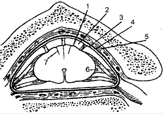
Fig. 105. Meninges of the spinal cord:
1 — pia mater of the spinal cord; 2 — subarachnoid space; 3 — arachnoid mater of the spinal cord; 4 — dura mater of the spinal cord; 5 — epidural space; 6 — denticulate ligament; 7 — intermediate cervical septum
The dura mater of the spinal cord is an elongated sac with thick, strong walls located within the vertebral canal, enclosing the spinal cord along with its roots and other meninges. The outer surface of the dura mater is separated from the periosteum lining the vertebral canal by the epidural space, which is filled with adipose tissue. The inner surface of the spinal dura mater is separated from the arachnoid mater by a narrow, slit-like subdural space traversed by numerous thin Connective Tissue septa.
Superiorly, the subdural space communicates with the corresponding space in the cranial cavity, and inferiorly, it ends blindly at the level of the second sacral vertebra.
The arachnoid mater of the spinal cord is a thin membrane located internal to the dura mater. It fuses with the latter in the region of the intervertebral foramina.
The pia mater of the spinal cord closely adheres to and fuses with the spinal cord. The arachnoid mater is separated from the pia mater by the subarachnoid space, which is filled with CEREBROSPINAL FLUID, totaling about 120–140 ml. In its lower regions, the subarachnoid space contains only the roots of the spinal nerves surrounded by fluid. In this area, below the level of the second lumbar vertebra, a lumbar puncture can be performed if necessary without the risk of damaging the spinal cord.
Last update: 08/08/2026
Editorial and Educational Adaptation: This material has been compiled based on the primary/original source text. The project team performed an editorial review, corrected technical inaccuracies, structured sections, and adapted the content for an educational format.
What was processed:
- elimination of formatting defects (OCR errors, structural breaks, corrupted characters);
- editorial organization of content;
- standardization of terminology in accordance with academic sources;
- verification of factual statements against the original source text.
All mentions of the author, publication year, and origin of the primary text have been preserved in accordance with the source.