Plant Physiology - Musienko M. M. 2001
Respiration
Catalytic Respiratory Systems
Oxidoreductases
The oxidizing apparatus of plants features specific characteristics. First, unlike that of animals, it is characterized by delocalization of the respiratory apparatus (Cell/35.html">Mitochondria, Peroxisomes, Cytoplasm, etc.). Second, it exhibits polyfunctionality, meaning the presence of catalysts with multiple properties. Finally, the plant oxidizing apparatus operates on the principle that the Organism contains not just one, but several Enzymes catalyzing the same or similar reactions. As is well known, Biological Oxidation of any substance involves the loss of electrons or electrons together with protons. Three types of oxidative reactions occur in plant Cells.
I. Reactions Involving the direct loss of an electron As a result of A change in its valence. Such reactions are catalyzed by oxidase enzymes, which contain metals in their active centers:
Class="center">
II. Through the abstraction of hydrogen ions from the oxidized substance by means of hydroxylase enzymes. Oxygen itself often acts as the proton acceptor in such reactions:

III. Direct oxidation by molecular oxygen with the participation of oxygenases or transferases:

In biological systems, all these pathways are interconnected, and the loss of an electron during oxidation is generally accompanied by the loss of hydrogen as well. For this reason, The oxidation of a particular substance in living systems is coupled with the reduction of another.
To determine the direction of electron flow, METABOLISM/2.html">THE CONCEPT OF the standard reduction potential (Eo) has been introduced. The reduction potential of the following reaction is conventionally taken as zero potential:

A measure of the electron-donating and electron-accepting capacity of a substance is the value of the redox potential expressed in volts (Table 9). All biological oxida-
Table 9. Standard redox potentials of biochemical systems
|
System |
E0’ (pH 7.0), V |
System |
E0’ (pH 7.0), V |
|
Oxygen/Water |
0.81 |
FMN/FMN H2 |
-0.12 |
|
Cytochrome a (Fe +3/Fe+2) |
0.29 |
Oxaloacetate/malate |
-0.17 |
|
Cytochrome c (Fe +3/Fe+2) |
0.22 |
Riboflavin (oxid./red.) |
-0.21 |
|
Cytochrome b (Fe +3/Fe+2) |
0.12 |
NAD+/NADH+H + |
-0.32 |
|
Ubiquinone (oxid./red.) |
0.10 |
Pyruvate/malate |
-0.33 |
|
Dehydroascorbate/ascorbate |
0.08 |
H/1/2H2 |
-0.42 |
|
Fumarate/succinate |
0.03 |
Ferredoxin (oxid./red.) |
-0.43 |
|
Succinate/ α-ketoglutarate |
-0.67 |
tive-reductive systems can be arranged in a specific series According to the magnitude of their redox potential.
Knowledge of the standard redox potentials of various biological systems allows us to predict the direction of electron flow from one redox system to another. Naturally, electron transfer proceeds from a system with a more negative redox potential to a system with a more positive potential. Energy must be expended to transfer electrons in the opposite direction.
Systems with a higher electronegative potential are characterized by a larger energy reserve. The reduction of a substance is coupled with a change in THE POSITION OF its energy levels. During reduction, along with the acquisition of an electron, there is an increase in the system's energy. Completely oxidized compounds are characterized by a minimal Free energy content. The amount of free energy that can be stored or released is directly proportional to the difference in redox potentials of the two systems participating in electron transfer.
The more negative the value of the reduction potential, the greater the capacity of a given substance to donate electrons (to be oxidized), i.e., to act as a reducing agent. Conversely, the more positive the reduction potential of a certain substance, the higher its capacity to accept electrons (to be reduced, i.e., to act as an oxidizing agent).
The universal oxidizing agent is O2, which has the maximum positive potential (+0.817 V), while various substances serve as Donors. All electrons are transported to O2 via a multi-component redox system. These listed features underlie the functioning of all groups of enzymes (without exception) that catalyze aerobic Respiration processes (Fig. 85). Since the oxidation of the electron and proton donor is coupled with the reduction of their acceptor, the enzymes catalyzing these reactions are called oxidoreductases. Oxidases are capable of transferring electrons exclusively to oxygen.
Dehydrogenases. The oxidation of organic substances in plant cells is carried out with the participation of dehydrogenases and oxidases. The Role of dehydrogenases is the activation and transfer of hydrogen (electrons) from respiratory substrates. According to their mode of action, they are divided into aerobic and anaerobic. Aerobic dehydrogenases transfer hydrogen directly to O2, whereas anaerobic ones transfer it to a specific acceptor, which is often another dehydrogenase rather than O2.

NAD is present in plant mitochondria in large quantities; for example, compared to cytochrome c, it is nearly 40 times more abundant.
Anaerobic dehydrogenases are Proteins, two-component enzymes whose Coenzymes are NAD+ or NADP+. Depending on the protein carrier to which the NAD or NADP coenzyme attaches, more than 150 pyridine dehydrogenases are distinguished. The substrate Specificity of the enzyme is determined by its protein component. The action of these enzymes is based on the ability of the pyridine ring, which is part of these dehydrogenases' coenzymes in the form of nicotinic acid amide, to accept and donate hydrogen.
Such a dehydrogenase, upon accepting or donating hydrogen or electrons, is capable of redox transformations. Oxidized dehydrogenase has an absorption maximum in the region of 260 nm, whereas the reduced form is characterized by the appearance of a second maximum at 340 nm; the Redox Potential of NADH is (-0.32 V).
Anaerobic dehydrogenase catalyzes The transfer of a hydrogen atom together with its bound electrons in the form of a hydride ion H- from the substrate molecule (AH2) to the positively charged fourth carbon atom of NAD(P)+, thereby forming NAD(P)H; the electron pair of H+ becomes the bonding pair of the newly formed covalent bond C4 → H. The substrate that has lost H from one of its atoms becomes positively charged AH+, which stabilizes by releasing H+ into the surrounding aqueous medium:
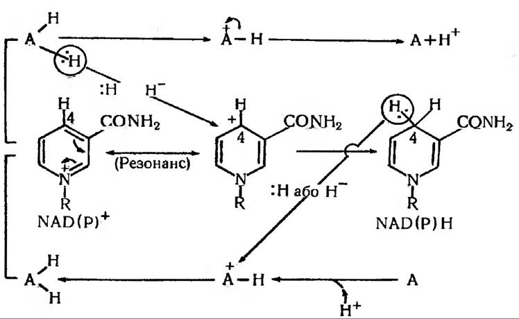
Thus, NAD(P)+ and NAD(P)×H+ differ by the presence or absence of a single hydride ion, which is equivalent to two electrons and one H+ proton. This readily explains why NAD and NADP are classified as hydrogen carriers and two-electron redox compounds. It should be kept in mind that NAD and NADP can accept hydrogen and electrons only when the substrate has a more negative potential value compared to them.
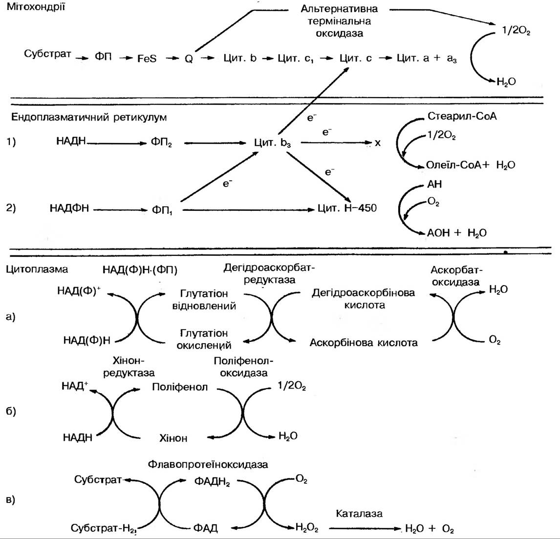
Fig. 85. Redox systems of plant cells
Pyridine dehydrogenases are widely distributed in both PLANT AND ANIMAL organisms.
Many NAD- and NADP-dependent dehydrogenases require the presence of divalent Metal Ions. For instance, the enzyme Alcohol dehydrogenase contains zinc ions. The oxidized and reduced forms of these coenzymes are capable of interconversion mediated by the enzyme NADP transhydrogenase.
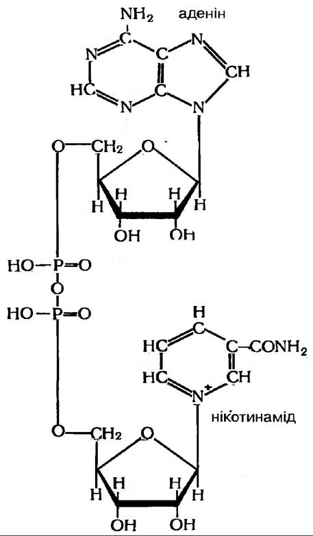
Despite their structural similarity, NAD and NADP dehydrogenases cannot substitute for one another: NAD dehydrogenases include malate, lactate, and ethanol dehydrogenases, whereas NADP dehydrogenases include glucose-6-phosphate dehydrogenase, isocitrate dehydrogenase, etc. It is worth noting the existence of a limited group of dehydrogenases that remain active even in the absence of coenzymes.
Aerobic dehydrogenases are also two-component enzymes, often referred to as flavin dehydrogenases (Flavoproteins). Their functional role is to intermediately transport protons—abstracted from the oxidative substrate or the reduced form of an anaerobic dehydrogenase—to O2 or to oxidase systems, such as polyphenol oxidase or cytochrome systems.
Their prosthetic group is a derivative of vitamin B2 (riboflavin), namely flavin adenine dinucleotide (FAD) and flavin mononucleotide (FMN). The Active Site of flavin dehydrogenases is the isoalloxazine ring, to which 2H (2H++2e-) attach:
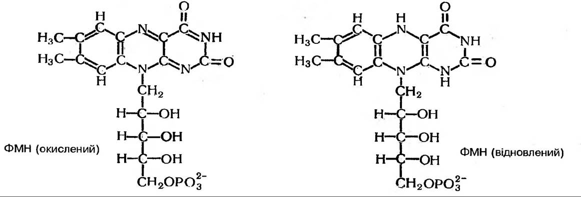
The prosthetic groups of flavin dehydrogenases are tightly bound to the protein carrier.
Almost all flavoproteins (FPs) contain metals (Fe, Cu, Mo, Mn). An example of a FAD-containing dehydrogenase is succinate dehydrogenase. Flavoproteins transfer hydrogen from NAD×H2 (occasionally from NADP×H2) to coenzyme Q, with FMN and FAD acting as intermediate acceptors that take up both protons and both electrons:
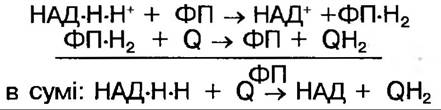
In their reduced form, FMN×H2 and FAD×H2 are colorless.
Some substrates are oxidized directly by flavoprotein enzymes, bypassing pyridine NUCLEOTIDES, as is the case with succinic acid. E’o for FMN and FAD is (-0.21 V). Depending on the bonding with other apoenzymes, it can vary (from +0.19 to -0.12 V) and sometimes even reaches (-0.34 V). Electron donors for aerobic dehydrogenases are anaerobic dehydrogenases, whereas Quinones, Cytochromes, and oxygen serve as acceptors.
Consequently, The primary function of dehydrogenases is to transfer the hydrogen or electron activated by them from a given compound to another acceptor possessing a higher oxidation potential.
Along with the direct dehydrogenation of substrate molecules, additional preliminary phosphorylation and Hydration of the oxidized compounds are frequently observed. Quite often, many dehydrogenases are linked together via intermediate Components of the Electron Transport Chain (ETC).
Oxidases. These are a group of enzymes that activate oxygen; in other words, they are aerobic dehydrogenases capable of transporting electrons from an oxidative substrate exclusively to O2. This process yields H2O, H2O2, or the superoxide oxygen anion. In the first case, 4e- are transferred to O2; In the second, 2e-; and in the third, 1e-. Examples of the first group of oxidases include cytochrome oxidase and polyphenol oxidase; of the second group, flavoprotein oxidases (such as amino acid oxidases); and of the third, Enzymes of the xanthine oxidase type. There are three mechanisms for introducing molecular oxygen into the oxidative process in a plant cell:
I — reduction of oxygen by monovalent or divalent donors. The enzymes catalyzing such reactions are oxidases. Their active centers typically contain iron or copper, meaning they possess a heme or flavin group.
II — reduction of molecular oxygen by divalent donors, coupled with the incorporation of the resulting reduction product into the oxidized molecule. Enzymes catalyzing these reactions belong to the group of mixed-function oxidases and are termed hydroxylases.
III — direct incorporation of oxygen into the molecule of the compound being oxidized. These enzymes are called transferases or oxygenases. It should be recalled that all the aforementioned mechanisms of oxygen incorporation into oxidative processes were postulated by the Bach-Palladin peroxide theory.
Thus, the group of oxygen-activating enzymes is quite extensive. However, the primary role within this group is played by iron-containing enzymes. These are two-component systems whose prosthetic group consists of iron-Porphyrins.
Cytochromes
In 1925, D. Keilin discovered a group of compounds in plant cells that came to be known as cytochromes. In their composition, they belong to iron-containing hemin derivatives. The iron-porphyrin group (heme) in cytochromes is tightly bound to the protein via the sulfur atoms of The amino acid Cysteine. (Incidentally, cytochromes in animal cells were first discovered by MacMunn as early as 1886.) D. Keilin believed that cytochromes transfer electrons from substrate molecules to O2. In cytochromes, iron can readily transition between the ferrous (divalent) and ferric (trivalent) states and vice versa. Cytochromes are strongly colored and, consequently, characterized by a distinct absorption spectrum. There are at least 4 classes of cytochromes—a, b, c, d—which differ from one another in The Nature of their prosthetic group; for instance, cytochromes a contain iron-formylporphyrins, cytochromes b contain iron-protoporphyrins, and d contain iron-hydroporphyrins (Fig. 86).
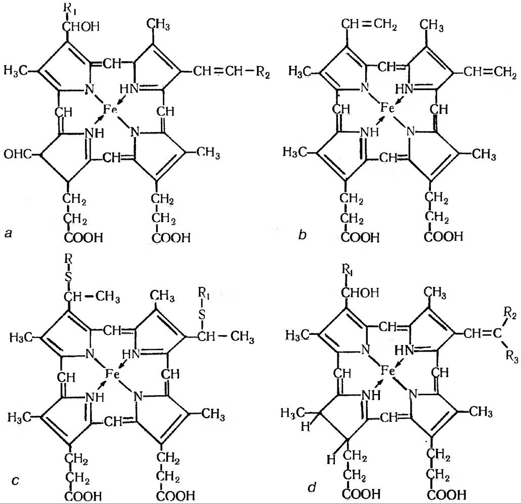
Fig. 86. Structure of cytochrome porphyrins a, b, c, and d
Cytochromes (up to 20 are known) have a relatively broad redox potential ranging from 0 to 0.6 V. The value of the redox potential depends on The structure of the prosthetic group, the apoprotein structure, and the mode of attachment of the apoenzyme to the prosthetic group. The structure of cytochrome c from animal organisms has been studied in the greatest detail. In plant cells, Two Types of cytochrome c have been found in mitochondria: c547 and c549 with an E0 of +0.235 V. Data regarding group b cytochromes in plant mitochondria are rather contradictory. Until the mid-1980s, it was believed that plant mitochondria contained three b-type cytochromes: b557 (E0 +0.075 V), b560 (E0 +0.045 V), and b566 (E0 +0.070 V). Subsequently, data were obtained indicating the presence of two additional b cytochromes in mitochondria, with absorption maxima around 558 nm.
Reduced cytochromes are oxidized through the action of the enzyme cytochrome oxidase (which is cytochrome a3, acting as the final link before O2). The reduction of oxidized cytochromes in living cells occurs via electrons transferred by dehydrogenases. The sequence of Electron transfer from one cytochrome to another is as follows:

Catalase and Peroxidase
The iron-porphyrin group also includes catalase and peroxidase, whose action is directed toward The conversion of hydrogen peroxide. Catalase decomposes it into water and molecular oxygen:

whereas peroxidase decomposes hydrogen peroxide with the release of active atomic oxygen:

Along with their ability to drive oxidation using peroxide oxygen, purified plant peroxidase preparations can function as specific oxidases. An increase in peroxidase activity has also been demonstrated in plant Tissues subjected to adverse environmental conditions. This prompts us to re-evaluate the Biological Role of this enzyme.
Copper-Proteins
Oxidative enzymes containing copper within their prosthetic group have also been found in plant tissues; these are the so-called copper-proteins, for example, polyphenol oxidase and ascorbate oxidase:
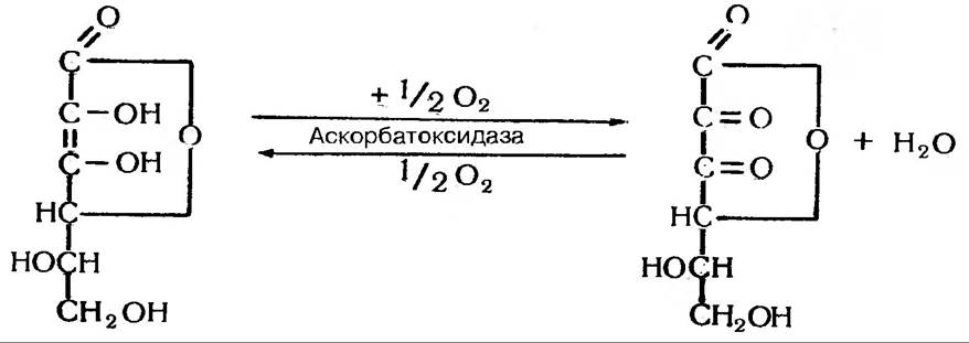
These are a group of enzymes that, in the presence of molecular oxygen, oxidize various phenols to form quinones; in other words, they catalyze the transfer of protons and electrons from phenols (hydroquinone, pyrocatechol, pyrogallol, etc.) to molecular oxygen.
Polyphenols are essentially the respiratory chromogens whose existence was predicted by V.I. Palladin. (Incidentally, mechanical tissue damage—such as cutting apples or potatoes—disrupts the coordination between the Oxidation and reduction phases, resulting in the accumulation of persistent colored pigments, causing the pulp to darken).
It belongs to the strictly specific phenol oxidases and primarily oxidizes ascorbic acid and certain compounds possessing a dual phenolic group. Plastocyanin is also among the copper-protein enzymes.
Oxygenases
In addition to oxidases that utilize O2 as an electron acceptor, plant cells also harbor oxygenases capable of activating O2 so that it can attach to Organic compounds. In this activated state, it directly binds to organic compounds. Simultaneously, either two atoms (Dioxygenases) or one atom of oxygen (hydroxylases) can be added. NADPH×H, FADH2, and Other Compounds serve as electron donors for them. They drive the hydroxylation of Amino Acids, phenols, sterols, and other endogenous compounds, as well as the detoxification of xenobiotics (compounds foreign to organisms, such as pesticides). Hydroxylases function according to the scheme:

where AH is an organic compound and DH2 is a hydrogen donor.
Dioxygenases can incorporate oxygen into A wide variety of organic compounds, such as:
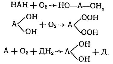
Thus, oxygenases are enzymes that activate oxygen with its subsequent incorporation into the substrate molecule. Unlike other groups of oxidative enzymes, oxygenases catalyze the direct interaction of oxygen atoms not with hydrogen, but with the carbon atoms of aromatic rings. Flavins and other oxidases, on the other hand, reduce oxygen molecules to form peroxides. The existence of mixed-function oxidases serves as yet another compelling proof of alternative oxygen activation pathways.
It should also be noted that recently, increasing weight has been given to the fact that the direct incorporation of oxygen into organic molecules is possible. This is one of the most widespread potential pathways for incorporating substances such as phenols, Lysine, lactates, and others into general metabolism.
Enzymes as intermediate hydrogen (electron) carriers. This is a rather large group of enzymes whose chemical nature classifies them as flavin derivatives. The specificity of this enzyme group lies in its ability to transfer electrons directly to oxygen. Functionally, this brings them close to oxidases. Their prosthetic group is FMN (flavin mononucleotide) or FAD (flavin adenine dinucleotide). Some flavoproteins accept hydrogen from the same substrate that undergoes oxidation (e.g., specific succinate dehydrogenase), whereas others accept it solely from reduced NAD.
The primary function of flavoproteins is the Oxidation of reduced pyridines (NAD and NAD×F dehydrogenases) and the transfer of electrons within the ETC toward oxygen. Potential electron acceptors include Other dehydrogenases, oxidized cytochrome, or ultimately O2 itself. Thus, flavoproteins are also capable of transferring electrons to oxygen, meaning they should essentially be considered oxidases as well. After all, their activity results in The formation of hydrogen peroxide, the oxygen of which is subsequently utilized by peroxidase (glucose oxidase is a prime example of such an enzyme). This is precisely why flavin enzymes and peroxidases constitute a coupled system.
There is still no consensus regarding which of the flavoproteins identified in the mitochondria of higher plants are involved in Electron Transport and what their exact localization is within The electron transport chain. Ferredoxins have also been discovered in both plant and animal organisms.
Some of them interact directly with flavin dehydrogenases, while others interact with anaerobic NAD dehydrogenase.
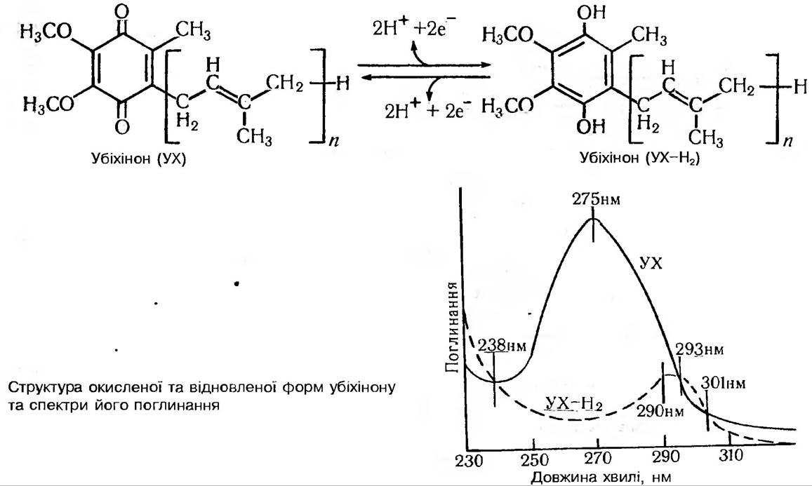
Coenzyme Q, or ubiquinone, is another intermediate hydrogen (electron) carrier.
This term refers to a group of compounds sharing the same quinone core but differing in the number of isoprenoid residues within the lateral lipophilic branch of the carbon chain. Ubiquinone is reduced to ubiquinol by simultaneously accepting two electrons and two protons. Consequently, ubiquinone Functions as a "two-electron redox compound" and an H2 "carrier." Ubiquinone is a relatively small molecule compared to the protein components of the terminal ETC segment. Furthermore, being a lipid, it is highly soluble and can therefore move freely within the lipid matrix of the mitochondrial membrane. Its concentration in the membrane is relatively high compared to cytochromes (5:1). These properties likely explain its role as an ETC electron collector, where electrons converge from various donors to be subsequently supplied to the cytochrome chain as needed. In addition to these enzymes, iron-sulfur proteins also participate in electron transport. They contain an Fe-S complex whose iron undergoes reversible oxidation and reduction.
Accessory enzymes
The enzyme systems involved in respiration also include A number of accessory enzymes, such as carboxylases. They catalyze the Cleavage of CO2 from various organic acids (keto acids, amino acids), resulting in the formation of compounds with shortened carbon chains. For example, the decarboxylation of pyruvate in this manner yields acetaldehyde, among other products.
Mention should also be made of another group of enzyme systems that do not directly participate in substrate oxidation or electron transport, but play an active role in preparing respiratory substrates. Recall that the oxidative capacity of dehydrogenase enzymes is restricted to compounds with relatively small, simple molecular structures (organic acids, alcohols, sugars). However, respiration utilizes quite complex organic polymers, proteins, and fats. Therefore, their utilization is preceded by structural modifications facilitated by hydrolytic complex enzymes. Next come enzymes whose activity aims to activate monomer molecules by converting them into phosphate esters. Further activation is achieved by altering the molecular chemical structure with the help of isomerases. We should also mention the group of transferases, which catalyze the cleavage and transfer of specific carbon chain fragments (transaldolases and transketolases).
The electron and hydrogen carriers of the ETC are organized into functional complexes within The inner mitochondrial membrane and, consequently, are in very close contact with the components of The Lipid Bilayer. Although they do not directly participate in electron transfer, lipids provide an optimal environment for ETC components, thereby significantly influencing its function. This Conclusion is supported by the observation that isolating Phospholipids from mitochondria drastically reduces electron transport activity, particularly at the cytochrome oxidase level. Adding extracted lipids back externally restores activity to its initial level. Moreover, the Lipid Composition of the inner mitochondrial membrane differs markedly from that of the outer membrane; for instance, diphosphatidylglycerol is localized exclusively in the inner membrane.
The pathway of electron and hydrogen transfer from one carrier molecule to another is a redox process. The driving force behind this transfer is a potential difference. Accordingly, the arrangement of individual ETC carriers, much like in Photosynthesis, is determined by the magnitude of their redox potential. At the beginning of the chain (Fig. 87) lies NAD, which possesses the most negative potential (-0.32 V), while at the end is oxygen with the most positive potential (+0.82 V).

Fig. 87. Sequence of electron transport in the Respiratory Chain
Enzymes of resistant respiration
Plant mitochondria differ from animal mitochondria in that they can utilize two distinct pathways for electron transport from substrates such as NADH and succinate to O2. If one of these pathways is inhibited by cyanides (KCN) that block cytochrome oxidase, The alternative pathway remains unblocked; hence, it is called the cyanide-resistant electron transport chain. Similarly, the oxidative process leading to the formation of this terminal chain is referred to as cyanide-resistant respiration, or cyanide-insensitive respiration.
It must be emphasized that these two chains are not entirely independent. In both cases, electron pairs enter the chain via internal and external NADH dehydrogenases and succinate dehydrogenase, after which they are transferred to ubiquinone. Only downstream of ubiquinone do their respective pathways diverge. In the normal chain, electrons are successively transferred via FP, b- and c-type cytochromes, and cytochrome oxidase to O2, whereas in the cyanide-resistant chain, electrons are transferred from ubiquinone to FP and then, via a cyanide-resistant terminal oxidase, also to O2.
The exact Nature of the terminal oxidase remains unelucidated. It is believed to be an iron-sulfur protein that is not a hemoprotein. The physiological significance of cyanide-resistant respiration is not fully understood. It is thought to be responsible for the Climacteric in fruits—that is, the respiration surge during pre-ripening. Evidence suggests that the climacteric is induced by Ethylene, leading to the view that ethylene acts as a stimulator of cyanide-resistant respiration. Furthermore, this type of respiration has been shown to function during the early Selection/3.html">Stages of development in certain seedlings, notably during seed imbibition.
Last update: 07/08/2026
Editorial and Educational Adaptation: This material has been compiled based on the primary/original source text. The project team performed an editorial review, corrected technical inaccuracies, structured sections, and adapted the content for an educational format.
What was processed:
- elimination of formatting defects (OCR errors, structural breaks, corrupted characters);
- editorial organization of content;
- standardization of terminology in accordance with academic sources;
- verification of factual statements against the original source text.
All mentions of the author, publication year, and origin of the primary text have been preserved in accordance with the source.